GI and Endocrine Parasites BSA (copy)
 Call Kai
 Learn
 Practice Test
 Spaced Repetition
 Match
 Flashcards
 Knowt Play
1/159

Earn XP
Description and Tags
160 Terms
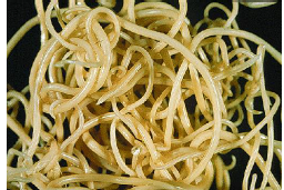
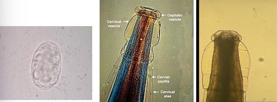
Toxocara canis

Toxocara canis
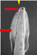
Toxocara canis

Male, finger-like projection
Toxocara canis
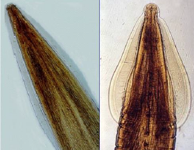
Identify parasites left and right
Left: Toxocara canis
Right: Toxocara cati
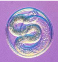
Toxocara canis

Dark brown egg, subglobular, thick rough pitted shell (golf ball)
Toxocara canis (egg)

Slightly ovoid, smooth thick colorless shell
Toxocara leonina (egg)
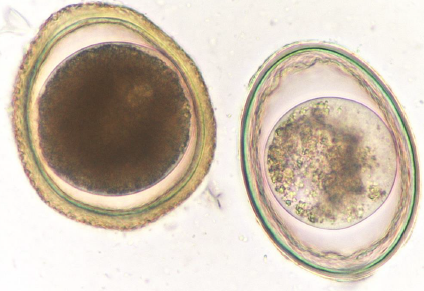
Identify parasites left and right
Left: Toxocara canis
Right: toxocara leonina

Ancylostoma

Ancylostoma

Ancylostoma
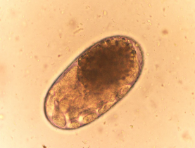
Eggs “strongyle type”, thin smooth shell
Ancylostoma caninum / Uncincaria
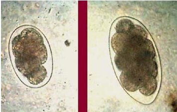
Identify parasites left and right
Left: Ancylostoma
Right: Uncinaria

Trichuris vulpis

Trichuris vulpis

Trichuris vulpis
Thick broad posterior end, long filamentous anterior end, embedded in mucosa (“Whipworms”)

Trichuris vulpis (egg)
Trichuris that infects pigs?
Trichuris suis
Trichuris that infects humans?
Trichuris trichiura
Trichuris that infects sheep, goats, cattle?
Trichuris ovis
Trichuris that infects camels, sheep, and goats?
Trichuris globulosa
Trichuris that infects canine?
Trichuris vulpis

Double pored/cucumber seed tapeworm
Dipylidium caninum

Eggs in egg capsule, (red arrow), egg sac contains up to 30 eggs
Dipylidium caninum (egg)

Thick broad posterior end, long filamentous anterior end, 2 sets genital organs + pore on each margin (red arrow).
Dipylidium caninum
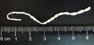
Proglottid: rice of grain or cucumber seed
Dipylidium caninum
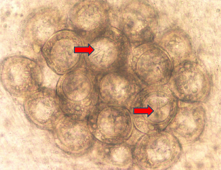
Arrows show hooklets of the hexacanth embryo
Dipylidium caninum

Dipylidium caninum

Taenia pisiformis

Cannot distinguish this species, all look the same
Taenia ova (egg)

Taenia pisiformis

Resistant stage for environmental transmission
Giardia spp (cysts)
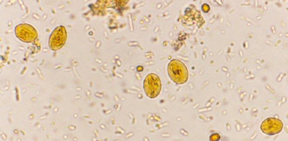
Giardia spp (cysts)
Giardia cysts, intestine, feces or environment, direct smear, with iodine, flotation
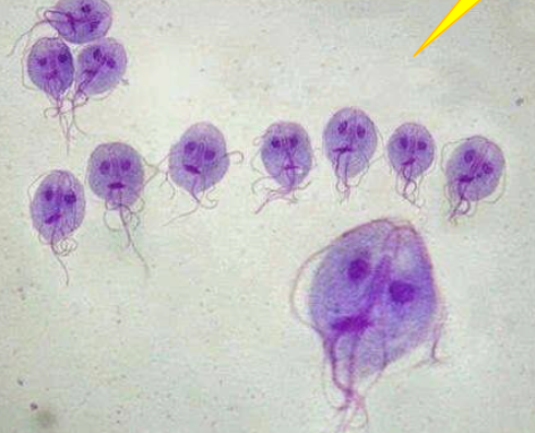
Motile stage in small intestine. Dorsal side convex, ventral side has a sucking disk. 2 anterior nuclei, 2 axostyles, 8 flagellae in 4 pairs
Giardia spp. (trophozoites)
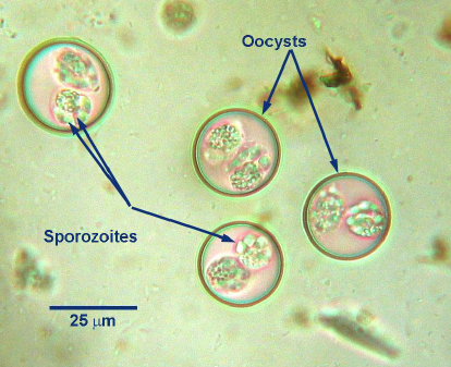
Sporulated (infective) oocysts in the environment
Cystoisospora spp.

Cystoisospora spp.
What does Cystoisospora spp. cause in animals?
Coccidiosis
Cystoisospora spp that infects cats?
C. neorivolta and C. felis
Cystoisospora spp that infects dogs?
C. canis and C. ohioensis
Cystoisospora spp that infects pigs?
C. suis
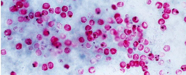
Cryptosporidium spp.

Are ovoid or spheroid, measure 5um and stain red with Acid fast stain
Cryptosporidium oocytes

Spirocerca lupi (Esophagus)
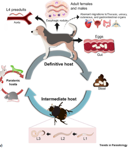
Definitive Canine host (Gray Arrow)
Intermediate Dung Beetle host (Blue Arrow)
Paratenic host (Red Arrow)
Life Cycle of Spirocerca lupi

Physaloptera rara

Chronic vomiting, especially in dogs and cats that consume insects or vertebrate prey. Affects stomach.
Physaloptera rara

Parasite of cats, occasionally dogs
Ollulanus tricuspis

Platinosum fastosum (fluke, cats)

Platinosum fastosum (fluke, cats)

Fasciola hepatica (liver fluke)
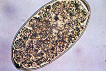
Fasciola hepatica (liver fluke)

Fasciola hepatica (liver fluke)
Common clinical signs of Fasciola hepatica (liver fluke)?
Anemia and submandibular edema

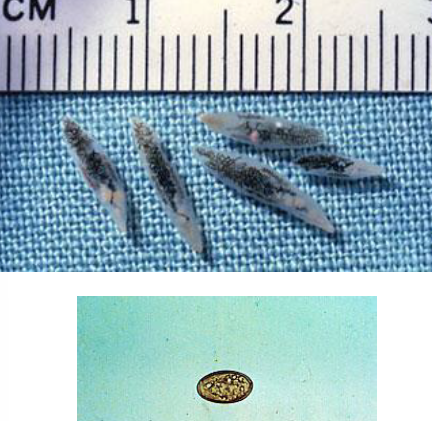
Dicrocoelium dendriticum

Dicrocoelium dendriticum

Lesions in the in the abomasum
Ostertagia ostertagi
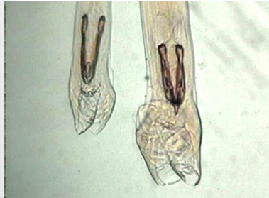
Male spicules
Ostertagia ostertagi
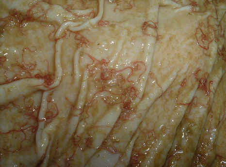
Barber’s pole worm
Haemonchus contortus
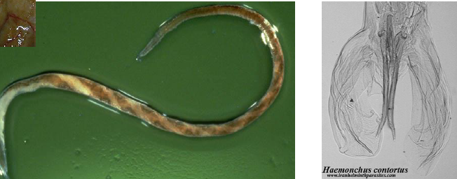
Haemonchus contortus
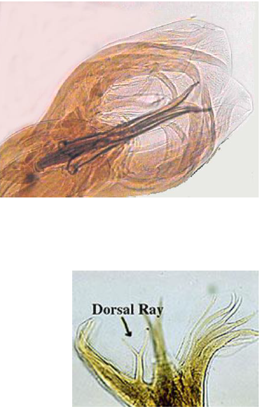
Bursa of males, dorsal ray, Y shaped
Haemonchus contortus

Haemonchus contortus (larvae)

Spiral of white and red along the body: the red is the blood-filled digestive tract, and the white is the egg-filled reproductive tract
Haemonchus contortus (adult female)

Vulva is protected by a flap of the cuticle
Haemonchus contortus

What do the arrows indicate for the Haemonchus and Ostertagia?
Dorsal ray. Shows the Bursa of males

HOTC (Haemonchus, Ostertagia, Trichostrongylus, Cooperia)
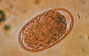
Ostertagia “strongyle-type” egg

Nematodirus

Nematodirus

L3 larva ingested while grazing
Trichostrongylus axei, stomach, horse, ruminants (T of HOTC)

Trichostrongylus axei, stomach, horse, ruminants (T of HOTC)

HOTC, strongyle type egg

Trichostrongylus axei, stomach, horse, ruminants (T of HOTC)

Identify top and bottom parasites
Moniezia expansa: Sheep (triangle)
Moniezia benedeni: Cattle (square)

Moniezia expansa / benedeni

Moniezia expansa / benedeni
Name two specific Eimeria that affect cattle
Eimeria bovis and Eimeria zuernii
What causes coccidiosis in cattle?
Eimeria spp.

Eimeria bovis and Eimeria zuernii
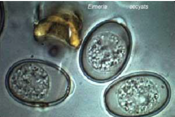
Eimeria unsporulated oocysts

Eimeria sporulated oocyst
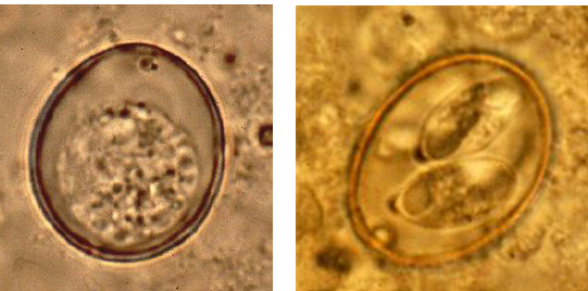
What is the difference between the left and right Eimeria?
Left: unsporulated
Right: sporulated oocyst

Eimeria life cycle

What are the stages of Eimeria in this picture?
Free sporozoite. Released from the ingested sporulated oocyst in the chicken's digestive tract.
Intracellular trophozoite. A sporozoite that has invaded a cecal epithelial cell.
Schizont (meront). The parasite undergoes multiple nuclear divisions (asexual schizogony / merogony).
Mature schizont with merozoites. Fully formed first-generation merozoites inside the host cell.
Released merozoites. Merozoites rupture the host cell to infect new epithelial cells (and undergo a second asexual generation, shown at 8, or differentiate into sexual stages).
Young macrogametocyte. A merozoite developing into a female sexual cell inside an epithelial cell.
Mature macrogamete. Contains prominent peripheral wall-forming bodies.
Second-generation schizont / merozoite. Represents additional asexual multiplication within host cells.
Microgametocyte. A male sexual cell undergoing multiple nuclear divisions.
Microgametes. Biflagellated, motile male gametes released from the microgametocyte.
Fertilization (Blue Arrow). A microgamete fuses with a mature macrogamete.
Zygote. Wall-forming bodies fuse to create a protective oocyst wall around the fertilized cell.
Unsporulated oocyst. Passed out of the chicken in the feces into the external environment.
Sporoblast stage. Inside the oocyst, the diploid sporont divides into four sporoblasts during exogenous sporogony.
Sporulated oocyst. The infective stage containing four sporocysts, each holding two sporozoites (eight sporozoites total).

Migrating larvae cause pneumonia and “milk spots” or “white spots” in liver
Ascaris suum

Identify parasite and intestine does it affect?
Ascaris suum (egg)
Small intestine
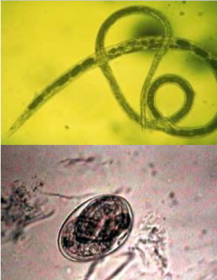
Identify parasite and which intestine does it affect?
Strongyloides ransomi
Small intestine

Strongyloides ransomi
Identify parasite and what intestine does it affect
Oesophagostomum spp
Large intestine
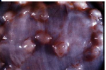
Thickening of large intestinal wall, catarrhal enteritis, nodule formation
Oesophagostomum spp

Oesophagostomum spp lifecycle
Oesophagostomum that infects cattle?
O. radiatum and venulosum
Oesophagostomum that infects sheep and goat?
O. columbianum and and venulosum
Oesophagostomum that infects pigs?
O. dentatum

See unsegmented pedicles, transversal grooves, trinagular spines
Sarcoptes scabiei

Notoedres cati

Has dorsal anus, no triangular spines
Notoedres cati
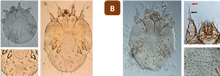
Which is Sarcoptes and Notoedres?
Left: Notoedres cati
Right: Sarcoptes scabiei

Ovoid body, 8 pairs of legs
Hint: copulatory suckers and pair of legs longer
Otodectes cynotis (male)

Ovoid body, 8 pairs of legs
Hint: genital aperture and pair of legs shorter
Otodectes cynotis (female)