OBS/GYNAE IMAGE REC
 Call Kai
 Learn
 Practice Test
 Spaced Repetition
 Match
 Flashcards
 Knowt Play
1/3

There's no tags or description
Looks like no tags are added yet.
4 Terms
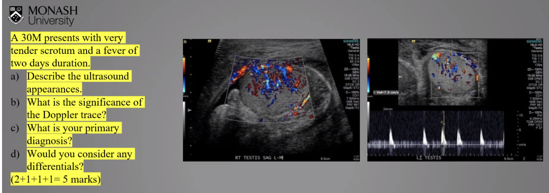
Describe the image
Ultrasound of the scrotum demonstrates increased vascularity (hypervascularity) of the right testis, consistent with orchitis. The epididymis appears enlarged, raising the possibility of associated epididymo-orchitis. The right scrotal wall appears thickened and oedematous, with a small right-sided hydrocele containing possible low-level internal echoes. Doppler assessment demonstrates increased vascularity within the right testicular parenchyma. Spectral Doppler demonstrates a high-resistance waveform with a sharp systolic upstroke and absent end-diastolic flow, which may reflect increased intratesticular resistance secondary to inflammation and oedema. The Doppler findings do not suggest active testicular torsion; however, intermittent or recently resolved torsion (torsion-detorsion) cannot be entirely excluded on ultrasound alone.

Describe the image
Ultrasound of the scrotum demonstrates marked scrotal wall thickening and oedematous changes, with multiple punctate echogenic foci demonstrating posterior dirty shadowing, highly suggestive of gas within the scrotal wall. A moderate hydrocele is also present, with anechoic fluid surrounding the testis. The testicular parenchyma appears homogeneous. The appearances are highly suspicious for Fournier's gangrene, given the presence of subcutaneous gas within the thickened, oedematous scrotal wall. Differential considerations include other gas-forming bacterial infections or surgical emphysema. Urgent clinical correlation is recommended.
Key point: The most important distinguishing feature of Fournier's gangrene on ultrasound is echogenic foci with posterior dirty shadowing within the thickened scrotal wall, representing gas. This is a highly characteristic finding and should raise strong suspicion for a necrotising soft tissue infection.

Describe the image
Transvaginal ultrasound demonstrates an enlarged, anteverted uterus measuring approximately 189 cc in volume, with heterogeneous myometrial echotexture and a characteristic 'Venetian blind' appearance, raising the possibility of underlying adenomyosis. A 40 × 39 mm solid, isoechoic mass is identified arising from the anterior fundal aspect of the uterus, with appearances most consistent with a subserosal fibroid with a broad base. It is difficult to determine whether the fibroid is pedunculated on the available images. The fibroid may exert mass effect on adjacent pelvic structures. Overall appearances are suggestive of uterine fibroid, with possible coexisting adenomyosis.

Describe the image
Axial ultrasound image of the orbits demonstrates a cystic structure located between the inferomedial aspect of the orbit and the adjacent nasal region, with appearances consistent with a dacryocystocele (lacrimal duct cyst). The appearance is typically benign and often resolves spontaneously. Both orbits should be assessed for symmetry and comparison. The presence of both ocular lenses should also be confirmed, as the lens is not clearly visualised on the provided image.