Physiology - Test 3
 Call Kai
 Learn
 Practice Test
 Spaced Repetition
 Match
 Flashcards
 Knowt Play
1/174

There's no tags or description
Looks like no tags are added yet.
175 Terms
Somatic motor
1 nueron
originates in CNS
always excitatory
neuromuscular junction
b/w motor nueron and skeletal muscle fiber
lots of ACh - nicotinic receptor
skeletal muscles
striated
multinucleated
voluntary control
cardiac muscle
striated
uninucleated
involuntary
smooth muscle
internal organs
influences movement of material into and out of body
Tendons
attach bone to muscle
made of collagen
origin of muscle
proximal end of limb
insertion of muscle
distal end of limb
fascicle
muscle fibers bound together
sarcolemma
muscle cell membrane
sarcoplasm
muscle cell cytoplasm
Myofibril
bundles of elastic protiens that carry out contraction
myosin
forms thick filaments - enzyme for ATP
actin
forms thin filaments
tropomyosin
protein polymer - binding site for actin
troponin
calcium binding - moves tropomyosin
cross bridges
mysoin heads of thick filaments bind to actin
sarcomere
pattern of thick/thin filaments - 1 repeat
contractile unit of myofibril
contraction
myosin and actin sliding over each other
sarcoplasmic reticulum
wraps around each fiber
concentrates and stores calcium
transverese tubules
extension of sarcolemma
dips into sarcoplasm
brings action potential closer to center of fiber
terminal cistrae
enlarged end region
triad
1 t-tubule + 2 flanking terminal cisternae
muscle tension
force created by contracting muscle
load
force opposing muscle contraction
contraction
creatoon of tension in a msucle
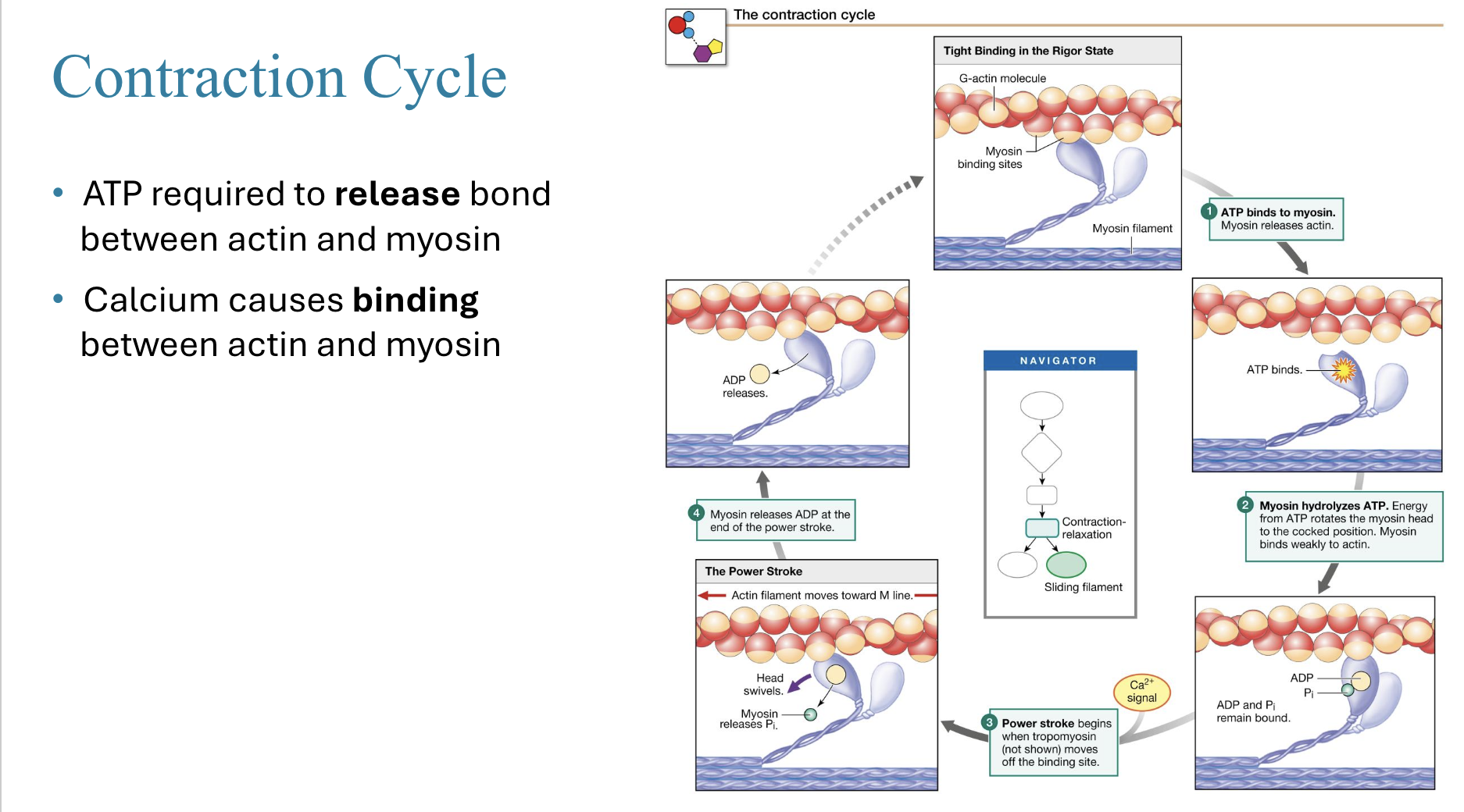
power stroke
calcium signal initiates
myosin cross bridges push actin filaments towards center of sarcomere
1 power stroke per contraction
steps for power stroke (5)
calcium concentration increases in cytosol
calcium binds to troponin
troponin pulls tropomyosin away from actin binding site (previously covered)
myosin binds to actin and performs power stroke
actin filament moves
relaxion
calcium concentration decreases - slow process (why summation is common)
Draw Excitation-contraction coupling and relaxation

Draw contraction cycle
Muscle twitch
single contraction-relaxation cycle
3 phases for muscle twitch
latent period
period of contraction
period of relaxation
latent period
events of excitation - contraction coupling (no muscle tension)
short delay b/w Action Potential and tension - caused by time required for calcium release and binding of troponin
period of contraction
cross bridges form - tension increases
period of relaxation
calcium goes back into SE - tension goes to zero
temporal summation
frequency of stimulation
more calcium releases before calcium used in first twitch can go back into SE
produces smooth and continuous contractions
size principal
number and type of motor neurons recruited
motor units with smallest muscle fibers recruited first (most excitable)
the largest motor units only recruited for very powerful contractions (least excitable)
phosphocreatine
backup energy source for muscles (creatine + ATP) - muscle cells have lots of creatine kinase
anaerobic glycolysis
glucose metabolized to lactate (2 ATP per glucose)
Unfused tetanus
maximum contraction and remains there
caused by increase in stimulus frequency
motor unit
1 neuron and its muscle fibers (all same fiber type) - 1 action potential effects all fibers
recruitment
involving other motor units to increase strength of contraction
asynchronously activated to prevent fatigue
low threshold neurons
control fatigue-resistant, slow-twitch muscle fibers
Heart
Pumps blood
capillaries
microscopic vessels where blood and interstitial fluid exchange materials
blood
cells and plasma
cardiovascular system
transports materials through body
blood vessels
hollow tubes transporting blood throughout the body
substances moved by cardiovascular system (3)
nutrients/water/gases that come from external environment
materials moving from cell to cell in body
wastes that cells eliminate
arteries
carry blood away from heart
(usually oxygenated)
Veins
bring blood to heart
(usually deoxygenated)
pulmonary circulation
takes blood to and from lungs
systemic circulation
takes blood to and from body
Atrium
upper chamber
receives blood entering heart
ventrical
lower heart chamber
pumps blood out of heart
septum
divides heart into left and right halves
keeps oxygenated and deoxygenated blood from mixing
right side job
receives blood from tissues
sends to lungs for oxygenation
left side job
receives oxygenated blood from lungs
pumps blood from heart to tissues
pulmonary arteries
pumps deoxygenated blood from right ventricle to lungs
pulmonary veins
pumps oxygenated blood from lungs to left atrium
aorta
transports oxygenated blood from left ventricle to tissues
superior vena cava
transport deoxygenated blood from veins from upper part of body to right atrium
inferior vena cava
transport deoxygenated blood from veins from lower part of body to right atrium
parallel circuits
blood entering and exiting both ventricles simultaneously
aortic sinus
pericardium
membranous sac around heart
myocardium
cardiac muscle
atrioventricular valves
2 sets
move blood between aorta and ventricles
closed during contraction
prevents blood from flowing back into atria
2 types of Atrioventricular valves
tricuspind (right AV valve)
mitral (Left AV valve)
semilunar valves
move blood from ventricles to arteries
prevent back-flow into ventricles
Chordae Tendinea
anchor the AV valve to prevent it from flipping inside out (cause of “broken heart syndrome”
ventricular contraction
semilunar valves - open
ventricles - contracted
chordae tendinae - tense
AV valves - closed
ventricular relaxation
semilunar valves - closed
ventricels - dilated
chordae teninae - relaxed
AV valves - open
anastomosis
alternative routes blood can take to the same capillary bed
diastole
when heart is relaxed
systole
when heart is contracting
coronary circulation
blood vessels feeding cardiac muscles encircling heart near base
blood circulation route (6)
right atrium
right ventricle
lungs
left atrium
left ventricle
body tissue
Autorhythmic cells
send signals for myocardial contractions (do not contract themselves)
self-generate action potentials
1% of cardiac cells
make up the sinoatrial and atrioventricular nodes
contractile cells
perform physical contractions of the heart
cardiac myocytes
contractile cells
striated fibers organize into sarcomeres
contractions cause blood to pump through heart
Skelatal muscle differences from Cardiac muscle (9)
Skeletal muscle:
multinucleated
fibers as long as whole muscle
3 subtypes (skeletal, smooth, cardiac)
fast glycolytic (aerobic mostly)
can perform summation
Action potentials between 1-2 ms
use neuromuscular junctions
muscle fibers are at optimal length for contraction
voluntary - only contract using somatic NS
Cardiac Muscle differences from skeletal muscles (9)
Cardiac Muscle:
only 1-2 nuclei
shorter, branched muscle fibers
2 sub types (contractile + autorhthmic)
only aerobic respiration (very mitochondria heavy)
Action potentials every 200-300ms
cannot use summation
use intercalated disks instead of neuromuscular junctions
fiber length shorter than optimal length
autonomic NS modifies rate of contractions
intercalated disks
cell junctions between cardiac myocytes
contain desmosomes and gap junctions
desmosomes
physically tie adjacent myocytes together
allows force to be transferred from cell to cell
gap junctions
allow electrical connection between myocytes
allow depolarization - contraction happen almost simultaneously through myocytes
autorhythmic cardiac fibers
make up SA and VA nodes
pacemaker potential
unstable membrane potential of autorhythmic cells
potential eventually depolarizes to threshold causing action potential
due to If cannels
If channel
causes pacemaker potential
inflow of Na+
(na+ flow higher than k+)
gradually close as cell depolarizes - different Ca+ channels open
actions of autorhythmic cell at threshold
different Ca++ channels open - steep depolarization
close at peak of action potential
K+ have opened and releases K+
Actions of autorhthumc cell at peak
Ca++ channels close
K+ channels open - leaves cell
pacemaker potential returns to lowest point
Actions of autorhthmic cells during repolarization
K+ open and K+ leaves cell

draw and label EC coupling in contractile cardiac muscle

steps of action potential in contractile cardiac cell
Na+ channels open
Na+ channels close
Ca++ channels open & fast K+ channels close
Ca++ channels close & slow K+ channels open
resting membrane potential
cardiac cell refractory period
same duration as action potential
summation not possible
plateau
there is a plateu in the repolarization period of cardiac contractile cells
caused by 2 things:
decreased permeability to K+
increased permeability to Ca++
helps slow action potentials, prevent summation and prevent tetanus
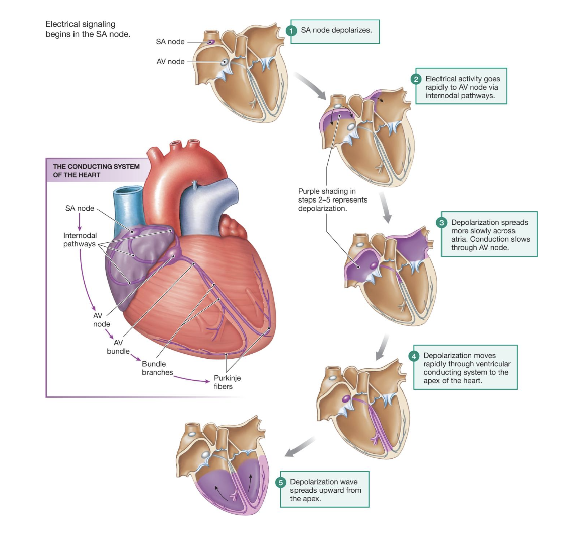
Conducting system of heart steps (5)
SA node depolarizes
electrical activity rapidly moves to AV node
depolarization slowly spreads across atria
depolarization rapidly spreads across ventricles to apex
depolarization wave spreads upward from apex
bradychardia
heart rate below 60 BPM
tachycardia
heart rate over 100 BPM