CSN BIO 224 Lab 1-4 Review for Lab Practical #1 (Fall 2019)
 Call Kai
 Learn
Learn Practice Test
 Spaced Repetition
 Match
Match Flashcards
 Knowt Play
1/331

There's no tags or description
Looks like no tags are added yet.
Name | Mastery | Learn | Test | Matching | Spaced | Call with Kai | Chat |
|---|
No analytics yet
Send a link to your students to track their progress
332 Terms
Right Atrium
What chamber is this?

Left Atrium
Which chamber is this?

Right Ventricle
Which chamber is this?

Left Ventricle
Which chamber is this?
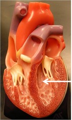
Epicardium
Which layer of the heart wall is this?

Myocardium
Which layer of the heart wall is this?

Endocardium
Which layer of the heart wall is this?

Aorta
Which great vessel is this?

Aortic Arch
Which great vessel is this?

Superior Vena Cava
Which great vessel is this?

Inferior Vena Cava
Which great vessel is this?

Pulmonary Trunk
Which great vessel is this?

Pulmonary Arteries
Which great vessel is this?

Pulmonary Veins
Which great vessel is this?

Aortic Semilunar Valve
Which valve is this?

Pulmonary Semilunar Valve
Which valve is this?

Bicuspid Valve (Mitral Valve) (Left Atrioventricular Valve)
Which valve is this?

Tricuspid Valve (Right Atrioventricular Valve)
Which valve is this?

Ligamentum Arteriosum
What is this structure?

Fossa Ovalis
What is this structure?

Right Auricle
What is this small, wrinkled protruding pouch-like structure?

Left Auricle
What is this small, wrinkled protruding pouch-like structure?

Interatrial Septum
What is this structure?

Interventricular Septum
What is this structure?

Trabeculae Carneae (Trabeculae=Latin for beam, Carneae=Latin for Flesh, feminism singular)
What are these structures? (Large muscular ridges and columns in the interior walls of the ventricles)

Papillary Muscles
What are these structures?

Chordae Tendineae
What are these structures?

Apex of Heart
What is this structure?

Opening of Coronary Sinus
What is this structure?

Coronary Sulcus (atrioventricular groove)
What is this structure? (The groove between the atria and ventricles in which the coronary arteries sit)

Anterior Interventricular Sulcus
What is this structure? (The groove between the left and right ventricles, below the arteries and veins)

Posterior Interventricular Sulcus
What is this structure? (The groove between the left and right ventricles, below the arteries and veins)

Right Coronary Artery
Name what the red arrows are pointing at?

Right Marginal Branch Artery
Name what the red arrows are pointing at?

Posterior Interventricular Artery
Name what the red arrows are pointing at?

Left Coronary Artery
Name what the red arrow is pointing at?

Anterior Interventricular Artery
Name what the red arrows are pointing at?

Circumflex Artery
Name what the red arrow is pointing at?

Small Cardiac Vein
Name what the red arrows are pointing at? (This vein loops around to the posterior side, to the right of the coronary sinus. DON'T FORGET!)

Middle Cardiac Vein
Name what the red arrows are pointing at?

Great Cardiac Vein
Name what the red arrows are pointing at?

Coronary Sinus
Name what the red arrow is pointing at?

(Sheep Heart) Aorta
What is #3?

(Sheep Heart) Superior Vena Cava

(Sheep Heart) Inferior Vena Cava

(Sheep Heart) Pulmonary Arteries
What are #2 and 6?
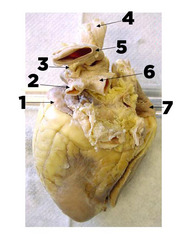
(Sheep Heart) Pulmonary Veins (from the left and right lung should be 4 openings into the left atrium)(this image is actually a real human heart could not find a good sheep heart pulmonary veins)
Name the Openings in the highlighted area

(Sheep Heart) Endocardium
Yellow arrow

(Sheep Heart) Myocardium
Name the layer indicated by the red arrow.

(Sheep Heart) Epicardium

(Sheep Heart) Interatrial Septum

(Sheep Heart) Interventricular Septum

(Sheep Heart) Bicuspid Valve

(Sheep Heart) Tricuspid Valve
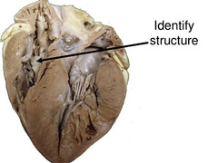
(Sheep Heart) Chordae Tendineae

(Sheep Heart) Pulmonary Semilunar Valve

(Sheep Heart) Aortic Semilunar Valve

(Sheep Heart) Opening of Coronary Sinus

(Sheep Heart) Right Atrium

(Sheep Heart) Left Atrium
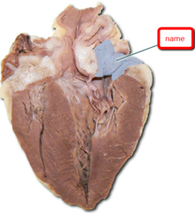
(Sheep Heart) Right Ventricle

(Sheep Heart) Left Ventricle
What is B?

The closing of the Atrioventricular Valves
While listening to the heart, you can hear the sound lubb-dupp. What causes the first sound LUBB?
(The) closing (of the) Semilunar Valves.
While listening to the heart, you can hear the sound lubb-dupp. what causes the second sound DUPP?
Atrial Depolarization
What is P?

Ventricular Depolarization
What is the QRS Complex?

Ventricular Repolarization
What is T?

The hidden event on the ECG.
The repolarization of the atria. It occurs at the same time as the QRS complex so it doesn't show on the ECG.
Red electrode
Negative - goes on right wrist
Black electrode
Positive - goes on left wrist
Green electrode
Ground - on the right wrist 2 inches above the red electrode
Any vessel that carries blood away from the heart.
Artery
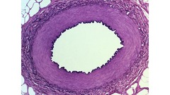
Blood moves slowest in which vessels? Why?
Capillaries. They are small, single-cell capacity and have lower pressure
Agglutinogens
Genetically determined antigens found on the surface of every individual's erythrocytes (RBC)
Agglutinins
Antibodies that will bind with antigens (clumping) in the presence of agglutinogens.
Blood moves fastest in which vessels? Why?
Arteries. They are smaller in diameter but have higher pressure
Any vessel that returns blood from a capillary network back to the heart.
Vein

Tunica Interna
What is B?

Considering a corresponding artery and vein which has a smaller diameter? Why?
Arteries, because they need higher pressure
Agglutination
The clumping of red blood cells, due to an antigen-antibody interaction.
Tunica Media
What is C?

Plethysmograph
An instrument used to measure changes in the size of an organ or the amount of blood flowing through it
What materials were used for blood typing?
■Alcohol prep pad
■Sterile lancets
■Plastic blood typing slide
■Toothpicks
■Blood typing antisera,
■Light box
Tunica Externa
What is D?

During each Systole of the heart the volume of blood in the finger increases or decreases?
Increases
What are the antiserum (antisera) used for blood typing?
■Anti-A
■Anti-B
■Anti-D (Rh)
Vein Tunica Externa
What is F?

During each Diastole of the heart the volume of blood in the finger increases or decreases?
Decreases
What materials were used for determining hematocrit?
■Sterile lancet
■Alcohol prep pad
■Heparinized capillary tube
Vein Tunica Media
What is G?

Volume Pulse
The change in blood volume with each cardiac cycle. It reflects the blood flow in peripheral circulation
Hematocrit
The percentage of packed red blood cells in the total blood volume, that also determines the oxygen carrying capacity of your blood is called the :_____________?
Name the 2 things volume pulse measures
Heart rate: which is done with frequency (frequency = the number of waves)
Amplitude: the height demonstrated in the graph
(Amplitude = the volume of blood that is going through the finger)
Vein Tunica Interna
What is H?

(Blood Type:) A +
When a sample of type unknown blood is exposed separately to Anti-A, Anti-B, Anti-D, where the sample clumps with Anti-A and, Anti-D (Rh) but not with Anti-B. What Blood Type is this?
40-50%
Hematocrit normal range for Men is __________?
(Blood Type:) A -
When a sample of type unknown blood is exposed separately to Anti-A, Anti-B, Anti-D, where the sample clumps with Anti-A, but not with Anti-B and Anti-D (Rh). What Blood Type is this?
What affects change in blood volume?
Temp, body position, physical activity, and emotional state. They all effect mean arterial blood pressure
Endothelium
What is the innermost layer?

36-45%
Hematocrit normal range for women is __________?