Unit 2 BIO Exam
 Call Kai
 Learn
Learn Practice Test
 Spaced Repetition
 Match
Match Flashcards
 Knowt Play
1/96

There's no tags or description
Looks like no tags are added yet.
Name | Mastery | Learn | Test | Matching | Spaced | Call with Kai | Chat |
|---|
No analytics yet
Send a link to your students to track their progress
97 Terms
Small size is efficient
More surface area relative to volume, allows for efficient use of diffusion (getting nutrients in and waste out).

Protein
The purple beans; acts as a gateway that allows larger molecules to pass though the membrane; receptors act as a lock (protein) and key (molecule)

Phospholipid bilayer
The orange circles and the yellow strings in-between; The bulk of the membrane, allows for small lipid soluble molecules to pass through (02 in and C02 out)

Cholesterol
Yellow grouped hexagons in phospholipid bilayer; Stiffen the membrane

Carbohydrate groups
Blue grouped hexagons on the outside of the membrane; Identify the cell
Passive Transport
Does not use ATP and relies on concentration gradient (high to low)
Diffusion
P.T.; The movement of molecules through the plasma membrane as a result of a random motion.
Osmosis
Diffusion of H20 though a semi-permeable membrane from high H20 conc. to low.
Diffusion through the lipid layer
O2 and Co2 diffuse freely through the plasma membrane
Diffusion through protein channels
Some polar and charged molecules (H20) diffuse through protein channels (some are always open some are gated)
Facilitated Diffusion
Diffusion through a protein channel with the help of a helper molecule (sugar enters through a protein channel with the help of insulin)
Factors of Diffusion speed
Molecular weight (small), temperature (hot), pressure (high), and conc. gradient (larger)
Endocytosis
Active transport; Moves an item into a cell
Exocytosis
A.T.; moves an item out of a cell
Sodium Potassium Pump
A.T.; Uses ATP to expel 3 sodium ions per every 2 potassium ions brought into the cell
Increased Cell Volume
Increase in H20 in cytoplasm by lowering the pump and allowing more sodium inside the cell (holds H20 in cell)
Decreased Cell Volume
Less H20 in cytoplasm by increasing the pump and expelling more sodium ions (pulls H20 out with it)
Receptor proteins
A signaling molecule approaches a receptor protein embedded in the cell membrane, binds to a specific receptor site in a lock-and-key fashion. This binding triggers a series of biochemical events that ultimately cause changes within the cell, once the signaling molecule detaches from the receptor site, the reaction stops.
Receptor proteins are highly specific for a particular molecule or a group of similar molecules.
Nucleus
Control center of the cell, contains DNA/genetic material;
A double membrane surrounds the nucleus, pore openings allow molecules to enter/exit, possesses chromosomes (individual pieces of genetic material), and has a nucleolus

Nucleolus
Makes ribosomes, center of nucleus
Ribosomes
Responsible for making specific proteins;
Free in the cytoplasm or membrane bound (attached to the ER)

Endoplasmic Reticulum
Smooth (makes lipids/fats) and Rough (Ribosomes are attached, makes proteins)
Tubular (hollow) and stacked
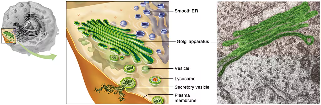
Golgi Apparatus
Receives substances from ER, then refine the proteins and lipids, place in a vesicle (round shipping sphere), Fed Ex of the cell;
Hollow, tubular, flattened + stacked
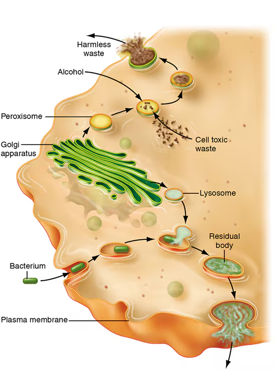
Lysosomes
Enzymes that break down Deloris (garbage disposal)

Peroximes
Enzymes that detoxify
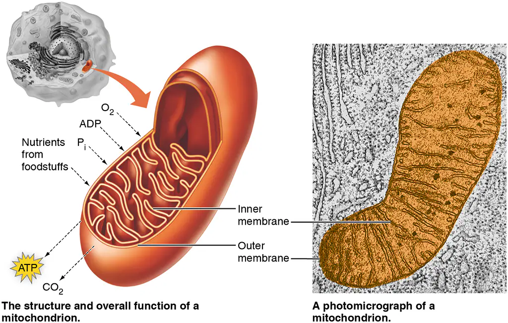
Mitochondria
Provides energy to the cell (creates ATP using food and 02), long term storage (fat: days-years) and short term (glycogen: hours);
Double membrane
Cytoskeleton
Supports the cell membrane from below, anchors organelles
Cillia
Move as a unit in a wave-like manner, moves items over the surface of cells
Flagella
Moves like a whip, propels cells forward (sperm cell)
Centriole
Paired organelles, only active during cell division, spindle fibers
Microvilli
Multiple folds in the cell membrane, high surface area
Metabolism
Chemical reactions that happen in the body in order to make ATP
Anabolism
Makes new organelles, repairs cells, makes new molecules, and stores ATP
Catabolism
Removes old/damaged organelles, breaks down large molecules, and releases stored ATP
Prep Step
To prepare for crabs, pyruvate enters the mitochondria and is converted to 2 CoA.
Citric Acid Cycle/CRABS
CoA is broken down by mitochondrial enzymes, and 2 molecules of ATP is produced
Glycoloysis
Enzymes break apart glucose; produces 2 ATP and pyruvate
Electronic Transport System
Energy from electrons used to produce ATP (32), H20, and C02
Anerobic pathways
Glycolysis happens in the cytoplasm and does not require 02 (pyruvate is converted into lactic acid instead)
Tissue
Groups of cells with a common function; Epithelia, connective, muscle, and nervous
Organ
Group of tissues with a common function
Epithelial Tissues
Line body cavities and cover surfaces
Glandular Epithelia
Epithelial cells adapted to make up glands (exocrine glands (to secrete out a duct to a surface) and endocrine glands (secretes into blood; hormones))
Basement Membrane
Structural support, attaches to epithelial layer to underlying tissues
Epithelial Tissue Classification
First name (number of layers), middle name (shape), last name (epithelia)
Simple Squamos
Found in capillaries (smallest blood cells) and areolas; function is diffusion
Simple Columnar
Found in most of the digestive tract, function is absorption
Simple Cuboidal
Found in kidney tubules ducts of glands, secretes and excretes
Stratified Squamas
Found in upper/outer layer of the skin (mouth, anal region, ect,), protection
Junctions
Holds cell together;
tight (holds 2 cell membranes tightly together),
adhesion/desmosomes (allow for stretching between cells (muscles, skin, stomach)
gap (protein tunnel/canals that connect to cells, rapid communication between cells (cardiac + smooth muscle))
Connective tissue
Support and connects body parts; contains living cells (fibroblasts: collagen, elastic, and reticular) and matrix (strong semi-solid liquid)
Fibrous Connective Tissue
Loose/areolar (cell = fibroblast, matrix = all fiber types, found in packing material around organs)
Elastic (Stretches)
Reticular (fine/delicate, found in soft organs)
Dense (Cell = fibroblast, matrix = collagen, found in tendons and ligaments)
Special Connective Tissue
Cartilage (chondrocyte), bone (osteocyte), blood (Red and white blood cells + platelets), fat (adipocyte), and muscle
Skeletal Muscle
Attached to bones, has multiple nucleuses, made up of protein fibers, voluntary + striated
Cardiac Muscle
Heart, cells are branched, one nucleus, involuntary, intercalated discs
Smooth Tissue
Surrounds hollow organs, cells are spindle like, involuntary and non-stirated
Nervous Tissue
Neuron, dendrites (extensions of the cell membrane that pick-up stimuli), cell-body (nucleus controls metabolism), axon (sends the signal to the next cell), and gilia
Glial Cell
Bodyguard; makes sure the needs of the cell is met (protection, insulation, nourishment)
Midsagittal
Cut me in half between my eyes
Frontal
Cut me in half using my ears
Transverse
Cut me in half like gojo
Anterior/Ventral
Towards the front
Distal
Farther from the trunk
Inferior
Away from the head
Lateral
Away from the middle of the body (midsagittal style)
Medial
Towards the middle (midsagittal)
Posterior
Towards the booty
Proximal
Towards the place of attatchement/trunk
Superior
towards the head
Integumentary System
Prevents dehydration and defends against microorganisms (has a cutaneous membrane)
Epidermis
Tissue: stratified squamous and dense connective tissue
Avascular; relies on diffusion from the dermis to get blood cells
Bone
Matrix - Calcium phosphate and collagen
Cells: Osteoblasts/cytes/clasts
Osteoblast
Builds bones by making new matrix
Osteocytes
maintenance of all the matrix live in the lacunae
Osteoclasts
remove bone matrix
Structure of Bone
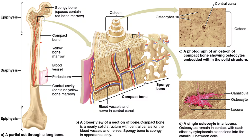
Spongy Bone
Contains red marrow, has stem cells that make red and white blood cells
Compact Bone
Type of dense bone tissue found on the outer surfaces of shafts of bones
Yellow marrow
Made up of primary fat, used to store energy.
Tendon
attaches muscle to bone
Ligament
Attaches bone to muscle
Osteon
A cylindrical structure in bone composed of layers of living bone cells and hard extracellular material, arranged like the layers of an onion. In the center is a central canal through which nerves and blood vessels pass.

Central Canal
In bone, the hollow central tube of an osteon that contains nerves and blood vessels.
Serous membrane
Covers organs + lines enclosed body cavities
Mucous membranes
Lines areas of body open to the outside
Synovial membranes
Lines freely movable joints
Cutaneous membrane
Skin
Fetal bone development

Bone Repair
1) Hematoma
2) Callus is than formed between the two broken ends of the bone
3) Osteoclasts arrive and remove the dead fragments of the og bone and blood cells
4) Osteoblasts that deposit osteoid matrix, converting calcium phosphate minerals into bone
Chondroblasts
Cartilage forming cells, live for 2-3 months
Axial Skeleton
Midline of the body (skull, ribs, sternum, and backbone)
Skull Anatomy
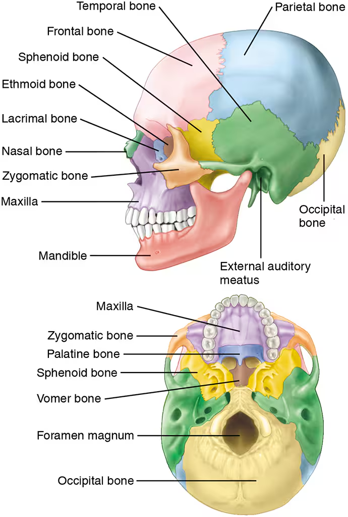
Spine Anatomy
Cervical (7), Thoracic (12), lumbar (5), Scrum (5), Coccyx (4)

Rib Anatomy
True (1-7), False (8-10), Floating (11-12).
Appendicular Skeleton
Appendages
Pectoral girdle
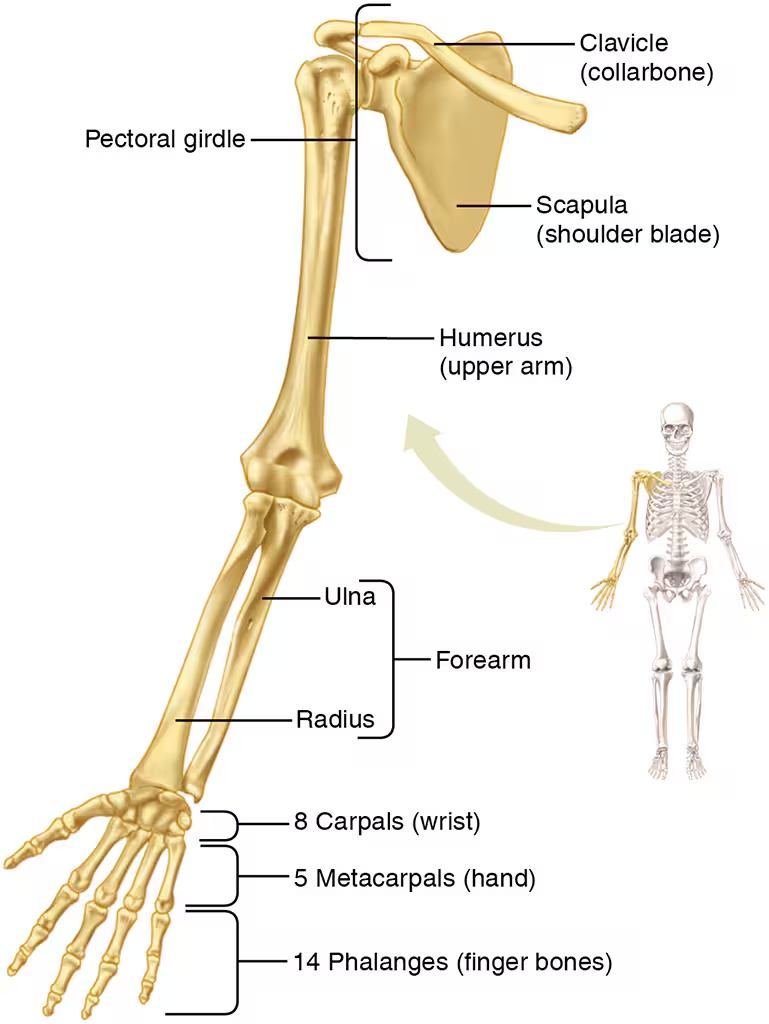
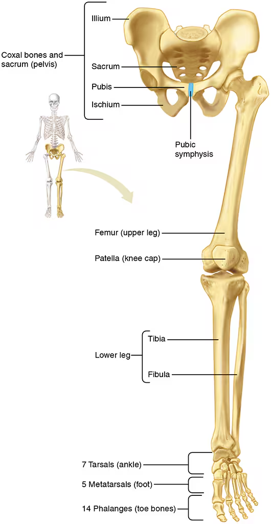
Pelvic Girdle