Hand Therapy
 Call Kai
 Learn
 Practice Test
 Spaced Repetition
 Match
 Flashcards
 Knowt Play
1/76

There's no tags or description
Looks like no tags are added yet.
77 Terms
What are the different assessment factors to consider?
Age
Gender
Occupation
Condition
Other pathology
Emotional and motivation
Cognitive ability
Socio-economic condition
Social support
What are the possible complications in hand injuries?
Bone deformities (malunions)
Delated union and non-union fractures
Bone infection (osteomyelitis)
Nerve or blood vessel damage
Osteoarthritis
What are the different precautions in hand evaluation?
Open wounds
Stage of wound healing
Sensations
Oedema
What is typically included in a hand assessment?
Hand dominance
Age
Work
ROM
Sensation
Dexterity
Functional use
Oedema
Scar assessment
ADL and IADL
What are the different upper limb standardised assessments?
Nine-Hole Peg Test
Box and Block Test
Jebsen-Taylor Hand Function Test
Smith Hand Function Test
Purdue Pegboard
Minnesota Manual Dexterity Test
Disabilities of the Arm, Shoulder and Hand (DASH)
Semmes-Weinstein Monofilament Test
Jamar Dynamometer and Pinchometer
What are the treatment modalities for oedema?
Elevation
Active range of motion
Contrast baths
Compression
Pressure garments
Manual oedema massage
What are the stages of wound healing and the intervention at each stage?
Acute inflammatory phase (0-6 days)
Rest/immobilise
No stress
Fibroplasia phase (5-36 days)
Mobilisation by applying light stress
Monitor effect of splint on swelling, pain and colour changes
Scar maturation phase (4 weeks - 2 years)
Mobilisation by applying more stress over a long period
Splints are designed to influence the remodelling of collagen and ensure the recovery of articular gliding and excursion
Wound contracture
Resist contractile forces even 6 months and excursion
Provide long-term splinting
What is the rationale for splints?
Pain relief
Support/position
Protection/immobilisation
Prevention/correction of deformity
Oedema control
Maintenance/restoration of ROM
Scar remodelling
What factors should be considered when making a splint?
Individual patient factors
Length of time splint will be used
Simplicity and cosmesis
Optimum sensation
Optimum function of the extremity
Efficient construction and fit
Easy application and removal
Splint exercise regime
Exact disease, injury or deformity
Goal of splinting regime
Most effective way to reach goal
Anatomic and material variables
What actions should be completed while making the splint?
Identify key joints
Revise purpose of the splint
Kinetic effect
Surface of splint application
Static or dynamic force
Use mechanical principles advantageously
Adapt for anatomic variables
What are the types of arthritis?
Osteoarthritis (OA): A degenerative joint disease characterised by the breakdown of articular cartilage, causing pain, stiffness and reduced joint function.
Rheumatoid arthritis (RA): A chronic autoimmune inflammatory disease in which the immune system attacks the synovial lining of joints, causing inflammation and progressive joint damage.
In which site does osteoarthritis develop?
1st CMC joint
DIP joints
PIP joints
What is the osteoarthritis quintet?
Heberden’s nodules
Mucoid cysts
OA of the 1st CMC joint
Trigger finger
Carpal tunnel syndrome
What is the initial treatment for OA?
Splinting
Improve stability
Prevent deformities
Decrease pain and improve function
Worn during the day only (night if pain)
Surgery: Excision arthroplasty/silastic joint replacement
If no improvement
What is the post-surgical treatment for OA?
POP splint
4-6 weeks
Scar and soft tissue massage
Gentle active and passive movement
Graded strengthening exercises
What is the treatment for OA of the DIP joints?
Arthrodesis of involved joints
K-wire fixation for 6 weeks
Protective splinting (from 10 days post operation)
Finger socks
Scar massage
Gentle active and passive motion of PIP and MP joints
Flexion against light sponge; putty exercises
What is the clinical picture of RA?
Wrist: Gradual radial deviation
MP joints: Gradual volar subluxation and ulnar deviation
PIP joints: Swan neck or Boutonniere deformity
Thumb: Boutonniere deformity
What is the treatment for RA?
Surgery: Synovectomy of the wrist
What are the types of brachial plexus injuries?
Neuropraxia (stretch)
Rupture
Avulsion
What are the brachial plexus nerves?
Axillary
Median
Musculocutaneous
Radial
Ulnar
What is the acute phase treatment for brachial plexus injuries?
Positioning
Splinting
Gentle exercises
What is the post-operative phase treatment for brachial plexus injuries?
Sling
4-6 weeks
One-handed technique education
Hand splint
Early passive mobility of distal joints
What is the rehabilitation phase treatment for brachial plexus injuries?
Roles
Work
Interests/hobbies
What is the general treatment for brachial plexus injuries?
Pain management
Splinting
ADL and IADL
Dexterity
Fine motor coordination
Hand-eye coordination
Strengthening
Exercises
Slings
Strengthening band
Putty
Sensory re-education
Assistive devices
Emotional and social factors
Patient education
Vocational rehabilitation
Hobbies and leisure
What is the treatment for neonatal brachial plexus palsy?
Passive movements
Sensory stimulation
Bilateral hand use
Possible wrist extension splint
Home programme:
ROM
Speed
Functional capacity
Mobility
Hand manipulation skills
What are levels of the Highest Scale for motor function?
M0: No contraction
M1: Trace of contractibility
M2: Joint motion, gravity eliminated
M3: Joint motion against gravity
M4: Joint motion with some resistance
M5: Joint motion with full resistance
What are levels of the Highest Scale for sensory function?
S0: No sensation
S1: Deep pain sensibility
S2: Some superficial sensibility, paraesthesia and pain
S3: Superficial sensibility, disappearance of paraesthesia and return of sweat
S3+: 2-point discirmination less than 12mm. Light touch near normal. Full use of hand.
S4: Normal sensibility.
What are the types of peripheral nerve injuries?
Neuropraxia
Axonotmesis
Neurotmesis
What factors affect the outcome of nerve repair?
Age
Mechanism of injury
Location of injury
Types of nerve
Associated injuries
Nerve gap size
Tension at repair site
Technique of repair
Timing of repair
What are the tests for nerve damage?
Tinel’s sign
Wrinkle test
Semmes and Weinstein Monofilament Test
Two-point discrimination test
Muscle strength
What does a proximal median nerve injury result in?
Weaker PIP and MP flexion
Absent DIP and PIP flexion of index and middle finger
Pronation with gravity
Sensation loss
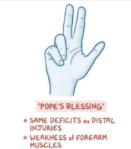
What does a distal median nerve injury result in?
Loss of intrinsic thumb function
Sensation loss

What is the treatment for a median nerve injury?
Surgery (neurotmesis)
Maintain ROM
Education
Splinting (preventative, functional)
Sensory and motor retraining
What does a distal radial nerve injury result in?
Triceps intact
No wrist extension
No finger extension
No thumb extension
Sensory loss

What is the treatment for a radial nerve injury?
Splinting:
Forearm-based static wrist extension splint
Forearm-based dynamic finger extension splint
Post-tendon transfer:
Immobilisation in cast
Shoulder motion retained
IP flexion/extension
Replace cast with thermoplastic splint
Individual joint mobilisation
Retrain function
What does a proximal ulnar nerve injury result in?

What does a distal ulnar nerve injury result in?

What is the treatment for an ulnar nerve injury?
Early:
Suture after diagnosis
Immobilisation in POP cast
Restrictive splint
Function and grasp
Sensory re-education
Post-reconstruction:
Immobilisation in POP
MP extension restriction splint
Scar management
Retraining of grasp
De-sensitisation
What are the methods for assessing oedema?
Water displacement: Volumeter
Circumferential figure-of-eight measurement: Tape measure
What are the treatment modalities for oedema?
Elevation
Exercise (AROM)
Massage
Compression
Contrast baths
Coban wrapping
Manual oedema mobilisation
Brazilian hand wrap
What are the causes of stiffness?
Joint tightness
Muscle tightness (instrinsic)
Muscle-tendon tightness (extrinsic)
Skin tightness
What are the treatment modalities for a stiff hand?
Splinting
Massage and heat
Heat pack
Wax bath
Passive stretching
Passive joint mobilisation
Scar management
Activities
What are the types of splints used to reduce stiffness?
Serial static splint (POP)
Dynamic splint
Static progressive splint
What is total end range time (TERT)?
The total time a joint is maintained at end range for.
What is daily end range time (DERT)?
The total time per day a joint is maintained at end range for.
What is the initial treatment for a severely injured hand?
Provide wound care
Rest (splinting/activity)
Gentle mobilisation
Manage oedema
Exercise, maintain joint mobility and prevent tendon gliding
Mobilise uninvolved joints
Consider psycho-social needs
What is the rehabilitative treatment for a compartment syndrome?
Splinting to maintain full extension
Scar massage
Gentle active and passive movements
Pressure garment to manage oedema
Functional use of hand
What is the intermediate phase treatment for a severely injured hand?
Static splint
Dynamic splint
Activities and exercise
Skin cover and scar management
Desensitisation
Skills retratining
Simple domestic activities
Psychological aspects
Patient education and supppport
Oedema control
Wound and pain management
AROM and PRO
What is the late phase treatment for a severely injured hand?
Exercise, strengthening activities
Work rehabilitation
What are the extrinsic flexor muscles?
Flexor digitorum profundus (FDP)
Flexor digitorum superficialis (FDS)
Flexor pollicis longus (FPL)
How is the action of FDP tested?
Action: DIP flexion

How is the action of FDS tested?
Action: PIP flexion

How is the action of FPL tested?
Action: IP flexion of thumb

What are the wrist flexor muscles?
Flexor carpi ulnaris (FCU)
Flexor carpi radialis (FCR)
What are the flexor tendon zones?

What is the common injury in flexor zone 1?
Jersey finger: Avulsion of FDP
What is the post-operative splinting for a flexor tendon injury?
Dorsal blocking splint
Forearm-based
Wrist in 20-30° flexion
MPs in 60° flexion
What is the protocol during phase 1 (0-4 weeks) following a flexor tendon repair?
Immbolisation
POP and thermoplastic splint
Least used protocol
Early passive mobilisation
Non-removable POP splint with dynamic traction
Wrist in 20-30° flexion
MCP in 70-80° flexion
IP in extension
Maintain flexion, allow extension
Most frequently used
Early active mobilisation
Removable thermoplastic splint
Which factors influence the protocol chosen following a flexor tendon repair?
Age
Zones injured
Quality of surgery
Patient’s residence
Comprehension and compliance
Time of OT referral
What are the splinting guidelines for early passive mobilisation following a flexor tendon repair?
Dorsal blocking splint
Elastic bands unattached at night and for alternating periods during the day
Crepe bandage wrapped lightly around splint
Active extension of fingers as far as allowed by splint
Maintain extension position for 5 seconds
Repeat 10 times every waking hour
What are the splinting guidelines for early active mobilisation following a flexor tendon repair?
Dorsal blocking splint
Strip on palmar aspect
Passive flexion of fingers
Maintain flexion position for 5 seconds
Repeat 10 times every waking hour
Fingers strapped into extension every other hour
Gentle active flexion after 2 weeks
Active place and hold:
Passive MP extension and wrist extension
Passive wrist flexion and MP + IP extension
What is the protocol during phase 2 (5-6 weeks) following a flexor tendon repair?
Week 5:
Splint removed for exercises
Scar massage commences
Active finger movement without resistance
Activities performed in soapy water
Splint remolded with more extension
Remove 3-4 times a day to mobilise and wash hand
Rubber traction removed
Paraffin wax and ultrasound
Finger socks and pressure gloves
Squeeze sponge in water
Week 6:
Adapted table games to encourage IP flexion (clamps)
Block joints proximally and flex fingers
Light to medium theraputic putty
What is the protocol during phase 3 (7-12 weeks) following a flexor tendon repair?
Week 11:
Dorsal blocking splint worn at night
Dynamic dorsal finger extension splint during day and night
Return to light duty
Therapy 1-2 times per week
Activities against resistance and requiring static grips
Scar management
Week 12:
Resume normal activities
No contact sport for another 4 weeks
What are the treatment modalities for a flexor tendon repair?
Passive motion (up to week 5)
Active motion (from 4 weeks)
Functional activities (after 6 weeks)
Therapeutic equipment
Manual oedema mobilisation
TENS
Splints
Post-operative flexor tendon POP
Dorsal extension blocking splint
What is the tendon glide exercise used for a flexor tendon repair?

Which assessments are used for a flexor tendon injury?
General observations
Oedema
ROM (from 6 weeks)
Muscle strength (from 8 weeks)
Sensation
Hand function (from 6-8 weeks)
Grasps (from 6-8 weeks)
What are the extensor tendon zones?

What are the extensor muscles?
Extensor digitorum communis (EDC)
Extensor pollicis longus (EPL)
What is the action of EDC?
Extend MP joints of medial 4 fingers
Assists in IP extension
What is the action of EPL?
Extends all joints of thumb
Assists in extension and abduction of wrist
What is the post-operative splinting for an extensor tendon injury in zone V-VII?
Wrist in 30-40° extension
MP in 30° flexion
PIP in extension
DIP free
What are the splinting protocols following extensor tendon repair?
Dynamic splinting with early passive motion
Static splinting with early active motion
What is the weekly protocol for dynamic splinting with early passive motion?
3-5 days:
Forearm-based dorsal dynamic extension splint
Wrist in 40-45° extension
MP joint in 0°
Worn 24 hours
Active MP joint flexion 10 times per waking hour
Oedema control
Scar management
Wound care
Splint adjustments
Positioning
Exercises (tenodesis)
2-4 weeks:
Palmar block removed (week 3)
4-6 weeks:
Traction removed (week 5)
Active finger extension
Extension against gravity
Exercises
6-8 weeks:
Splint removed
Resistance training
Full flexion at 8 weeks
What is the weekly protocol for static splinting with early active motion?
0-4 weeks:
Static forearm-based palmar splint
Wrist in 40-45° extension
MPs in 20-30° flexion
Gentle strap over MPs
Exercises
Passive MP extension
Active MP extension
4-6 weeks:
Splint shortened to free MPs
More MP flexion and tendon excursion
6-8 weeks:
Splint removed
Resistance training
Full flexion at 8 weeks
What are the classifications of fractures?
Acute
Simple or compound
Stable or unstable
Intra-articular
With associated injuries
Chronic
Malunion
Non-union
With associated joint contractures
Pathological
Colles fracture
What are the methods of stabilising fractures?
Conservative
Static splinting
DIP extension splint
Burkhalter splint
Dynamic splinting
Buddy splint
Surgical