Acquired Brain Injury: Stroke
 Call Kai
 Learn
 Practice Test
 Spaced Repetition
 Match
 Flashcards
 Knowt Play
1/25

There's no tags or description
Looks like no tags are added yet.
26 Terms
Objective #1
Describe the functional organization of the nervous system using appropriate terminology.
Nervous System Divisions (2)
1) Central Nervous System (CNS)
contains brain & spinal cord
lacks regenerative properties!!
w strokes/TBI: neurons cannot regenerate!!!
2) Peripheral Nervous System (PNS)
cranial + spinal nerves

Divisions of the CNS (4)
tiered structure throughout!!!
1) Cerebrum:
a) bilateral cerebral hemispheres
b) diencephalon
2) Brain stem (origin of SC):
a) midbrain
b) pons
c) medulla oblongata
3) cerebellum
4) spinal cord

Neurons: structure + communication
cell body (soma): contain’s cell nucleus + majority of organelles
dendrites: extend from soma and receive input from other neurons via synapsis
axon: single process that emerges from axon hillock
sends output impulse to other cells
synaptic communication:
synapses allow for unidirectional communication b/w neurons
electrical signal converted into —> chemical signal
release various neurotransmitters to excite/inhibit post-synaptic cell

Functional Organization of CNS
organized w/ bilateral symmetry
cereBRUM: crossed representation —> contralateral functions
cereBELLUM, brain stem, SC —> primarily ipsilateral
multiple specific tracts originate or terminate within various cortexes:
efferent/motor maps: motor homunculus
somatosensory maps: sensory homunculus
retinotopic: occipital lobe
topographic: right parietal cortex

Objective #2
Identify the primary cerebrovascular vessels (ACA, MCA, PCA) and the role of the Circle of Willis.
Vascular Supply
Internal carotids + vertebral arteries ascend to skull + converge to form:
Circle of Willis!!!
arterial circle formed around pituitary stalk/gland
provides collateral circulatory pathway in cerebral circulation

Great Cerebral Vessels
major terminal branches of INTERNAL CAROTID:
Anterior cerebral artery (ACA)
Middle cerebral artery (MCA)
major branches of VERTEBRAL ARTERIES:
posterior cerebral arteries (PCA)
basilar artery
thrombi forming or traveling to ANY of these WILL cause ischemia (stroke)!

Acquired Brain Injury
An injury to the brain that is neither genetic nor congenital
vascular insults: strokes
TBIs
infections
autoimmune + inflammatory conditions
Objective #3
Compare and contrast the impact of various types of strokes, including TIA, ischemic, and hemorrhagic.
Types of Stroke (2)
stroke: sudden onset neurological impairment due to vascular insult
Ischemic:
blockage due to a thrombosis or embolus —> blood flow blocked + unable to reach target tissues
hypoperfusion due to reduced circulation or small vessel disease
Hemorrhagic:
rupture of an aneurysm or vessel within brain tissue or surrounding space
exposes neural tissue to blood + its products
failure of blood brain barrier = cascading sequence of destructive events

Ischemia Types
Stroke:
sudden onset
focal involvement of the CNS
vascular cause
lack of rapid resolution
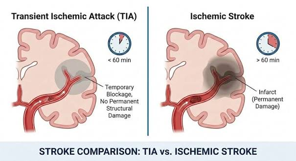
Transient Ischemic Attack (TIA):
lasts about 5-15 minutes
ischemic event where symptoms resolve rapidly (b/c of spontaneous resolution of cause)
Ex: embolus becomes dislodged restoring blood flow
24 hours is a commonly used boundary b/w TIA and stroke
Ischemic Strokes (2)
follow stroke characteristics from previous flashcard (sudden onset, etc.)
Hypoxic ischemia:
interruption of blood flow to the brain deprives cells of glucose + oxygen
leads to ischemic death of brain tissue of all cell types, resulting in chronic lesions (infarction)
irrecoverable + produce scarring within CNS
Vascular Ischemia:
blood flow is obstructed by a thrombus or embolus, preventing delivery of oxygen and glucose
tissue death will occur within minutes to hours (irreversible)
Objective #4
Differentiate the functional impact of various stroke syndromes, including ACA, MCA, and PCA territories.
Anterior Cerebral Artery (ACA) Infarction
territory impacting the MEDIAL aspect of the frontal and parietal lobes
frontal lobe —> limbic system —> personality changes + disinhibited, impulsive behaviors
secondary motor area —> apraxia = impaired motor planning
MEDIAL portions of motor + sensory cortexes —> contralateral to injury —> weakness on one side of body (hemiparesis)
will produce contralateral hemiparesis + sensory loss of the leg, apraxia + personality changes!!

Middle Cerebral Artery (MCA) infarction
territory impacting the lateral aspect of the frontal, parietal + temporal lobes (trunk, UE, face)
will produce contralateral hemiparesis + sensory loss of the arm, apraxia
if dominant hemisphere is involved, aphasia may occur —> inability to produce speech
if non-dominant hemisphere is involved, neglect of environment may occur
profound multi-sensory lack of awareness
not respond to visual, auditory or tactile stimuli on non-dominant side
note: dominant hemi. is usually left

Posterior Cerebral Artery (PCA) infarction
territory impacting the occipital + temporal lobes
will produce contralateral homonymous hemianopia
“same-sided half blindness”
right lesion = no visual info from left side of environment

Objective #5
Describe the clinical manifestations of hemorrhagic strokes (ICH and SAH).
Hemorrhagic Stroke: Intracranial hemorrhage (ICH)
primarily a result of chronic HTN + underlying vascular disorders
metabolic effects of blood disturb the function of surrounding brain tissue, as well as produce focal edema
release of blood into tissues of cerebrum

Hemorrhagic Stroke: ICH — secondary effects
sequence of ICH injury:
hematoma ruptures white matter tracts; expands for up to 24 hours
cerebral edema develops as product of inflammation (rupture of BBB) in surrounding tissue for up to 2 weeks
edema produces mass effect —> shifts cerebral tissues + collapsing arterial vessels

Hemorrhagic Stroke: Subarachnoid hemorrhage (SAH)
starts w/ BAD headache
caused by rupture to vessels within the subarachnoid space
cerebral dysfunction occurs b/c of increased intracranial pressure + from toxic effects of subarachnoid blood
vasospasm is a complication
can impede blood flow + provoke ischemic stroke
vasospasm = spastic contraction of smooth muscle lining

Cerebral Aneurysm (can lead to SAH)
focal dilatation of the vessel wall; thin walled + at risk for rupture
major cerebral vessels lie in the subarachnoid space —> rupture results in SAH
some aneurysm may occur within neural tissues themselves

Objective #6
Describe basic medical and surgical management for the various types of strokes.
Hemorrhagic Stroke Medical/Surgical Management
surgical clipping: open or endovascular removal of aneurysm (pictured)
cerebellar/lobal hemorrhage: decompression + evacuation
for subA + intracranial hemorrhages
deep hemorrhage: no surgical options, only medical management

Ischemic Stroke Medical/Surgical Management
*time = brain loss!!!*
prevention through anti-platelet therapies + management of artherosclerotic disease
intravenous thrombolysis
IV administration of recombinant tissue plasminogen activator (rTPA) —> anticoagulant to break apart clots
must be administered within 4.5 hours of onset of symptoms to reduce disability + mortality
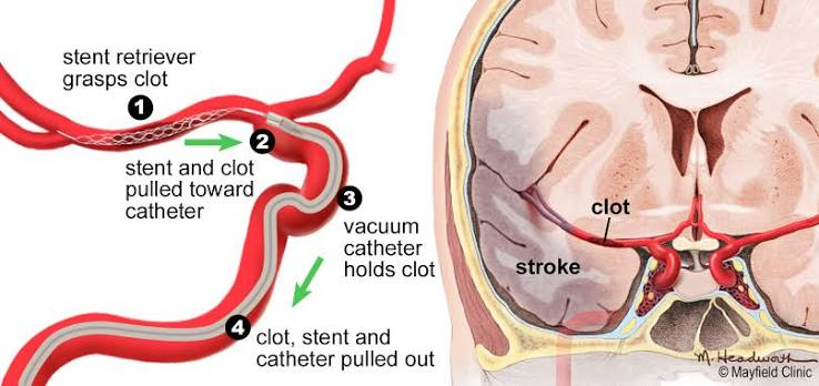
clot retrieval (pictured)
thrombectomy w/ stent or coil retrievers
may be used in conjuction with rTPA
Ischemic Stroke Medical/Surgical Management (cont.)
carotid endarterectomy (pictured)
surgical removal of thrombus from a stenotic carotid artery in neck
decompressive craniectomy (stroke w/ significant edema)
used to prevent cerebral herniation after large hemispheric strokes
reduce intracranial pressure —> resecting portion of skull beneath skin flap to accommodate cerebral edema
