Osmoregulation evolution (All but Kidneys)
 Call Kai
 Learn
 Practice Test
 Spaced Repetition
 Match
 Flashcards
 Knowt Play
1/8

There's no tags or description
Looks like no tags are added yet.
9 Terms
Drivers of Osmoregulatory Organ Evolution
Osmoregulatory organs became more complex as ocean osmolarity changed, animals moved onto land, respiration shifted, and body plans became more complex.
All animals need to gain water and lose some water to remove water soluble wastes.
Osmoregulatory organs need access to body water and solutes, plus a route to the external environment.
Osmoregulation evolutionary differences in different species
Invertebrates: Less complex regulation of water & solutes & less sophisticated osmoregulatory organs. Most are Osmoconformers
Marine invertebrates: Blood and tissues osmolarities the same as ocean osmolarity in which they reside but the solutes are not the same.
Brackish Invertebrates: much more adaptable to changes in osmolarity but are less common.
Freshwater Invertebrates: have blood that is hypertonic to freshwater
Terrestrial Invertebrates: have to deal with desiccation
Main evolutionary innovations
Osmoregulatory systems evolved from simple solute loss toward direct blood access, faster filtration, selective recovery, and temporary urine storage.
Early systems mainly secreted specific solutes, and water followed passively down an osmotic gradient.
Direct access to blood improved regulation because blood better reflects whole body conditions than interstitial fluid.
Development of a size but not solute-specific filtration barrier. The advantage here is to remove any solute under a certain size, without the need for a specific transporter.
Ultrafiltration increased the speed of removing water and small solutes from the body.
Reabsorption and secretion allowed urine composition to be adjusted as conditions changed.
Use of membrane transporters to control blood osmolarity.
Shared design of osmoregulatory organs
Most osmoregulatory organs use a tube or vacuole that exchanges solutes and water with body fluids and opens to the environment.
The core structure is usually a membrane bound space connected to the outside.
Simple organs rely more on secretion, while complex organs add filtration barriers and pressure driven flow.
A size selective barrier can remove many small solutes at once without needing a separate transporter for each one.
Transport proteins across membranes remain central across both simple and complex systems.
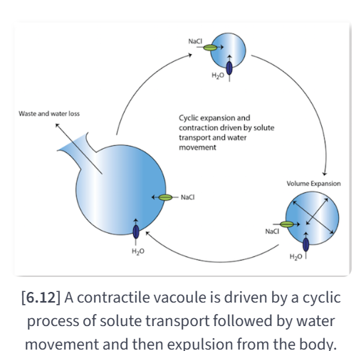
Contractile Vacuoles - Sponges
Contractile vacuoles fill with solutes and water by transporter activity, then empty cyclically to expel filtrate.
Proton pumps create an H+H^+H+ gradient that helps move solutes into the vacuole.
As vacuole osmolarity rises, water enters passively through aquaporins.
The vacuole likely empties when internal pressure rises enough to force fluid outward.
Contractile vacuoles lack blood access, ultrafiltration, and reabsorption, so their control is relatively limited.
Nephridia, metanephridia and protonephridia - Annelids, Arthropods, & Mollusks
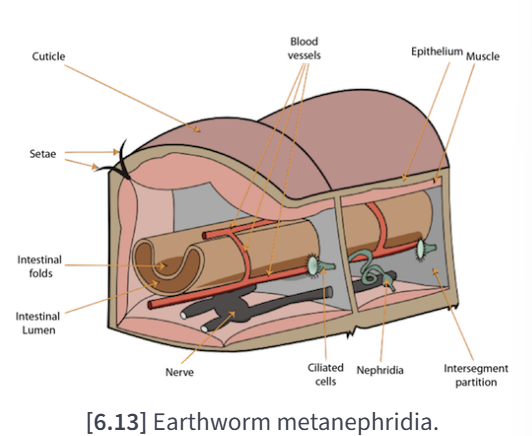
Nephridia introduced tubular flow and reabsorption, with cilia generating pressure or suction to move filtrate through ducts.
Metanephridia use a ciliated funnel to draw coelomic fluid into a duct that opens outside the body.
Saccate Metanephridia: Crustaceans
Excretory gland similar to a metanephridia
Filters blood through a barrier (no proteins & large carbs) -> Funnel (Rudimentary filtration barrier)
Fluid -> Urine by selective solute reabsorption
Cells lining the duct reabsorb useful solutes and help convert filtrate into urine.
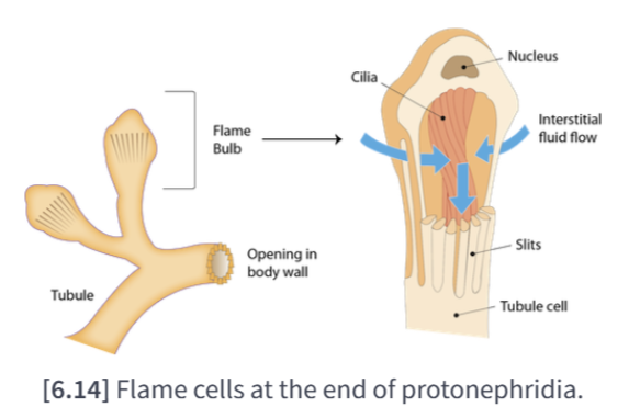
Protonephridia are dead end tubes where flame cells or solenocytes drive fluid through small size selective openings.
These systems shifted from osmotic filling toward mechanically driven movement of fluid.
Green gland, or coxal gland - Horseshoe crabs & Arthropods
Green glands added direct blood filtration by pressure, making removal of small solutes faster and more efficient.
Podocytes near blood vessels create a filtration barrier connected directly to blood.
Blood pressure drives ultrafiltration, which rapidly removes water and small solutes.
The tubule then modifies the filtrate by reabsorption and secretion.
A bladder adds temporary urine storage, which supports more flexible water use.
Saccule -> Labyrinth -> Tubule -> Bladder

Malpighian tubules - Insects, Arthropods, & Tardigrades
Malpighian tubules draw solutes from hemolymph into gut-linked tubes, and water follows passively once an osmotic gradient is established.
These tubules branch from the digestive tract and collect wastes from the hemolymph.
Specific transporters move solutes such as Na+Na^+Na+, K+K^+K+, and amino acids into the tubule.
Water then enters passively, making the system function more like a contractile vacuole than a pressure filter.
Because there is no filtration barrier, only solutes with suitable transport routes are readily lost.
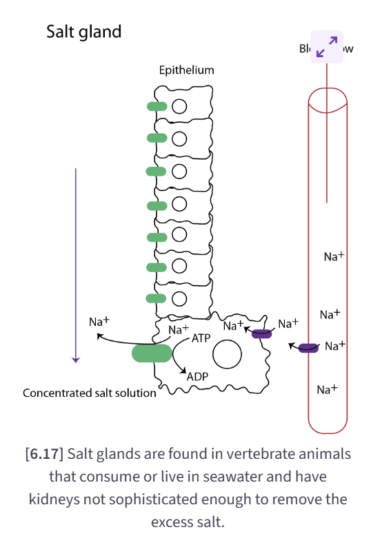
Salt glands - Sharks, rays, skates, seabirds, & some reptiles
Salt glands excrete excess salt with minimal water loss because they move solutes strongly while limiting water permeability.
Salt glands occur in some marine vertebrates whose kidneys cannot remove enough salt on their own.
They are built from epithelial cells rich in transporters but have aquaporin channels on only one side along their length.
This setup allows strong salt secretion while reducing passive water loss.
Their size tends to increase with the animal's salt load.