A&P Lab 8 - Nerves and Spinal Cord
 Call Kai
 Learn
 Practice Test
 Spaced Repetition
 Match
 Flashcards
 Knowt Play
1/75

There's no tags or description
Looks like no tags are added yet.
76 Terms
Dendrites
short branches extending from cell body

Cell Body (Soma)
contains large nucleus and nucleolus

Axon Hillock
where axon attaches to cell body and where action potential gathers before propagating down axon
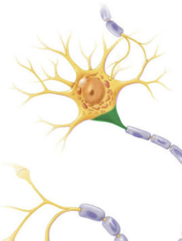
Myelin sheath (Schwann cell)
forms around axons to insulate axon for rapid propagation of action potentials

Node of Ranvier
areas between schwann cells where action potentials can more rapidly jump

Axon terminal
where a neuron meets up with other neurons to pass action potentials down the chain

Spinal cord
extension of brain into vertebral column where spinal nerves emergge from and pass through intervertebral foramen

Meninges
protective membranes of spinal cord

Conus medullaris
tapered, conical end of cord at L1/L2 (light green)

Cauda equina
nerve roots extend below conus medullaris, “horse tail” (dark green)
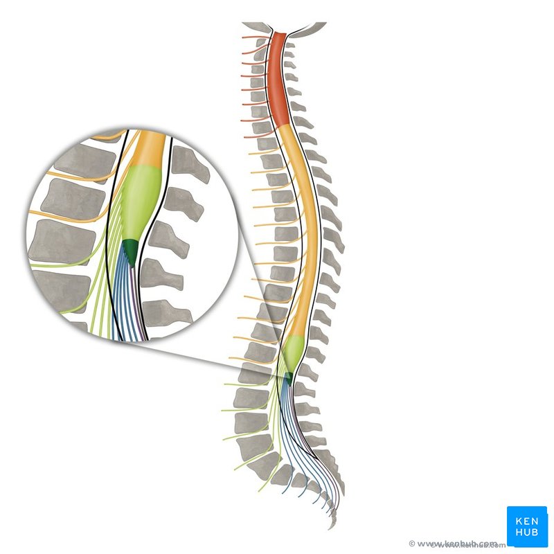
Filum terminale
thin threads of fibrous tissue at end of conus medullaris

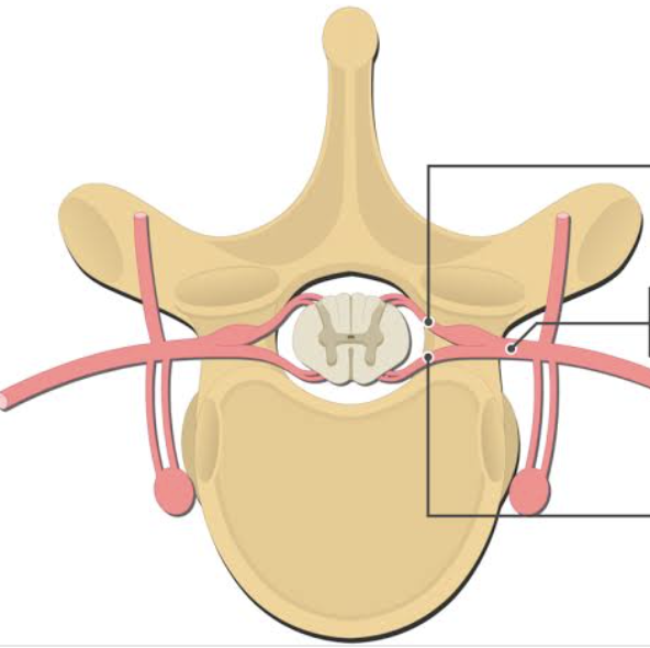
Dorsal root
axons of sensory neurons (top indication)

Dorsal root ganglion
contain cell bodies of sensory neurons in dorsal root

Ventral root
axons of motor neurons (bottom indication)
Posterior median sulcus
grooves dividing spinal cord left/right

Anterior median fissure
grooves dividing spinal cord left/right

Central canal
middle hole, contains cerebrospinal fluid

White matter parts
anterior, posterior, lateral white columns
Anterior white column
Posterior white column
Lateral white column
Gray matter parts
posterior, anterior, lateral gray horns
Gray commissure
Anterior gray horn
somatic motor nuclei, CNS to skeletal muscles
Posterior gray horn
somatic and visceral sensory nuclei, receptors to CNS
Lateral gray horn
visceral motor nucleus, CNS to other effectors
Epidural space
Dura mater
Arachnoid mater
Subarachnoid space
Pia mater
8 cervical spinal nerves (CSN 1-8)
12 thoracic spinal nerves (TSN 1-12)
5 lumbar spinal nerves (LSN 1-5)
5 sacral spinal nerves (SSN 1-5)
Cervical plexus nerve(s)
phrenic nerve
Phrenic nerve

Brachial Plexus Nerve(s)
axillary, musculocutaneous, radial, median, ulnar
Lateral cord
Musculocutaneous nerve

Median nerve (1/2)
Medial cord
Median nerve (1/2)
Ulnar nerve

Posterior cord
Axillary nerve

Radial nerve

Lumbosacral plexus nerve(s)
obturator, femoral, gluteal, sciatic, tibial, common fibular
Femoral nerve

Obturator nerve

Gluteal nerve

Sciatic nerve

Tibial nerve

Common fibular nerve

Patellar reflex
curling of toes, present in healthy adults
Calcaneal (achilles) reflex
plantar flexion when calcaneus/achilles is hit
Plantar (babinski) reflex
spreading of toes without plantar flexion, normal in infants, pathological in adults
Withdrawl (flexor) reflex
removal of body part away from pain stimulus
Crossed extensor reflex
stimulation of opposite limb to maintain balance away from painful stimulus
Phrenic nerve muscle innervation
diaphragm
Axillary nerve muscle innervation
deltoid
Musculocutaneous nerve muscle innervation
biceps brachii
Radial nerve muscle innervation
all forearm extensors
Median nerve muscle innervation
lateral flexors of forearm
Ulnar nerve muscle innervation
medial flexors of forearm
Obturator nerve muscle innervation
adductors
Femoral nerve muscle innervation
quadriceps
Gluteal nerve muscle innervation
gluteus maximus
Sciatic nerve muscle innervation
hamstrings
Tibial nerve muscle innervation
gastrocnemius (1) and tibialis posterior (2)
Common fibular nerve muscle innervation
tibialis anterior
Plantar reflex characteristics
polysynaptic, ipsilateral
Calcaneal (achilles) reflex characteristics
monosynaptic, ipsilateral
Plantar (babinski) reflex characteristics
polysynaptic, ipsilateral
Withdrawal (flexor) reflex characteristics
polysynaptic, ipsilateral
Crossed extensor reflex characteristics
polysynaptic, contralateral