supporting ventilation
 Call Kai
 Learn
 Practice Test
 Spaced Repetition
 Match
 Flashcards
 Knowt Play
1/70

There's no tags or description
Looks like no tags are added yet.
71 Terms
SpO₂ vs. EtCO₂
These are two important measurements used to evaluate respiratory function. They measure DIFFERENT things.
Measurement | SpO₂ | EtCO₂ |
|---|---|---|
Full name | Peripheral capillary oxygen saturation | End-tidal carbon dioxide |
Measures | Oxygenation | Ventilation |
Normal range | 95–100% | 35–45 mmHg |
Equipment | Pulse oximeter | Capnograph |
Reflects | Oxygen delivered to blood | CO₂ eliminated from lungs |
Response | Delayed (30–60 seconds) | Almost immediate |
SpO₂ — Pulse Oximetry
What does it measure?
Percentage of hemoglobin molecules carrying oxygen.
Shows how well oxygen is being delivered from the lungs into the bloodstream.
Normal range: 95–100%.
Some patients with COPD may have a target of 88–92%.
Common uses:
Postoperative monitoring.
Respiratory distress.
Sedated patients.
Patients receiving oxygen.
Patients at risk for hypoxemia.
Limitations — when can the reading be inaccurate?
Poor perfusion (shock or hypothermia).
Patient movement.
Nail polish or artificial nails.
Carbon monoxide poisoning (can give falsely high readings).
EtCO₂ — Capnography
What does it measure?
The amount of CO₂ in exhaled air at the end of expiration.
Reflects how effectively the patient is ventilating and eliminating CO₂.
Normal range: 35–45 mmHg.
Provides real-time feedback.
Common uses:
Procedural sedation.
Anesthesia.
Mechanical ventilation.
Confirming endotracheal tube placement.
Monitoring CPR effectiveness.
Detecting hypoventilation, hyperventilation, and airway obstruction.
Important clinical points:
A sudden absence of the waveform may indicate tube dislodgement, obstruction, or apnea.
During CPR, EtCO₂ greater than 10–20 mmHg during compressions indicates adequate circulation, according to the lecture.
EtCO₂ may not accurately correlate with PaCO₂ in severe ventilation-perfusion mismatch, such as ARDS or pulmonary embolism.
What do changes in EtCO₂ and SpO₂ mean?
Finding | Possible Meaning | Nursing Action |
|---|---|---|
SpO₂ decreases | Hypoxemia or decreased oxygenation | Check oxygen source, reposition, suction, encourage deep breathing, titrate O₂ |
EtCO₂ increases | Hypoventilation or CO₂ retention | Stimulate patient, check airway patency, evaluate ventilation |
EtCO₂ decreases | Hyperventilation or poor perfusion | Assess circulation, ventilation, and possible tube dislodgement |
EtCO₂ absent | No exhaled CO₂ detected | Immediately assess airway and circulation |
HIGH-YIELD TAKEAWAY:
SpO₂ tells you whether the patient is oxygenating.
EtCO₂ tells you whether the patient is ventilating.
EtCO₂ changes more quickly than SpO₂.
Memory trick: SpO₂ = OXYGEN. EtCO₂ = EXHALED CO₂.
Respiratory Physiotherapy
Purpose: Breathing exercises improve ventilation, promote gas exchange, and strengthen respiratory muscles.
Commonly used after surgery or in patients with chronic respiratory illnesses.
Diaphragmatic (Abdominal) Breathing
Purpose:
Encourages use of the diaphragm instead of accessory muscles.
Promotes deeper inspiration.
Slows respiratory rate.
Reduces work of breathing.
Indications:
Post-thoracic surgery.
Post-abdominal surgery.
Anxiety control.
Prevention of atelectasis.
Technique:
Place one hand on the abdomen and one hand on the chest.
Slowly inhale through the nose.
Focus on making the abdomen rise.
Exhale gently through pursed lips.
Caution: In moderate-to-severe COPD, diaphragmatic breathing may worsen dyspnea due to hyperinflation.
Pursed-Lip Breathing (PLB)
Purpose:
Prolongs exhalation.
Prevents bronchiolar collapse.
Helps prevent air trapping.
Promotes CO₂ elimination.
Indications:
COPD.
Dyspnea.
Anxiety.
Shortness of breath with activity.
Technique:
Inhale slowly through the nose.
Purse lips as if blowing out a candle.
Exhale slowly.
Make exhalation approximately 3 times longer than inhalation.
Teaching cue: "Smell the roses, blow out the candle."
Airway Clearance Techniques
Purpose: Mobilize secretions, maintain airway patency, and improve gas exchange.
Commonly used for:
COPD.
Cystic fibrosis (CF).
Bronchiectasis.
Pneumonia.
Huff Coughing
Purpose: Moves secretions from smaller airways into larger airways so the patient can cough them out.
Technique:
Take a deep diaphragmatic breath.
Hold for 2–3 seconds.
Exhale forcefully with an open mouth, as if fogging a mirror.
Repeat 3–5 cycles until mucus is cleared or the patient becomes fatigued.
Clinical benefit:
Especially effective in COPD and emphysema.
Helps clear secretions.
Helps prevent airway collapse.
Chest Physiotherapy (CPT)
Includes three main techniques:
1. Postural Drainage
Uses gravity to drain secretions from affected lung lobes.
Requires specific patient positioning.
Perform before meals or several hours after meals.
2. Percussion
Rhythmic striking of the chest wall using cupped hands.
Helps loosen mucus so it can be coughed out.
Avoid in patients with rib fractures, bleeding disorders, or osteoporosis.
3. Vibration
Gentle shaking applied during exhalation.
Helps move mucus into larger airways for expectoration.
Airway Clearance Devices
Examples include:
Flutter valve.
Acapella.
TheraPEP.
High-frequency chest wall oscillation vest.
Nursing Management of Respiratory Physiotherapy
Assess patient tolerance and contraindications.
Recent surgery.
Trauma.
Head injury.
Unstable hemodynamics.
Administer bronchodilators and hydration before therapy as appropriate.
Assess lung sounds before and after therapy.
Evaluate sputum characteristics.
Monitor for complications.
Possible complications:
Rib fractures.
Hypoxemia.
Bruising.
Nursing tip: Bronchodilators before chest physiotherapy can help open the airways and improve secretion mobilization.
Oxygen Therapy
Purpose: Oxygen therapy treats hypoxemia and hypoxia.
It increases the partial pressure of oxygen (PaO₂) in the alveoli, allowing more oxygen to diffuse into the bloodstream.
Oxygenation Goals
General goal: SpO₂ ≥ 92%.
Chronic CO₂ retainers: SpO₂ ≥ 88%.
General PaO₂ goal: greater than 80 mmHg.
COPD PaO₂ goal: greater than 60 mmHg.
Low-Flow Oxygen Systems
Low-flow systems deliver a variable FiO₂, meaning the exact oxygen concentration received can vary depending on the patient's breathing pattern.
Device | Flow Rate | Approximate FiO₂ | Important Nursing Considerations |
|---|---|---|---|
Nasal cannula | 1–6 L/min | 24–44% | Comfortable; allows eating and talking |
Simple face mask | 6–12 L/min | 35–50% | Minimum 6 L/min to prevent CO₂ rebreathing |
Partial/non-rebreather mask | 10–15 L/min | 60–90% | Keep reservoir bag inflated |
nasal cannula
Commonly used for patients needing low levels of supplemental oxygen.
Assess nares for irritation.
Humidify oxygen when flow is greater than 5 L/min
Simple Face Mask
Must receive at least 6 L/min.
Prevents rebreathing of excessive CO₂.
Remove for meals.
Partial/Non-Rebreather Mask
Provides a higher oxygen concentration.
Keep the reservoir bag inflated.
Monitor closely for bag collapse, which indicates inadequate flow.
High-Flow Oxygen Systems
High-flow systems provide a more precise FiO₂ regardless of the patient's breathing pattern.
Device | Oxygen Delivery | Important Information |
|---|---|---|
Venturi mask | 24–50% | Ideal for COPD patients who need controlled FiO₂ |
High-flow nasal cannula | Up to 60 L/min | Provides warm, humidified oxygen |
Trach collar/T-piece | Humidified O₂ | Delivers oxygen directly to tracheostomy |
Important distinction:
Low-flow = variable FiO₂.
High-flow = fixed/precise FiO₂.
Combustion
Oxygen supports fire.
Keep oxygen away from flames, heat, and smoking.
Avoid oil-based products around oxygen equipment.
Use "No Smoking" signage
Oxygen Toxicity
Prolonged exposure to FiO₂ greater than 60% for more than 24 hours can cause alveolar damage.
Oxygen free radicals damage cells.
Alveoli can become inflamed and fill with fluid.
Nursing interventions:
Use the lowest effective FiO₂.
Monitor ABGs and respiratory status.
Report new cough, chest pain, or dyspnea.
Absorption Atelectasis
High oxygen concentrations can wash nitrogen out of the alveoli.
Nitrogen normally helps keep alveoli open.
Without enough nitrogen, alveoli may collapse.
Nursing interventions:
Encourage coughing and deep breathing (C&DB).
Encourage incentive spirometry (IS).
Titrate oxygen as prescribed.
Suction if mucus is obstructing the airway
CO₂ Narcosis
Some patients with chronic COPD and CO₂ retention may have altered respiratory drive.
Excessive oxygen administration can worsen CO₂ retention in susceptible patients.
Nursing interventions:
Carefully titrate oxygen.
Monitor respiratory status and ABGs.
Follow the prescribed oxygen saturation target
Infection
- Moist oxygen devices can harbor bacteria.
Nursing interventions:
Maintain device hygiene.
Use sterile or disposable devices as appropriate.
Replace equipment according to policy.
management and safety of oxygen therapy
Nursing Management of Oxygen Therapy
Assess baseline vital signs, SpO₂, and mental status.
Monitor response to oxygen frequently.
Adjust oxygen flow to achieve the ordered SpO₂ target.
Collaborate with respiratory therapy (RT).
Check tubing for kinks and ensure connections are secure.
Teach patients proper oxygen use and signs of hypoxia.
Home Oxygen Safety
Review the prescribed flow rate. Never change it without a provider's order.
Clean nasal cannula weekly.
Replace nasal cannula every 2–4 weeks.
Keep oxygen tanks at least 5 feet from open flames.
Store oxygen tanks upright.
Avoid wool or synthetic blankets that can create static.
Notify the electric company if dependent on an oxygen concentrator.
Encourage mobility and travel using portable oxygen concentrators when appropriate.
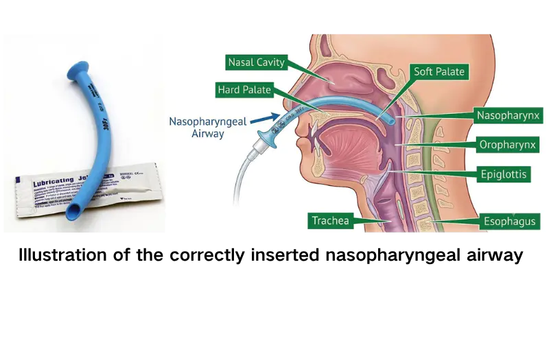
Nasopharyngeal Airway (NPA)
What is it?
Flexible tube inserted through the nostril.
Maintains ventilation in conscious or semiconscious patients.
Nursing considerations:
Measure from the tip of the nose to the earlobe.
Lubricate before insertion.
Insert gently toward the ear.
Position the bevel toward the septum
Oropharyngeal Airway (OPA)
What is it?
Hard, curved tube that prevents the tongue from obstructing the airway.
Used ONLY in unconscious patients.
Nursing considerations:
Measure from the corner of the mouth to the angle of the jaw.
Insert upside down, then rotate 180°.
NEVER use in a conscious patient.
Why? It can trigger the gag reflex and cause vomiting or aspiration.

Endotracheal Tube (ETT)
Purpose: Used for short-term mechanical ventilation or airway protection.
Indications:
Respiratory failure.
Apnea.
Airway obstruction.
Secretion management.
Aspiration risk.
Nursing responsibilities:
Confirm bilateral breath sounds and chest rise.
Maintain cuff pressure at 20–30 cm H₂O.
Document insertion depth.
Provide oral care every 2–4 hours.
Use chlorhexidine according to agency protocol.
Reposition and secure the tube according to policy.
Prevent ventilator-associated pneumonia (VAP).

Tracheostomy
What is it?
A surgically created opening through the anterior neck into the trachea.
Purpose:
Long-term ventilation.
Relief of airway obstruction.
Main components:
Outer cannula.
Inner cannula.
Flange.
Cuff, if present.

Chest Drainage System Components
1. Collection Chamber
Collects drainage from the pleural space.
Monitor and document drainage amount, color, and consistency.
2. Water-Seal Chamber
Acts as a one-way valve.
Allows air to leave the pleural space but prevents air from flowing back in.
Tidaling reflects pressure changes during breathing.
3. Suction-Control Chamber
Regulates the amount of suction.
Wet suction = water column.
Dry suction = dial regulator.
Nursing Responsibilities During Chest Tube Insertion
Position patient with the arm raised and HOB approximately 45°.
Maintain sterile setup.
Monitor for respiratory distress.
Provide analgesia and reassurance.
Apply an occlusive dressing after insertion.
Confirm placement with chest X-ray (CXR).
Nursing Management of Chest Drainage
Monitor drainage:
Record color, consistency, and amount.
Assess hourly initially.
Report drainage greater than 100 mL/hr or sudden bright-red output.
Maintain the drainage system:
Keep the drainage system BELOW chest level.
Avoid dependent loops and kinks.
Do NOT routinely milk or strip the tubing.
Encourage:
Coughing and deep breathing.
Range-of-motion exercises.
Mobility as appropriate.
Assess for subcutaneous emphysema:
Feels like crackling or bubbling under the skin.
May indicate an air leak.
Chest Tube Emergencies
If the chest tube becomes disconnected from the drainage system:
Place the distal end of the tube into sterile water, approximately 2–3 cm deep.
This temporarily re-establishes a water seal.
Reconnect to the drainage system as soon as possible.
If the chest tube is accidentally pulled out:
Apply an occlusive dressing taped on THREE sides.
Notify the provider immediately.
Monitor for respiratory distress.
If the water-seal chamber stops tidaling:
Assess for kinks.
Check for dependent loops.
Consider lung re-expansion.
CXR can help confirm lung re-expansion.
Important: Never routinely clamp a chest tube.
Chest Tube Removal
Premedicate for pain.
Tube is removed during the Valsalva maneuver.
Apply an occlusive dressing.
Obtain a post-removal CXR.
Types of Chest Surgery
Procedure | Description | Common Indications | Nursing Considerations |
|---|---|---|---|
Thoracotomy | Surgical incision into chest wall | Lung cancer, trauma, cardiac surgery | High pain risk; teach splinting and IS |
Lobectomy | Removal of one lung lobe | Tumor, bronchiectasis, infection | Chest tubes post-op; promote lung expansion |
Pneumonectomy | Removal of an entire lung | Extensive malignancy | No chest tube or gravity drainage; position on operative side |
Wedge/segmental resection | Removal of part of a lobe | Small lesions, biopsy | Encourage deep breathing and pain control |
Decortication | Removal of fibrous pleural layer | Empyema | Chest tubes post-op |
VATS | Minimally invasive thoracoscopy | Biopsy, nodule resection | Less pain, shorter stay, faster recovery |
pre/post op nursing care
Preoperative Nursing Care
Assess cardiopulmonary status.
Review CXR, ECG, ABGs, and pulmonary function tests (PFTs).
Teach breathing exercises.
Teach splinting.
Teach incentive spirometer use.
Provide reassurance and manage anxiety.
Postoperative Nursing Care
Manage pain aggressively (PCA or epidural as prescribed).
Monitor vital signs and oxygenation.
Assess drainage and lung sounds.
Encourage ambulation and range of motion.
Prevent DVT and pneumonia.
Assess incision for infection or dehiscence.
Memory trick:
Lobectomy = one lobe removed.
Pneumonectomy = entire lung removed.
VATS = minimally invasive approach.
Noninvasive Ventilation (NIV)
Noninvasive ventilation provides respiratory support without inserting an endotracheal tube.
Two important types are CPAP and BiPAP.
CPAP — Continuous Positive Airway Pressure
How it works:
Provides one continuous pressure throughout the respiratory cycle.
Keeps alveoli open.
Common uses:
Sleep apnea.
Heart failure.
Nursing considerations:
Monitor tolerance.
Assess skin integrity around the mask.
Monitor cardiac status.
Some patients may have difficulty exhaling against the pressure.
Contraindications:
Vomiting risk.
Altered level of consciousness.
Cardiac or respiratory arrest.
Acute MI.
Shock.
Maxillofacial trauma.
BiPAP — Bilevel Positive Airway Pressure
How it works:
Provides higher inspiratory pressure (IPAP).
Provides lower expiratory pressure (EPAP).
Helps improve ventilation and CO₂ removal.
Common indications:
COPD exacerbation.
Acute CHF.
Post-extubation respiratory support.
Contraindications:
Vomiting risk.
Altered level of consciousness.
Facial trauma.
CPAP vs. BiPAP
CPAP | BiPAP |
|---|---|
One continuous pressure | Two pressure levels |
Keeps alveoli open | Supports inhalation and exhalation |
Commonly used for sleep apnea | Commonly used for COPD exacerbation |
Supports oxygenation | Helps improve ventilation and CO₂ removal |
Memory trick:
CPAP = Continuous pressure.
BiPAP = TWO pressure levels.
Nursing Management of NIV
Assess level of consciousness.
Assess hemodynamic stability.
Ensure proper mask fit.
Patient must be able to remove the mask independently.
Do NOT use restraints to keep the mask on.
Provide frequent mouth and skin care.
Relieve pressure points.
Elevate HOB 30–45° to reduce aspiration risk.
If the patient's condition worsens, notify the provider for possible intubation evaluation.
Mechanical Ventilation
Mechanical ventilation supports or takes over breathing when a patient cannot adequately ventilate independently.
Types of Ventilators
Negative-Pressure Ventilation
Encases the chest or body.
Creates subatmospheric pressure to help air enter the lungs.
Example: iron lung.
Rarely used today.
Positive-Pressure Ventilation
Forces air into the lungs.
Exhalation is passive.
Can be volume-controlled or pressure-controlled.
Mechanical Ventilation Modes
Mode | How It Works |
|---|---|
Volume Control (VC) | Delivers a preset tidal volume; pressure varies |
Pressure Control (PC) | Delivers a preset pressure; tidal volume varies |
Assist-Control (AC) | Delivers set breaths; patient can trigger additional full-volume breaths |
SIMV | Allows spontaneous breaths between machine-delivered breaths |
Pressure Support (PS) | Augments the patient's spontaneous breaths |
Important distinctions:
Volume control = set volume, variable pressure.
Pressure control = set pressure, variable volume.
Assist-control = patient can trigger additional supported breaths.
SIMV = patient can breathe spontaneously between machine breaths.
Pressure support = helps the patient's spontaneous breaths.
PEEP — Positive End-Expiratory Pressure
What is PEEP?
PEEP is the pressure that remains in the lungs at the end of exhalation while the patient is receiving mechanical ventilation.
Purpose:
Prevents alveoli from collapsing between breaths.
Improves oxygenation.
Increases surface area available for gas exchange.
Decreases work of breathing.
Improves lung compliance and functional residual capacity (FRC).
PEEP Settings:
PEEP Level | Typical Use |
|---|---|
3–5 cm H₂O | Normal physiologic PEEP |
5–10 cm H₂O | Common therapeutic range |
10–20+ cm H₂O | May be used for severe alveolar collapse, such as ARDS |
Important nursing consideration:
Higher PEEP can increase intrathoracic pressure and decrease venous return to the heart. This can cause hypotension.
Memory trick: PEEP = Prevents alveoli from collapsing.
Endotracheal Intubation
Purpose: Establishes an airway for patients who cannot maintain adequate ventilation or protect their airway.
Steps of Intubation
Preoxygenate with 100% oxygen for 2 minutes.
Rapid sequence intubation (RSI) may be performed using a sedative and paralytic.
Insert the endotracheal tube using a laryngoscope.
Confirm placement with EtCO₂ and auscultation.
Secure the tube and document insertion depth.
Obtain a chest X-ray.
Nursing Responsibilities
Monitor SpO₂ continuously.
Assess bilateral chest rise.
Auscultate breath sounds.
Monitor for tube dislodgement.
Maintain cuff pressure at 20–30 cm H₂O.
Provide oral care every 2–4 hours.
Elevate HOB 30–45° to help prevent VAP.
Assess pain, anxiety, and sedation needs.
Use a sedation assessment tool such as the RASS scale.
Collaborate with respiratory therapy for ventilator adjustments.
HIGH-PRESSURE VENT ALARM
Means the ventilator is meeting increased resistance when delivering a breath.
Possible causes:
Secretions.
Kinked tubing.
Patient biting the tube.
Bronchospasm.
Obstruction.
Nursing actions:
Assess the patient first.
Check tubing for kinks.
Assess for secretions.
Suction if indicated.
Check for biting or obstruction.
Insert a bite block if appropriate.
LOW-PRESSURE VENT ALARM
Means the ventilator is detecting a loss of pressure, often due to a leak or disconnection.
Possible causes:
Disconnected ventilator tubing.
Cuff leak.
Accidental extubation.
Nursing actions:
Assess the patient.
Check all connections.
Assess cuff integrity.
If the tube is out, manually ventilate and call for help.
APNEA ALARM
Possible causes include disconnection, respiratory fatigue, or excessive sedation.
Assess the patient.
Stimulate the patient if appropriate.
Initiate manual ventilation if needed.
HIGH RESPIRATORY RATE OR HIGH VOLUME ALARM
Possible causes include pain, anxiety, or hypoxia.
Reassure the patient.
Assess sedation needs.
Evaluate oxygenation
Suctioning an ETT or Tracheostomy
Purpose: Removes secretions and maintains airway patency.
When Should the Nurse Suction?
Do NOT suction routinely. Suction only when clinically indicated.
Indications include:
Coarse breath sounds.
Visible secretions.
Decreased SpO₂.
Increased peak inspiratory pressures (PIP).
Suctioning Procedure
Assess the patient for signs of retained secretions.
Preoxygenate with 100% oxygen.
Insert the suction catheter WITHOUT applying suction.
Apply suction while withdrawing the catheter using a rotational motion.
Limit suctioning to 10 seconds or less.
Reoxygenate after each pass.
Limit suctioning to no more than 3 passes per episode.
Potential Complications
Hypoxemia.
Mucosal injury.
Dysrhythmias.
Nursing priorities:
Preoxygenate.
Limit suction duration.
Monitor SpO₂ and cardiac rhythm.
Reassess the patient after each pass
Barotrauma / Volutrauma
Excessive pressure or volume damages alveoli.
Can cause alveolar rupture and pneumothorax.
May lead to subcutaneous emphysema.
Nursing considerations:
Monitor respiratory status.
Assess breath sounds.
Monitor for sudden hypotension.
Watch for subcutaneous emphysema.
Oxygen Toxicity
Excessive FiO₂ can cause free-radical damage to the lungs.
Use the lowest effective FiO₂.
Ventilator-Associated Pneumonia (VAP)
Pneumonia that develops in a patient receiving mechanical ventilation.
Prevention:
Oral care.
Elevate HOB 30–45°.
Subglottic suctioning when indicated.
Follow infection-prevention protocols.
Hypotension
Positive-pressure ventilation increases intrathoracic pressure.
This decreases venous return to the heart.
Reduced venous return can decrease cardiac output and blood pressure.
GI Ulcers and Venous Thromboembolism (VTE)
Provide prescribed prophylaxis.
Encourage early mobility when appropriate.
Ventilator Asynchrony
Occurs when the patient is not breathing in coordination with the ventilator.
May be related to discomfort, anxiety, or ventilator settings.
Nursing interventions:
Assess comfort and sedation.
Collaborate with RT.
Evaluate ventilator settings
weaning definition
Gradually reducing ventilator support so the patient can resume independent breathing.
Weaning Readiness Criteria
Criteria | Target |
|---|---|
SpO₂ | ≥ 92% |
PEEP | ≤ 5–8 cm H₂O |
FiO₂ | ≤ 40% |
pH | ≥ 7.35 |
Hemodynamics | Stable |
Consciousness | Stable/appropriate |
Cough | Strong |
Secretions | Minimal |
Other steps:
Perform a spontaneous awakening trial (SAT).
Then perform a spontaneous breathing trial (SBT) lasting 30–120 minutes.
Formal readiness assessment every 24 hours.
Weaning Process & Post-Extubation Nursing Care
Ventilator support is gradually reduced.
Example from the lecture:
SIMV → Pressure Support Ventilation (PSV) → CPAP
Post-Extubation Nursing Care
Provide humidified oxygen.
Monitor for stridor.
Encourage coughing.
Encourage incentive spirometry.
Monitor oxygenation and respiratory effort.
Important: Stridor after extubation can indicate upper airway narrowing or obstruction and needs prompt assessment.
Tracheostomy Care
Nursing Management
Initial Care
Maintain airway patency.
Ensure the tracheostomy tube is secure.
Keep an emergency replacement kit at the bedside.
The kit should include an obturator and spare tracheostomy tube.
Suctioning
Use sterile technique.
Preoxygenate.
Limit suctioning to 10 seconds or less.
Inner Cannula Cleaning
If disposable: remove and replace with a new inner cannula.
If reusable: clean with sterile saline.
Stoma Care
Clean with mild soap and water.
Dry thoroughly.
Replace ties if soiled.
Humidification
Prevents airway dryness.
Helps prevent thick secretions.
Tracheostomy Emergency: Accidental Decannulation
If the tracheostomy is LESS than 7 days old:
Do NOT blindly reinsert the tube because a false passage may develop.
Call for emergency assistance.
Maintain oxygenation and ventilation.
The lecture describes covering the stoma with a sterile dressing and ventilating over the mouth and nose.
If the tracheostomy is MORE than 7 days old:
Use a sterile obturator to insert the replacement tracheostomy tube.
If unsuccessful, ventilate through the stoma with a bag-valve mask.
Always:
Monitor SpO₂.
Monitor heart rate.
Assess level of consciousness.
Maintain the airway until it is secured
Patient Teaching for a Permanent Tracheostomy
Teach the patient to:
Perform routine cleaning.
Follow the prescribed tube-change schedule.
Maintain stoma hygiene.
Perform suctioning as instructed.
Use humidification.
Use a protective shower cover to prevent water from entering the tracheostomy.
Keep a spare tracheostomy kit at home.
Recognize signs of infection or airway obstruction.
Provide writing materials.
Use communication boards.
Introduce speaking valves when appropriate and after cuff deflation.
Support psychosocial adjustment and body-image concerns.
Sudden Hypotension or Tachycardia During Mechanical Ventilation
Possible causes:
Excessive PEEP → decreased venous return.
Barotrauma → tension pneumothorax.
Ventilator disconnection → hypoxemia.
Sedation effects.
Shock.
Nursing actions:
Assess chest rise and symmetry.
Auscultate breath sounds.
Unilateral absence of breath sounds may indicate pneumothorax.
Assess the ventilator circuit for disconnection or obstruction.
Monitor BP, HR, and oxygenation.
If pneumothorax is suspected:
The lecture directs the nurse to disconnect from the ventilator and manually ventilate with a bag-valve mask.
Notify the provider immediately.
Prepare for possible chest tube insertion.
If tension pneumothorax is suspected:
Emergency needle decompression may be required.
This is followed by chest tube insertion.
Why is this dangerous?
Increased intrathoracic pressure can decrease venous return and cause obstructive shock.
Accidental Extubation or Decannulation
Recognize the emergency:
Sudden ventilator pressure loss or alarm.
Patient coughing or speaking.
Respiratory distress.
Visible displacement of ETT or tracheostomy tube.
Nursing priorities:
Stay with the patient.
Call for help immediately.
Restore oxygenation and ventilation.
Activate the emergency airway team when needed.
Continuously monitor SpO₂, HR, and level of consciousness.
If an ETT is accidentally removed:
Manually ventilate with a bag-valve mask and 100% oxygen.
Prepare for rapid re-intubation.
Activate the emergency airway team.
If a tracheostomy is accidentally removed:
Less than 7 days old: Do NOT blindly reinsert. Cover the stoma with a sterile dressing and ventilate over the mouth and nose as described in the lecture.
More than 7 days old: Use a sterile obturator to insert a replacement tube. If unsuccessful, ventilate through the stoma with a bag-valve mask.
Ventilator Alarms and Malfunctions
f you cannot identify or correct the cause of a ventilator alarm within 30 seconds, disconnect from the ventilator and manually ventilate with 100% oxygen.
Alarm | Possible Cause | Immediate Nursing Response |
|---|---|---|
High pressure | Secretions, kinked tubing, biting, bronchospasm | Assess obstruction, suction if needed, check tubing |
Low pressure | Disconnection, cuff leak, extubation | Check connections, assess cuff, evaluate tube placement |
Apnea | Disconnection, fatigue, sedation | Assess patient, stimulate, initiate manual ventilation if needed |
High RR/high volume | Pain, anxiety, hypoxia | Reassure patient, assess sedation and oxygenation |
High pressure = obstruction or resistance.
Low pressure = leak or disconnection.
Apnea = patient may not be breathing adequately.
MOST IMPORTANT: Assess the patient before assuming the ventilator is the problem.
Chest Tube Emergencies
If the chest tube becomes disconnected:
Submerge the distal end 2–3 cm in sterile water.
This temporarily maintains the water seal.
Reconnect to the drainage system as soon as possible.
If the chest tube is pulled out:
Apply an occlusive dressing taped on THREE sides.
Notify the provider immediately.
Monitor for respiratory distress.
If the water-seal chamber stops tidaling:
Assess for kinks.
Check for dependent loops.
Consider lung re-expansion.
Obtain CXR confirmation as appropriate.
Refractory Hypoxemia
Definition: Hypoxemia that persists despite oxygen therapy.
Possible causes:
ARDS.
Pneumothorax.
Pulmonary embolism (PE).
Sepsis.
Nursing actions:
Escalate FiO₂ and PEEP according to protocol.
Notify the provider.
Prepare for possible intubation and mechanical ventilation.
Ensure IV access.
Anticipate ABGs and CXR.
Prepare for possible transfer to critical care.
ABCO₂: Emergency Respiratory Assessment
Use ABCO₂ to organize your assessment when a patient is experiencing respiratory distress.
Letter | Meaning | Nursing Action |
|---|---|---|
A | Airway | Is it patent? Suction, reposition, or insert an airway adjunct if needed |
B | Breathing | Assess rate, depth, effort, and symmetry |
C | Circulation | Monitor BP, HR, and skin color; address oxygenation before hypotension worsens |
O | Oxygen delivery | Verify equipment, oxygen source, and FiO₂ |
2 | Two steps ahead | Anticipate escalation: RT, provider, or intubation |
Emergency Management: Key Takeaways
Always assess the PATIENT first, equipment second.
If in doubt, BAG THE PATIENT.
Manual ventilation is an immediate intervention when adequate ventilation cannot be maintained.
Recognize early signs of respiratory failure:
Rising EtCO₂.
Falling SpO₂.
Accessory muscle use.
Altered level of consciousness.
Activate rapid response or code early when oxygenation or ventilation cannot be maintained.
Document interventions and patient outcomes
KNOW THESE NUMBERS
Topic | Value |
|---|---|
Normal SpO₂ | 95–100% |
COPD SpO₂ target in lecture | 88–92% |
Normal EtCO₂ | 35–45 mmHg |
Nasal cannula | 1–6 L/min |
Simple face mask minimum | 6 L/min |
Non-rebreather | 10–15 L/min |
ETT cuff pressure | 20–30 cm H₂O |
Suction duration | ≤ 10 seconds |
Maximum suction passes | 3 per episode |
HOB for NIV/VAP prevention | 30–45° |
Chest tube drainage to report | >100 mL/hr |
PEEP common therapeutic range | 5–10 cm H₂O |
KNOW THESE CLINICAL CONNECTIONS
Decreased SpO₂ = assess oxygenation and ventilation.
Increased EtCO₂ = possible hypoventilation or CO₂ retention.
Decreased EtCO₂ = possible hyperventilation or poor perfusion.
Absent EtCO₂ waveform = immediately assess airway and circulation.
COPD exacerbation = BiPAP may help improve ventilation and CO₂ removal.
CPAP = one continuous pressure.
BiPAP = two pressure levels.
High-pressure ventilator alarm = obstruction or resistance.
Low-pressure ventilator alarm = leak or disconnection.
PEEP = keeps alveoli open.
Kussmaul respirations = compensation for metabolic acidosis.
Chest tube disconnected = distal end in sterile water temporarily.
Chest tube pulled out = occlusive dressing taped on three sides.
New tracheostomy decannulation = do not blindly reinsert.
Sudden hypotension + unilateral absent breath sounds during ventilation = suspect pneumothorax.
THE BIGGEST PRIORITY RULES
Assess the patient before troubleshooting equipment.
Airway and breathing come before routine interventions.
Do not ignore a sudden change in EtCO₂ or a flat waveform.
Do not routinely suction an intubated patient—suction when indicated.
Do not routinely clamp chest tubes.
If ventilation cannot be maintained, call for help and prepare to manually ventilate.