Reproductive System (Male) - Lecture Notes
 Call Kai
 Learn
Learn Practice Test
 Spaced Repetition
 Match
Match Flashcards
 Knowt Play
1/54

There's no tags or description
Looks like no tags are added yet.
Name | Mastery | Learn | Test | Matching | Spaced | Call with Kai | Chat |
|---|
No analytics yet
Send a link to your students to track their progress
55 Terms
Components in Male Systems:
Gonads: Testes
Gametes: Sperm
Sex Hormones: Testosterone
Accessory Reproductive Organs: Ducts, glands, and external genitalia
Male reproductive
Testes are the primary reproductive organ for males
Accessory:
Ducts and tubules leading from testes to penis
Male accessory glands
Penis

Scrotum Characteristics
Provides cooler environment
Needed for sperm development and maturation
Ridge seam at midline - raphe
Marks location of fusion of two embryonic tissue
Scrotum wall
Thin layer of superficial fascia internal to skin
Layer of smooth muscle, dartos muscle, internal to fascia

Scrotum Characteristics: Spermatic Cord
Multilayered structure traveling from abdomen to testis
Blood vessels and nerves supplying each testis
Originates in inguinal canal
Tube like passageway through inferior abdominal wall
Spermatic cord wall consists of three layers
Spermatic Cord Layers
External spermatic fascia: Formed from aponeurosis of external oblique
Cremaster muscle and cremasteric fascia: Formed from extensions of internal oblique and its aponeurosis
Internal spermatic fascia: Formed from fascia deep to abdominal muscles
Scrotum Characteristics (continue)
Testicular artery:
Branch from abdominal aorta
Resides within spermatic cord
Pampiniform plexus:
Plexus of veins surrounding testicular artery
Pre-cools arterial blood prior to reaching testes
Scrotum response to temperature changes
With hot temperature
Relaxation of dartos muscle
Testes move inferior (away from body), cools testes
Cremaster muscles relaxes to allow testes to move inferiorly
With cold temperature
Contraction of dartos and cremaster muscles
Testes and scrotum pulled closer to body
Helps conserve heat
Testes
Small organs housed within the scrotum
Produce sperm and androgens
Covered anteriorly and laterally by a serous membrane
Tunica vaginalis
Derived from abdominal peritoneum
Outer parietal layer and inner visceral layer
Separated by cavity filled with serous fluid

Testes: Tunica albuginea
Thick fibrous capsule covering the testis
Deep to visceral layer of tunica vaginalis
Testes: Mediastinum testis
Thickening of tunica albuginea projecting into interior testis
Blood vessels, ducts, lymph vessels, nerves enter or leave through here
Testes: Septa
Internal projections of tunica albuginea
Subdivide internal space into 250 lobules
Testes: Seminiferous tubules
Extremely convoluted and elongated; up to 4 per lobule
Nondividing support cells, sustentacular cells
Nourish developing sperm
Release hormone inhibin when sperm count high
Inhibits FSH secretion and regulates sperm production
Contain dividing germ cells continuously producing sperm

Testes: Blood-testis barrier
Protects developing sperm from material in blood
Protects sperm from body’s leukocytes
Formed from tight junctions between sustentacular cells
Testes: Interstitial spaces and cells
Interstitial spaces:
Spaces surrounding seminiferous tubules
Interstitial cells:
Reside in interstitial spaces
Stimulated to produce androgens by luteinizing hormone
Most common androgen: testosterone
Majority released from interstitial cells; small amounts secreted by adrenal cortex
Hormonal regulation
Hypothalamus secretes GnRH
FSH and LH stimulate spermatogenesis and androgen production
Increased testosterone has multiple effects
Sustentacular cells release inhibin
Testosterone stimulates libido and development of secondary sex characteristics

Spermatogensis
Spermatogonia: Stem cells
Primary Spermatocyte: Undergoes meiosis I
Secondary Spermatocyte: Undergoes meiosis II
Spermatids: Mature into sperm
Occurs in the seminiferous tubule

Spermatogonia
Primordial germ cells from which all sperm develop
Diploid cells near base of seminiferous tubule
Surrounded by cytoplasm of sustentacular cell
Divide by mitosis into new spermatogonium and primary spermatocyte
Clinical View: Sexually Transmitted Infections
Groups of infectious diseases transmitted via sexual contact
STIs transmitted because symptoms go unnoticed
Condoms help prevent spread but not 100% effective
A leading cause of pelvic inflammatory disease in women
Can lead to blockage of tubes and infertility
Clinical View: Chlamydial
Most frequently reported in U.S.
Due to Chlamydial trachomatis bacterium
Most people are asymptomatic
May have symptoms of vaginal discharge, painful urination, back pain
Treated with antibiotics
Clinical View: Genital Herpes
Caused by herpes simplex virus
Cyclic outbreaks of blister formation in genital and anal regions
No cure; antiviral medications lessen severity and length
Clinical View: Gonorrhea
Caused by bacterium Neisseria gonorrhoeae
Spread by sexual contract or from mother to baby during delivery
Treatment with antibiotics; untreated may cause epididymitis in males
Clinical View: Syphilis
Cause by bacterium Treponema pallidum
Spread sexually via contact with syphilitic sore, chancre, or in utero
Treatment with antibiotics
Clinical View: Paternal Age Risks for Disorders in the Offspring
Males over 45 years of age
4 times more likely to pass on genetic mutation than men in their 20s
Risk increases with age
Bipolar disorder, schizophrenia, autism, depression, attention deficient hyperactivity disorder
Maternal age not correlated with an increase in these disorders
Age of both sexes carries risk of development problems
Duct System: Epididymis
Head on superior testis surface
Body and tall on posterior surface of testis
Internally contains long duct of the epididymis
Lined with pseudostratified columnar epithelium with stereocilia
Stores sperm until they are fully mature and motile
Duct System: Ductus deferens (vas deferens)
Sperm enter after leaving epididymis
Thick-walled muscular tube in the male reproductive system
Transports sperm from the epididymis to the ejaculatory duct
Enlarges and forms ampulla
Unites with proximal seminal vesicle to form ejaculatory duct
Duct System: Ejaculatory Duct
Each duct is between 1 and 2 cm long
Epithelium of pseudostratified ciliated columnar epithelium
Conducts sperm and a component of seminal fluid toward urethra
Opens into prostatic urethra
Duct System: Urethra
Transports semen from ejaculatory ducts to outside of body
3 components
Prostatic urethra: extending from bladder through prostate gland
Membranous urethra: continuing through urogenital diaphragm
Spongy urethra: extending through penis
Duct System
Epididymis → Ductus Deferens → Ejaculatory Duct → Urethra

Accessory Glands
Seminal Vesicles: Fructose for sperm energy
Prostate Gland: Produces PSA
Bulbourethral Glands: Lubrication
Semen: Sperm + seminal fluid
Clinical View: Benign Prostatic Hyperplasia
Noncancerous enlargement of prostate gland
Common in older men
Due to hormonal changes in aging males
Large nodules forming within prostate and compressing urethra
Frequent urinating at night, frequent and painful urination
Can be treated with drugs
May require surgical removal of prostatic enlargement
Clinical View: Prostate Cancer
One of most common malignancies in men over 50
Forms hard, solid nodules with prostate
Asymptomatic early on, later urinary symptoms
Screening with digital rectal exam, PSA test
Treatment: radiation therapy or surgical removal
Penis
Forms external genitalia with scrotum

Penis: Root
Internally attached portion of penis
Dilated internal to body surface
Forms bulb and crus of penis
Bulb attaching penis to bulbospongiosus muscle
Crus attaching penis to public arch
Penis: Body, Glans & Prepuce
Body: Elongated movable portion
Glans:
Tip of penis
Contains external urethral orifice
Prepuce (foreskin):
Circular fold of skin
Skin attached to raised edge of the glans
Penis: 3 Cylindrical Erectile Bodies
Unsheathed by tunica albuginea
Provides attachment to skin over penile shaft
Paired corpora cavernosa
Located dorsolaterally & terminate in shaft of penis
Corpus spongiosum
Ventral to corpora cavernosa in midline
Contains spongy urethra and continues within the glans
Anatomy of Penis: Cross Section

Phase of male sexual response: Excitement phase
Erectile bodies of penis composed of venous spaces surrounding artery - blood fills it
Erectile body becomes rigid - erection
Parasympathetic control (nitric oxide release)
Blood unable to leave erectile bodies until excitement over
Near the end of the phase, heart rate, blood pressure, and respiratory rate increase
Phase of male sexual response: Orgasm
Intense pleasure, tension release, expulsion of semen
Ductus deferens move sperm toward urethra
Accessory glands secrete seminal fluid components:
Combine with sperm to form semen
Internal urethral sphincter of bladder contracted
Ensures no urine enter urethra
Ejaculation at end of orgasm:
Occurs due to sympathetic control
Sex Differentiation: Genetic Sex
Genotypic sex based on sex chromosomes inherited
Determined at fertilization
XX → genetic female
XY → genetic male
Sex Differentiation: Phenotypic Sex
Refers to at he appearance of the individual’s internal and external genitalia
Ovaries and female external genitalia, phenotypic female
Testes and male external genitalia, phenotypic male
Apparent seventh week of development
Sex-determining region Y (SRY) gene
Located within larger testis-determining factor (TDF) region on Y chromosome
In genetic male with Y chromosome
SRY gene expressed
Produces proteins to stimulate androgens
Androgens initiate male phenotypic development
If Y chromosome is absent, or lacks or has an abnormal SRY gene, female phenotypic sex results
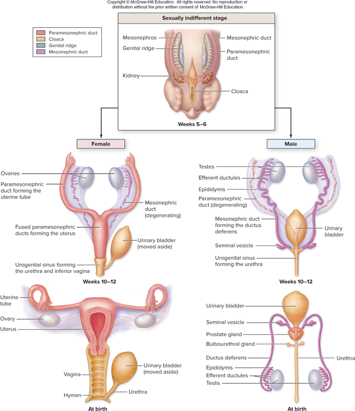
Duct and gonad development: Genital Ridges
Form in fifth week of embryonic development
Form from intermediate mesoderm
Will form gonads
Medial to developing kidneys
Duct and gonad development: Primordial germ cells
Migrate from yolk sac to genital ridges between weeks 5 and 6
Will form future gametes
Duct and gonad development: Mesonephric ducts
Form most of male duct system
Connect mesonephros to developing urinary bladder
Duct and gonad development: Paramesonephric ducts
Form most of female duct system
Include uterine tubes, uterus, superior vagina
Appear lateral to mesonephric ducts
Duct and gonad development
Only one remaining in fetus
Female:
Development of paramesonephric ducts
Degeneration of mesonephric ducts
Male:
Development of mesonephric ducts
Degeneration of paramesonephric ducts
Puberty
Increased GnRH, FSH, LH secretion
Females: Breast development, menarche
Males: Testicular and penile growth, voice deepens
Clinical View: Intersex Conditions (Disorders of Sex Development)1
Discrepancy between genotype and external genitalia
True gonadal intersex - very rare
Individual with both ovarian and testicular structures
Ambiguous external genitalia
Genetic male or genetic female
Pseudohermaphroditism
Genetic sex and phenotypic sex do not match
Clinical View: Intersex Conditions (Disorders of Sex Development)2
46 XY intersex:
Genetic male with external genitalia resembling a female
From reduction of lack of male hormones during development
46 XX intersex
Genetic female with external genitalia resembling male
Clitoris enlarged and labia partially fused
May result if fetus exposed to excessive androgens
May result from congenital adrenal hyperplasia
Fetus’s adrenal glands producing excessive androgens
The areolar glands are ___
sebaceous glands
What do testes produce
androgens
spermatozoa
A serous membrane, the tunica ___around each testis has a parietal and visceral layer
vaginalis
The labia majora of the female are homologous to the ___ of the male
scrotum
Name of the process where spermatids become mature spermatozoa
spermiogensis
All the stages in which the cells are haploid
spermatozoon
secondary spermatocyte
spermatid