NEU C61 Midterm #2
 Call Kai
 Learn
 Practice Test
 Spaced Repetition
 Match
 Flashcards
 Knowt Play
1/217

There's no tags or description
Looks like no tags are added yet.
218 Terms
Embryonic Brain Development
Brain organization emerges within weeks of conception
At birth, the brain is only ~20–28% of its adult volume
Most of early development (cell proliferation, migration, connectivity etc.) - is genetically programmed, not learned
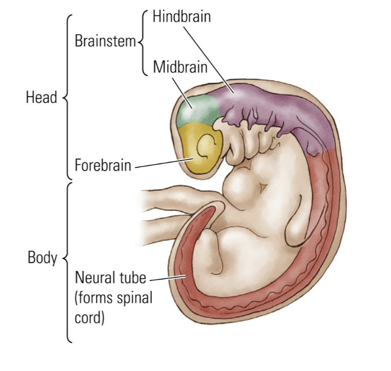
Embryonic Brain Development Timeline
Day 1: fertilization; sperm + egg = zygote
Day 2: Rapid cell division begins
After ~1 week: blastocyst, with thousands of cells and an embryonic disk
Cell fate also begins to be determined. Cells become progenitor/precursor cells headed toward nervous system, heart, skin, etc
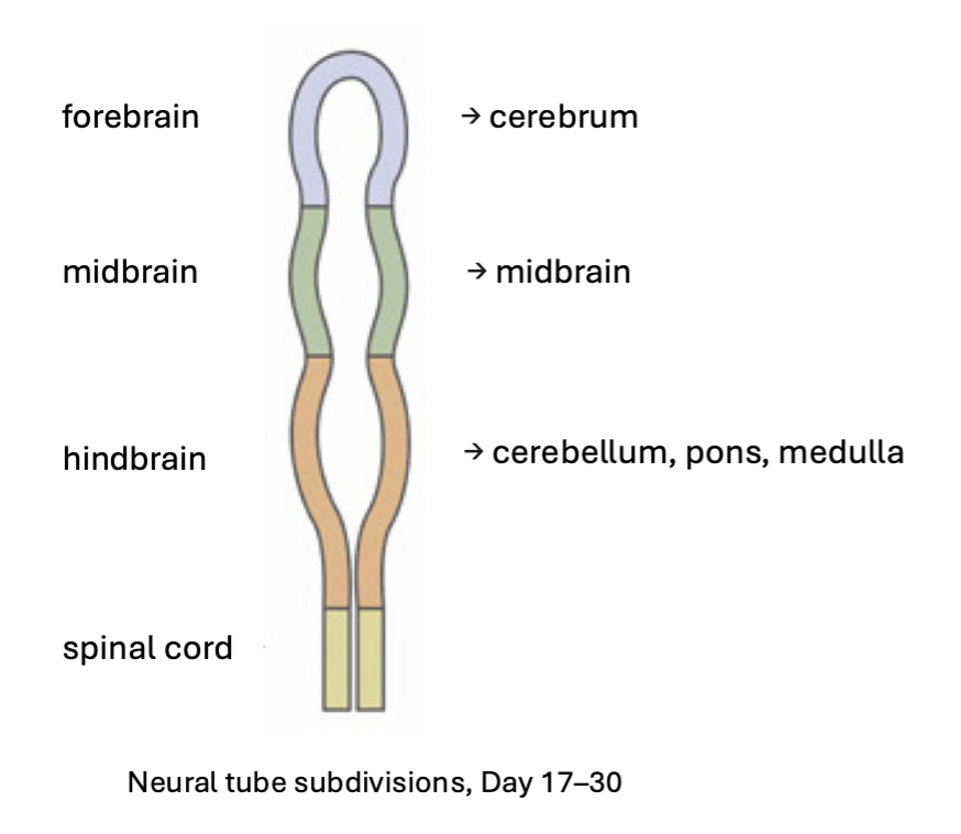
Day 17-30: Neural tube forms, which eventually becomes the nervous system

Neural Tube Segments
Hollow center of the tube becomes the ventricles (lined with ependymal cells, which make and distribute CSF)
Genetics
~3 billion base pairs total
~21,000 protein-coding genes
Less than 1% of the genome codes for protein!
Most of the rest is regulatory (transcription factors) that control when/where genes turn on by enhancing or blocking RNA polymerase
Every cell has the same DNA; different cells express different genes. Cell type = which genetic program is being expressed
Proliferation and Differentiation of Brain Cells
Stem cell → nervous system progenitor (nervous system in general) → neural precursor cells (neurons specifically) → differentiate into neurons or glia
A stem cell divides asymmetrically: one daughter stays a progenitor (cell that divides to produce more specialized cells), one differentiates

Terminally Differentiated Meaning
Once mature, they never divide again. This is because neurons have many processes and would need to retract them to divide, which is metabolically impractical.
How is the cortex built?
Built by radial glial cells, glia with long processes that divide to produce neurons and astrocytes
Each cell migrates between the ventricular surface and the pial surface (outer surface), then returns and divides
Symmetric division: two new radial glia (the stem cell pool expands)
Asymmetric division: one radial glial cell + one neural precursor, which can divide again into a neuron and a glial cell

What are transcription factors?
Transcription factors are proteins that bind DNA and control RNA polymerase
Which combination of transcription factors is present determines which genes get transcribed
This is how one genome produces thousands of distinct cell types
Mechanism of symmetric or asymmetric division
Based on proteins (e.g. notch and numb which are antagonistic and sit at opposite ends of the cell. Angle of the mitotic spindle determines whether both daughters get both proteins (symmetric) or one gets Notch and the other Numb (asymmetric))

Migration of Neurons
New neural precursors climb the radial glial processes toward the marginal zone, "like climbing a tree."

“Inside-Out” Cortex
The adult cortex has six layers (layer 1 has no cell bodies, mostly neuronal processes). Deep layers form first, later cells migrate past them to form outer layers. Layer 6 is deepest and forms first; layers 1–2 are most superficial and form last.

Neuron Polarity
Migrating neurons initially have no axon or dendrites; they form on arrival.
Semaphorin 3A is secreted at the cortical surface (near layer 1). It repels axons and attracts dendrites. This cue gives the pyramidal neuron its polarity
It's a developmental signal; the gradient goes away once polarity is established.

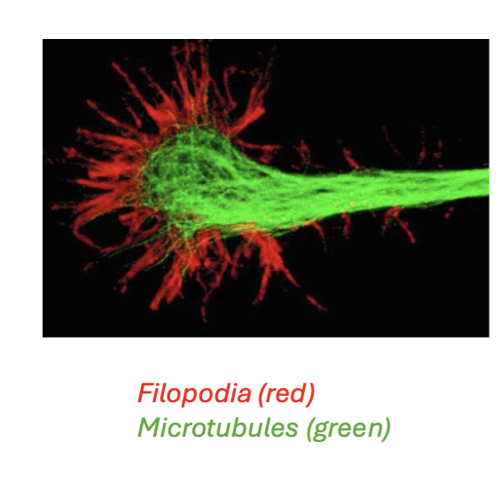
What is a growth cone?
The tip of a growing axon. Made of microtubules and actin filaments that push out filopodia (red), finger-like projections with chemical receptors that "sniff" for cues. Microtubules (green) form the stiffcore structure of the growing axon and act as a transport highway. Both are polymers built by adding monomers.
Fasciculation
Axons bundle together via cell adhesion molecules (CAMs). A pioneer axon does the searching while others follow. It’s kinda like they’re piggybacking on the earlier axons, and a whole bunch of axons are guided by just a few pioneers finding the way.
Laminin: Major protein in extracellular matrix, helps tissues survive, guides cell movement, and gives structure to organs
Integrin: extracellular receptors for laminin

Chemical Guidance Cues
Growth cones follow gradients of secreted molecules
Chemoattractants (e.g., netrin) pull the growth cone toward the source
Chemorepellents (e.g., slit) push it away
Both soluble (diffuse from a point) and contact- based (physically bump into another
cell, then is attracted/repelled) cues exist

Cajal and Sperry
Santiago Ramón y Cajal (the Nobel winner discussed alongside Camillo Golgi, famous for stains and drawings) first described the growth cone in chick embryos
Roger Sperry asked: is axonal connectivity genetically fixed, or shaped by experience
Sperry’s Frog Experiment Background
Background: the optic nerve carries visual information from retina → thalamus → visual cortex. Visual (and most sensory) systems are topographic: neighboring points in the visual field map to neighboring neurons, giving a continuous spatial map.
Why frogs: amphibians can regenerate a cut optic nerve (humans can't; severing it causes permanent vision loss). Frogs also have a stereotyped behavior: striking at a fly with their tongue, which requires accurate visual localization. Perry wanted to know if eye-to-brain map is hardwired or shaped by experience

Sperry’s Frog Experiment Manipulations
Rotate the eye 180° (nerve intact): the frog is permanently confused. It strikes 180° opposite the fly's true position, for life
Cut the optic nerve, no rotation and let the nerve regrow: the frog recovers accurate strikes
Cut the nerve AND rotate the eye, then let it regrow: the axons still grow back to their original targets in the tectum, so the frog makes the same 180° misdirected strikes as in #1. The wiring is genetically specified, not shaped by experience.
Chemoaffinity Hypothesis
Neurons carry distinct chemical “address tags” that guide axons back to the same targets every time. his is genetically programmed, it does not adapt to whatever the eye's actual orientation is. It is a gradient

Neurotrophin
A protein promoting neuron survival and axon growth
What is NGF (nerve growth factor)?
Discovered by Rita Levi-Montalcini and Stanley Cohen, the first neurotrophin. They won the Nobel Prize for discovering it
How Synapses First Form
Axon and dendrite filopodia touch; the presynaptic side builds machinery (active zone, vesicles) and the postsynaptic side clusters receptors.
Example, neuromuscular junction: the motor neuron releases agrin, which binds MuSK receptors on the muscle, causing acetylcholine receptors to cluster at the synapse.

Overproduction During Development
Neurons are overproduced during development; only those that successfully connect and receive enough neurotrophin survive
Losers undergo apoptosis (programmed cell death)
This “pruning” of synapses refines neural circuits and makes them more specific

What is neuroplasticity?
The brain's capacity to change with experience. Unlike development, this is activity- and experience-dependent, not purely genetic
What is Hebbian Plasticity?
Neurons that fire together wire together. Fire out of sync, lose their link. Co-active connected neurons strengthen; out-of-sync ones weaken.
NMDA Receptors
Both AMPA and NMDA receptors are glutamate receptors.
At rest, the NMDA channel is blocked by a magnesium ion. Opening requires both: (1) glutamate binding and (2) postsynaptic depolarization (e.g., from AMPA receptor activation) to expel the Mg²⁺ block. So it's both ligand- and voltage-gated.
AMPA receptors are fast glutamate-gated channels, always available
When it opens, calcium enters, triggering plasticity: up- or down-regulating AMPA receptors in the postsynaptic membrane
Result: NMDA receptors only pass current when pre- and postsynaptic activity coincide (Hebbian plasticity)
Play a key role as coincidence detectors

Long-Term Depression (LTD)
Weak NMDA receptor activation triggers rapid removal of postsynaptic AMPA receptors
Fewer AMPA receptors → that synapse has less and less influence → eventually eliminated
This is the same logic behind “use it or lose it” at the level of a single synapse
Low firing rate between pre- and post-synaptic cell

Synaptic Pruning Across Childhood
Synapse density rises steeply after birth, peaks in early childhood, then declines through adolescence
Net loss of synapses causes measurable thinning of cortical gray matter
Pruning proceeds caudal → rostral: prefrontal cortex is the last region to mature

Axon growth and synaptogenesis in human cortex
Synaptogensis: Biological process by which neurons form new synapses, based on the image you can tell that they take a while to grow

Why do we prune synapses at all?
Activity-dependent stabilization: synapses that are used are strengthened and kept
Activity-dependent elimination: synapses that are rarely or never used are weakened and removed
Microglia (the brain's immune cells) physically engulf and clear away weak, “tagged” synapses (complement/C1q signaling)
Net effect: circuits are sculpted down to the connections actually supported by experience
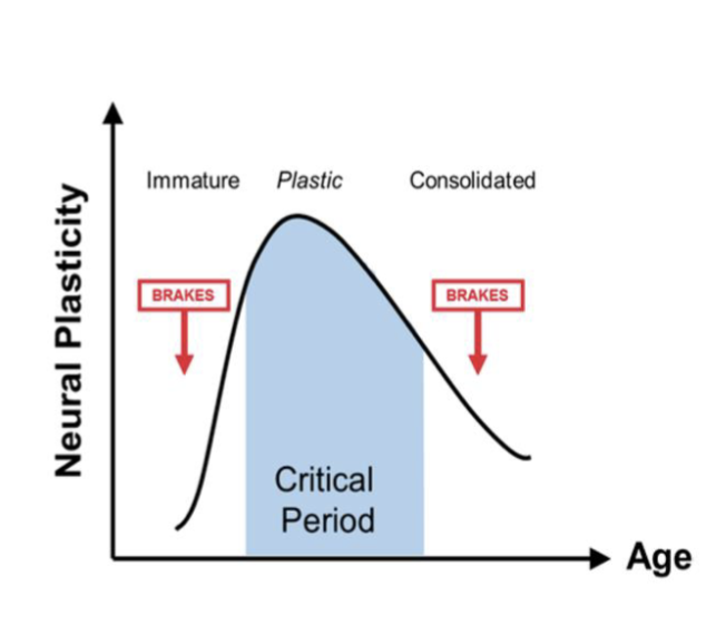
Critical Periods in Development
Critical Period: a developmental window when a circuit is unusually plastic — shaped easily by
experience, then much less so afterward

Why do critical periods end?
No single answer, several mechanisms likely:
Axon growth itself ceases in the relevant circuit
Excitatory synaptic transmission matures (NMDA receptor subunits change)
Patterns of cortical activation become more constrained
Inhibitory interneurons mature and dampen further rewiring
Understanding why plasticity turns off is key to therapeutics for traumatic brain injury in adulthood!
Epigenetics
Factors that change gene expression without changing DNA sequence. Example: DNA methylation- addition of a methyl group onto histones → blocks expression of that gene. Methyl groups can be inherited from parents. Also can be experience dependent
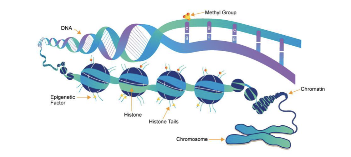
Example of Epigenetics
Low maternal licking & grooming → methyl groups added → some hormone receptor genes not expressed → high anxiety pups
High maternal licking & grooming → no methyl groups → low anxiety pups

Marr’s Three Levels of Analysis
Computational: Why/Goal
Algorithmic: What Rules/Mental Processes are Used to Achieve It?
Implementational: How/What Physical Processes Does it?
Marr’s Three Levels of Analysis Bird Flight Example
Computation (Flight): Why fly? Cooperate in a flock, compete for food, hunt (if a hawk)
Algorithm (Flapping): How? Spatial learning/mapping, reinforcement learning, cost-benefit computation
Implementation (Feathers): What? Wings, muscles, rain areas, circuits, cells, molecules
Correlation
A and B occur together, Example: fMRI: Region X is active during behavior Y
Necessity
Without A, B doesn’t happen. Example: Inhibition/Damage: Remove region X → behavior Y is lost
Sufficiency
Whenever A, B happens. Example: Stimulation: Activate region X → behavior Y occurs
Four Trade Offs When Understanding the Brain
Spatial resolution: How precise does the tool identify the location of a signal? Smallest unit you can resolve (single neuron vs. neighborhood vs. whole cortex).
Temporal resolution: How fast you can record/control activity (milliseconds for action potentials vs. seconds for blood-based signals).
Coverage: How much of the brain you can access (ten neurons vs. whole brain).
Invasiveness: How much damage is required (an MRI causes none; recording from a mouse brain usually means going through the skull).
X-Ray Photography
High-energy, short-wavelength light, passes through soft tissue, absorbed by bone and dense structures
Differential absorption creates contrast: bone is bright; soft tissue is faint
First time in history it was possible to see inside a living human body
Cons: poor soft-tissue contrast for brain; ionizing radiation damages DNA
Type of structural brain imaging
CAT Scan (CT Scan)
3D X-Ray Images
Non-invasive
Very high exposure to radiation
Usually used in medical settings for injury or disease diagnostics
Type of structural brain imaging
MRI (Magnetic Resonance Imaging)
Strong magnet aligns hydrogen protons in the body's water; a radio pulse knocks them out of alignment
As protons relax back, different tissues emit signal at different rates, creating anatomical contrast
Strengths: excellent soft-tissue contrast; no ionizing radiation (unlike X-ray/CT)
Limitations: expensive, slow, requires holding still in a strong magnetic field
Type of structural brain imaging
EEG (Electro-encephalography)
Type of functional brain imaging
Electrodes on the scalp measure summed electrical fields from millions of neurons firing in coordination
Signal attenuates to nanovolts crossing skull and tissue — only massive, synchronous activity is detectable
High temporal resolution (~ms); very poor spatial resolution (~cm)
Cheap, portable, completely non-invasive - genuine clinical and research advantages
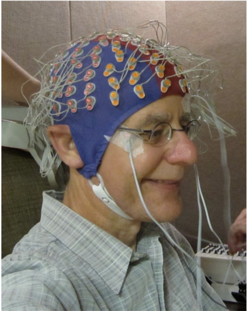
EEG Brain Waves

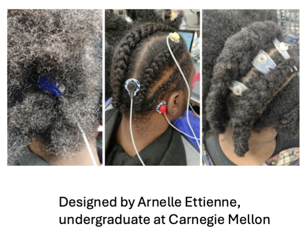
Racial Bias in EEG Studies
Standard electrodes on unbraided textured hair produce high impedance (more than 12× the typical threshold)
Participants excluded or asked to chemically alter/shave hair
Only 5 of 81 EEG studies surveyed included any Black participants
Solution: Sevo electrode (Etienne et al., 2020), designed by an undergraduate who noticed the problem because it was her own hair.
Flexible wing-shaped clips side under cornrows; reduced impedance by 95%
ECoG (Electrocorticography)
Like EEG, but placed under the skull, directly on the cortical surface
Signal no longer filtered by skull and tissue, much better spatial resolution (~mm vs. ~cm for EEG)
Used clinically for pre-surgical epilepsy mapping
Enables research on language, memory, and speech in awake human patients
Highly invasive, only performed on patients who require surgery for clinical reasons
Type of functional brain imaging

fMRI (Functional MRI)
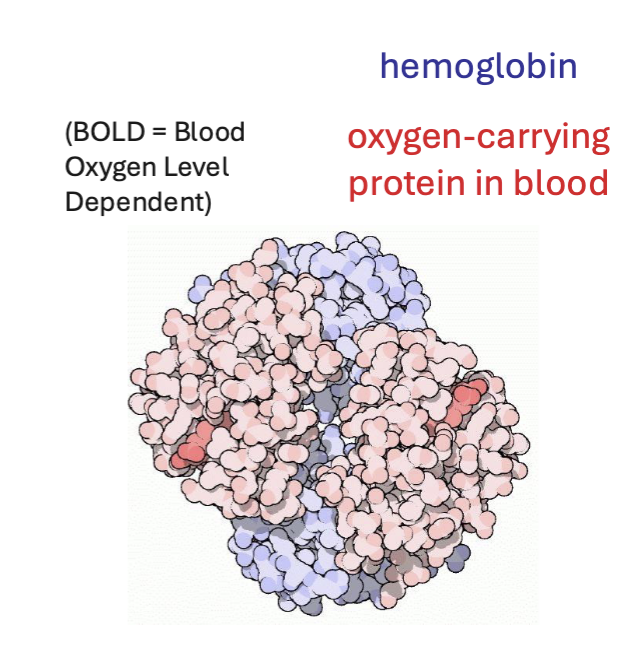
Neural activity creates metabolic demand → local increase in oxygenated blood → detectable change in MR signal
Oxygenated and deoxygenated hemoglobin have different magnetic properties
Whole brain coverage; spatial resolution ~1-3 mm (each voxel = 1 million neurons)
Temporal resolution: 4-6 second hemodynamic response, measuring blood flow, not neural activity directly
Type of functional brain imaging
Electrophysiology
Gold-standard for temporal resolution millisecond precision
Directly captures action potentials (“spikes”)
High spatial and temporal resolution, at the level of neurons
Can isolate single neurons or small populations
But coverage is sparse (you record from tiny fractions of neurons in specific parts of the brain)
Invasive requires electrode implantation → primarily used in animals (and rare clinical cases in humans)
Tradeoffs: High precisions but low coverage, neurons recorded can change across days, makes longitudinal studies more challenging
Type of functional brain imaging

In-vivo Calcium Imaging
In vivo fluorescent imaging of neural activity
Uses an engineered molecule called GCaMP: GFP + calmodulin (a calcium-binding protein)
Calcium entering during an action potential makes GCaMP fluoresce ~10x brighter
Light shone through a window or lens lets you watch hundreds to thousands of neurons flash as the animal behaves
~100ms temporal resolution (slower than electrodes but still very good)
Requires genetic modification, not used in humans currently
Main benefit is for longitudinal tracking across days
Type of functional brain imaging

Example use of in-vivo calcium imaging of taste-responsive cells
Calcium imaging found neurons selective for specific tastes (sweet, bitter, salty, sour), plus a "salty hotspot" where ~40% of cells responded to salt.

Transcranial Magnetic Stimulation (TMS)
A magnetic field induces an electrical current in underlying cortex, briefly disrupting activity in ~1–2 cm patch of brain. Modern method to establish causality.
Easily reversible
Can be done in healthy participants
Applied at a specific moment during a specific task (high temporal resolution)
Used for treatment-resistant OCD and smoking cessation
Limitation: Surface cortex only; stimulating a region also disrupts its connections

Deep Brain Stimulation (DBS)
Another method to establish causality
Surgically implanted electrodes deliver continuous electrical stimulation to deep brain structures (highly invasive)
Most established use: for Parkinson’s disease - remarkably effective for controlling tremor
Also used for treatment-resistant depression, epilepsy, OCD
Limitation: blunt (stimulates all cells and fibers
nearby without much specificity)

Optogenetics
A technique that uses light to control the activity of genetically defined neurons
Combines genetics + optics + neuroscience
Allows millisecond-precision control of neural firing
Introduce light-sensitive proteins (opsins) into specific neurons (via viral vectors or transgenic animals)
Implant a fiber optic cable to deliver light into the brain (very invasive)
Can control neural activity by switching light on and off

Optogenetics Example
Activating neurons in the subfornical organ (controls water balance/thirst) makes a mouse lick for water; when the light goes off, licking stops.

What’s the difference between low and high contrast structural MRIs?
MRIs with contrast use a dye injected via IV that highlights tissues
Causes specific structures like tumors or blood vessels to light up brighter
Good for detecting very small tumors, early stages of cancer, inflammation, etc.
Non-contrast MRIs are best for structural injuries like lesions in the brain, joint problems etc.

General Framework for All Sensory Systems
A stimulus in the environment (light, touch, odor, sound, etc.)
Acts on receptor cells (multiple types per system), which change their membrane (receptor) potential. Some receptor cells are neurons, some are not.
They communicate with the first neuron in the system: afferent neurons, which carry information from the periphery toward the spinal cord/brain via action potentials.
Afferent/relay neurons pass information to higher-order pathways (sometimes with stops along the way, sometimes straight to cortex).
Cortex is generally responsible for representing the sensation/perception
Sensations are constrained to what can be detected by receptors & what can be interpreted by the brain
Vision
Dominate primate sense
1/3 of our cerebral cortex is dedicated to vision
100M photoreceptors per eye (~100 megapixels)
No other sense comes close to vision's spatial resolution and bandwidth
Diurnal
Organisms that evolved in well-lit environments where vision provides an extraordinary information advantage. Includes humans and most primates
Vision vs Camera
Vision
Makes approximations of the world
Uses context, prior knowledge, expectation
Camera
Captures what is physically there
Faithful representation of input
No interpretation
Examples
Color context illusion: a center patch looks bluish-gray on one background and yellowish on another, but when the surround is removed they are the same gray
Anatomy of the Eye
Where light becomes a signal

What does the lens and cornea and pupil do in the eye?
Focus incoming light onto the retina + controls how much light enters
What does the retina do in the eye?
Light-sensitive sheet lining the back of the eye, where phototransduction happens
What does the fovea do in the eye?
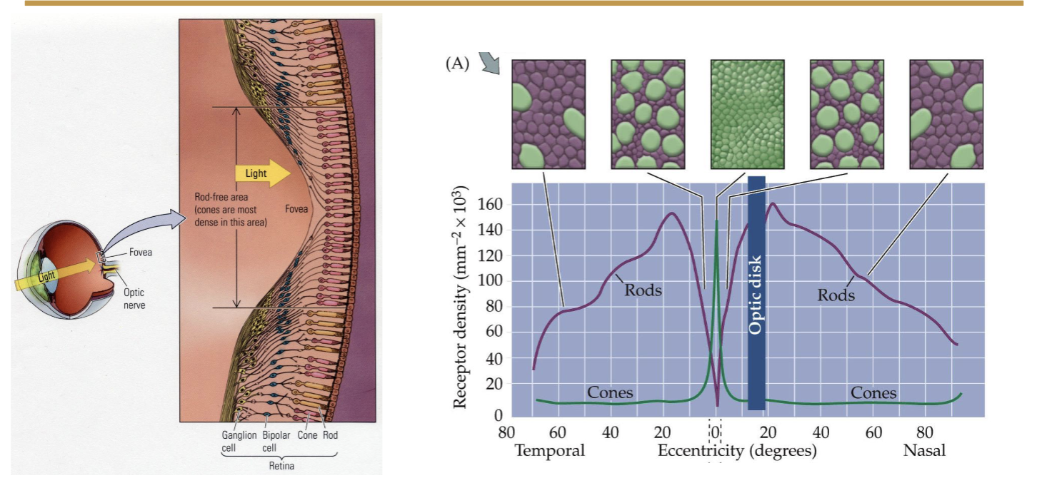
High-acuity center because of high photoreceptor density. Enables sharpness and color vision.
What does the optic disc do in the eye?
Where the optic nerve exits. Location of blind spot because no photoreceptors are here.
What two types of photoreceptors does the retina contain?
Rods and cones
Rod
High sensitivity, specialized for night vision (can detect single photons of light)
Achromatic (without color, black and white)
Not present in fovea: low acuity (sharpness)
~120 million per eye
Contains photoreceptor protein called rhodopsins
Concentrated in the periphery

Cone
Cone = Colorful
Low sensitivity, specialized for day vision
~6 million per eye
Chromatic: 3 types of pigments (S, M, and L)
Contains photoreceptor protein called cone-opsins
Concentrated in fovea: high acuity (sharpness)
Distribution of Rods and Cones in the Retina Images
Structure of Rods and Cones

Structure of a Rhodopsin Molecule

Phototransduction
Photon of light passes through retinal layers until it hits rhodopsin/opsin in rod or cone
Rhodopsin changes shape (isomerization)
G-protein cascade activates (signal amplification)
4. Na⁺ channels close
5. Photoreceptor hyperpolarizes
Bipolar cells receive graded (analog) signal and pass signal to ganglion cells
Receive and integrate input from bipolar cells
Generate action potentials
9. Axons form the optic nerve → brain
One single photon is enough to trigger this entire cascade: one of the most sensitive detection systems in biology

Electromagnetic Spectrum

How Trichromatic Vision Works
Three cone types: Short (S), Medium (M), Long (L)
Color perception comes from downstream visual areas comparing the outputs of all three
Your brain computes ratios: how much is L firing vs. M vs. S?
Colorblindness is usually due to a missing or altered M or L cone

What is color?
Primary quality:
A wavelength of light, e.g. 650 nm for red
Measurable by a spectrometer
Exists objectively in the world
Secondary quality:
The experience of ‘red’
Constructed through perception
Subjective (only exists as experience)
Cannot be objectively measured
What is qualia?
The subjective, felt quality of an experience. Example: What is it like to be a bat? - Thomas Nagel, 1974. You can know everything about bat echolocation - every neuron, every circuit —-and still not know what it feels like from the inside. That inner felt quality is a “quale.”
Layered structure of the retina
Ganglion Cells: Fire action potentials, send output via optic nerve
Bipolar Cells: Excitatory cells, first level of processing
Rod and cone photoreceptor cells: light sensitive, but hyperpolarized
Light reaches the front first but signal actually starts from the back first

What are receptive fields?
For any sensory nerve cell, the region of space that when stimulated, changes a neurons firing rate. Example in touch: each neuron's axon terminals sit in a patch of skin; touching that patch drives it, while another patch doesn't.
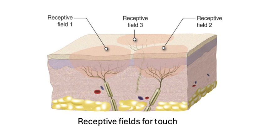
Visual receptive fields
Patches of the visual field (left and right halves) that a given neuron responds to.

Center-Surround Receptive Fields
Retinal Ganglion Cells (RGCs) have center-surround receptive fields that enable contrast detection. A circular center plus a surround ring. Cells are on-center or off-center.
On-center cell: fires when light hits the center (and not the surround)
Off-center cell: the opposite; likes light in the surround, quiets with light in the center.

Center-Surround Receptive Fields Detect Edges
On-center and off-center cells both fire at a baseline rate when the entire receptive field is either completely light or completely dark
Increase or decrease firing rate if there is a difference between center and surround because they are detecting contras

Contralateral Connectivity
Each hemisphere of the brain processes visual information from the opposite (contralateral) side of the visual field
At the optic chiasm, axons from half of each retina cross to the opposite hemisphere
Result: left visual field → right hemisphere; right visual field → left hemisphere
Contralateral organization is a general principle of the nervous system

How do we actually see?
Retinal Ganglion Cells (RGCs): Retinal; Detects contrast and edges
Lateral Geniculate Nucleus (LGN): Thalamus; Receives input from RGCs, maintains center-surround fields, separates input from each eye (layered), relays information to primary visual Cortex (V1), refines visual signals: adjusts contrast, sharpens boundaries, state switching: shifts your brain between different processing modes (asleep, awake, drowsy, etc):
Primary Visual Cortex (V1): Visual Cortex of the Occipital Lobe; Contains retinotopic map: neighboring retinal areas → neighboring V1 neurons; Orientation-selective neurons: detect edges at specific angles (Hubel & Wiesel), Direction-selective neurons: detect motion

Ratio of Rods and Cones to RGCs in the retina?
Cones: One to One Convergence
Rods: Many to One Convergence

What was Hubel and Wiesel’s Experiment?
Recording from V1 of cats while shining lights and objects in the visual field. They happened to slide in a glass slide whose edge cast a line, and a neuron fired like crazy. They'd discovered orientation selectivity and direction selectivity. Hubel and Wiesel shared the 1981 Nobel.
Orientation Selective
A V1 neuron has a particular receptive field location and preferred line orientation. Across V1, neurons cover all orientations (0-360°). Firing is greatest when a bar is aligned with the preferred angle, and drops as it rotates away (nothing at perpendicular).
Why important? The world is full of edges/lines; they allow us to delineate and distinguish objects (e.g., the outlines of people, chairs, tables).

How V1 builds orientation selectivity
Because of the retinotopic map, several neighboring LGN cells with center-surround fields lined up in a row can converge onto one V1 neuron. For example, a row of on-center fields flanked by off-center fields makes an elongated "bar-shaped" receptive field.
These oriented V1 neurons are called simple cells. A line of the right orientation placed in the center of the field (the "burger patty") drives the cell; if the light falls on the flanking regions ("buns"), or if the line is mis-oriented, it doesn't.
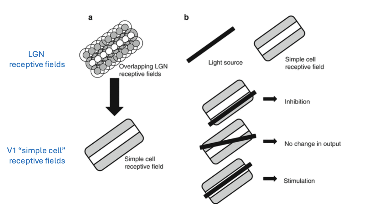
Binocular Vision
Two eyes at the front of the face give a large region of overlapping visual field. Humans, cats, and predators have large overlap; rabbits, chickens, fish have eyes on the sides of the head (monocular-type vision, minimal overlap). Enables depth perception

Binocular Disparity
Your two eyes are separated by ~6cm. Each gets a slightly different view of the world
The brain computes the difference between these two views (binocular disparity)
Large disparity = nearby object. Small disparity = distant object.
This gives you stereopsis: 3D depth perception from two 2D images
Requires the brain to combine input from both eyes

LGN Neurons are monocular
LGN has six layers, alternating layers respond to left or right eye

Binocular neurons
Are in visual cortex (V1), integrate input from both eyes

Ocular Dominance
Most V1 neurons respond to input from both eyes, but with a preference for one (ocular dominance). Organized in alternating columns
In a normal animal, the distribution is roughly balanced, each eye gets about equal representation
Neurons tuned to each eye are organized in alternating columns - ocular dominance columns

Monocular deprivation alters ocular dominance
Suggests that the input from each eyes compete during development
Eye closing does not alter receptive fields in retina or LGN, so changes must be taking palce in the cortext


Ocular Dominance Columns can be induced artificially

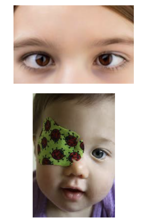
Amblyopia
Strabismus (misaligned eyes) during early life leads to pronounced visual defects (“amblyopia”).
Strabismus induced amblyopia can be treated either with surgery or sometimes simply covering the healthy eye for several hours a day
This implies that there is active competition between the eyes for representation in the brain.
Dorsal Stream
The “where” pathway in the brain, projects through the parietal lobe to represent location
and motion. Responsible for spatial location, Motion, Guiding action

Ventral Stream
The “what” pathway in the brain, projects through the temporal lobe to represent form (color,
shape) and object/face recognition. Responsible for Object identity · Shape · Faces · Meaning

Area MT/V5
Area after V1. Middle Temporal Visual Area, Moving objects in the world are highly important and salient: predators, prey, social signals, catching objects etc.
A dedicated system for motion is evolutionarily important for survival
Area MT/V5: neurons respond to direction and speed of motion (not to shape, color, or identity)
MT receives strong input from V1 and from direct subcortical routes

Area MT/V5 Sensitive to Motion Coherence

Patient LM: Akinetopsia (dorsal stream lesion)
Bilateral damage to area MT/V5 following a stroke
• Visual acuity: normal
• Color vision: normal
• Object recognition: normal
• Motion perception: completely absent
"The world appeared as a series of frozen snapshots”