1. Neurobiology 3 and 4
 Call Kai
 Learn
Learn Practice Test
 Spaced Repetition
 Match
Match Flashcards
 Knowt Play
1/84

Earn XP
Description and Tags
W2 Southard
Name | Mastery | Learn | Test | Matching | Spaced | Call with Kai | Chat |
|---|
No analytics yet
Send a link to your students to track their progress
85 Terms
What are the three layers of the meninges?
1. Dura
2. Arachnoid
3. Pia

What are the three spaces of the meninges?
1. Epidural
2. Subdural
3. Subarachnoid

What are the characteristics of the dura mater (pachymeninges)?
- tough and fibrous
- outer layer
- 2 key infoldings separate parts of the brain
- encompasses venous sinuses
- function = protection

What are the two infoldings of the dura mater?
- Faux cerebri (between the cerebral hemispheres right and left)
- Tentorium cerebelli (between cerebrum and cerebellum)

What are the characteristics of the arachnoid mater (Leptomeninges)?
- spider web appearance
- forms granulations that reabsorb CSF
- location of small arteries
- function = cushioning and absorption of CSF
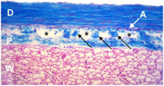
What are the characteristics of the pia mater (leptomeninges)?
- delicate membrane
- follows the gyri and sulci of the brain
- function = barrier, blood vessel penetration

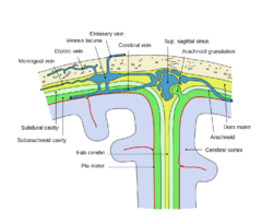
What do the meninges look like all put together?

What is the epidural space?
between bone and dura

What is the subdural space?
potential space between arachnoid and dura mater

What is the subarachnoid space?
space between arachnoid and pia mater- contains CSF
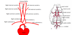
What vessels provide blood flow to the brain? (general)
Brain Arterial Supply: The brain receives 15–20% of cardiac output despite comprising only ~2% of body weight.
Blood is supplied by two sets of paired arteries:
Internal Carotid Arteries
Vertebral Arteries
oin along the ventral surface of the medulla to form the basilar artery.

What is the Circle of Willis?
a ring of arteries at the base of the brain that connects the brain's anterior and posterior circulations

What parts of the brain does the circle of Willis supply?

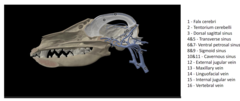
What are the parts of the venous drainage of the brain?
https://skfb.ly/orwWW
What is the dorsal spinal artery?
it supplies the dorsal white matter and grey matter
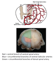
What is the ventral spinal artery?
supplies the ventral and lateral white and grey matter

What are the two major veins of the spinal cord?
1. dorsal spinal vein
2. ventral spinal vein

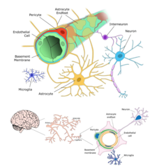
What is the function of the blood brain barrier?
protect the neural tissue from pathogens and toxins in the bloodstream.
What are the components of the blood brain barrier?
1. endothelial cells with tight junctions
2. basement membrane
3. pericytes- contractile cells
4. astrocyte foot processes
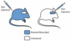
How was the blood brain barrier discovered?
intravenous injections of dyes into mice stained all tissues except the brain (no dye in the CSF)
The choroid plexus and meninges were stained
NOT the brain itself!
What is cerebrospinal fluid?
- clear, colorless fluid
- fills the ventricular system and subarachnoid space
- Produced by choroid plexus from blood
produced from plasma but very different in quality and content
What are the functions of cerebrospinal fluid?
1. protection/ shock absorption
2. homeostasis
3. clearance of waste products
What is the choroid plexus?
specialized ependymal cells that form papillary structures containing many blood vessels
located in lateral, third, and fourth ventricles
use a variety of pumps and channels to form CSF from the blood
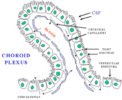
Where is cerebrospinal fluid produced?
in the lateral, third, and fourth ventricles
Describe the flow of CSF?
1. lateral ventricles
2. interventricular foramen
3. third ventricle
4. mesencephalic aqueduct
5. fourth ventricle
6. medial and lateral foramens
7. subarachnoid space

Spinal tap???
either in cranial or lat. dorsal
SITES
Cerebellomedullary cistern (cisternal tap)
Lumbar cistern (lumbar tap)
• Risks:
brain herniation (sudden pressure shift forces nervous tissue out)
• Safe volume: ~0.5 mL/kg per day
• Sterile technique should always be used
What are spinal nerves?
they arise from the spinal cord

What structures do the spinal nerves innervate?
structures caudal to the head
where are the cell bodies of the spinal nerves located?
in the dorsal root ganglia (sensory) or ventral horn (motor)
True/false- spinal nerves are mixed (sensory and motor)
TRUE
How are the spinal nerves numbered?
by the corresponding spinal cord segment (the # varies by species)
They may also combine to form plexuses and named nerves (the sciatic nerve is formed from spinal nerves L6, L7, and S1)
What are cranial nerves?
they arise from the brain
What structures do cranial nerves innervate?
structures in the head and neck
True/false- cranial nerves are mixed (sensory and motor)
FALSE- they can be sensory, motor, or mixed (sensory and motor)
Where are the cell bodies of cranial nerves located?
in the nuclei within the brain or ganglia outside the brain
How many cranial nerves are there?
12 pairs (designated I-XII)
The dorsal and ventral roots connect with the _________________
spinal cord

The spinal nerve splits into ______________________
dorsal and ventral branch/ramus

The dorsal branch is sensory/motor to the _________________
back
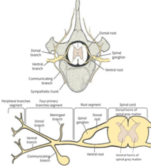
The ventral branch is sensory/motor to the ______________________________
ventral body wall, limbs, and visceral organs

each sensory spinal nerve innervates a strip of skin called a __________________________
dermatome:
an area of skin in which sensory nerves derive from a single spinal nerve root
it also detects sensations in muscle, organs, and connective tissue

where are the cell bodies for sensory neurons located?
in the dorsal root ganglia
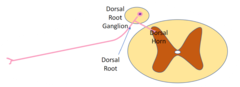
where are the cell bodies for motor spinal nerves located?
in the ventral horn

Spinal motor nerve branches combine to innervate specific ______________
muscles

What is the structure of the peripheral nerves?
1. the axons can be myelinated or unmyelinated
2. connective tissue (endoneurium, perineurium, epineurium)
3. Schwann cells (myelinate axons)
4. blood vessels

What is the endoneurium?
wraps around individual axons

What is the perineurium?
wraps around bundles of axons
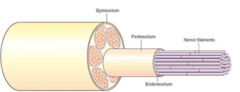
What is the epineurium?
wraps around nerves

What are the characteristics of peripheral nerves?
1. white
2. solid and do not stretch (differentiate nerves from arteries)
3. typically run in the fascia between muscle bodies
4. may parallel the blood vessels (neurovascular bundle)
What are the divisions of the peripheral nervous system?

What is the function of the autonomic nervous system?
regulates involuntary physiologic processes (heart rate, blood pressure, respiration, digestion, sexual functioning)

The autonomic nervous system is composed of the _________________________
sympathetic, parasympathetic, and enteric systems

What is the function of the somatic nervous system?
conscious sensation and voluntary movement
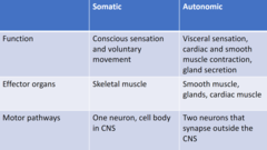
What are the effector organs of the autonomic nervous system?
smooth muscle, glands, and cardiac muscle

What are the effector organs of the somatic nervous system?
skeletal muscle

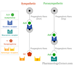
What are the motor pathways of the autonomic nervous system?
two neurons that synapse outside of the CNS
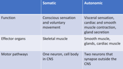
What is the motor pathway of the somatic nervous system?
One neuron, cell body in CNS

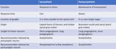
What are the differences between the sympathetic and parasympathetic nervous systems?
Cholinergic receptors bind _________________
Ach/ acetylcholine
nicotinic and muscarinic receptors
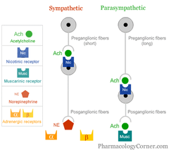
What is the difference between preganglionic and postganglionic neurons?
Preganglionic:
The cell bodies of the preganglionic neurons are in the brainstem or spinal cord of the central nervous system (CNS).
Postganglionic:
The cell bodies of the postganglionic neurons are in autonomic ganglia located peripherally.
Axon terminal of preganglionic neurons synapse on dendrites and cell bodies of postganglionic neurons.
What are muscarinic receptors?
They are postganglionic parasympathetic
activated by the molecule acetylcholine
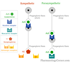
What are nicotinic receptors?
Preganglionic and somatic
activated by acetylcholine

Adrenergic (alpha, beta) receptors bind to ______________________
norepinephrine

What are alpha and beta receptors?
postganglionic sympathetic

What are the functions of the sympathetic nervous system?
1. fight, flight, fright response
2. activated in times or stress
3. increased heart rate and respiratory rate
4. increased blood flow to muscles
5. decreased digestive processes
6. innervates nearly every tissue in the body
7. ganglia located in a chain adjacent to the spinal cord
The motor pathways of the sympathetic nervous system involves ____________ neurons
TWO
What what are the characteristics of the preganglionic neuron in the motor pathways of the SNS?
- short axon
- cell bodies in intermediolateral cell column
- synapses in sympathetic ganglion
- neurotransmitter = Ach, receptor = nicotinic

What are the characteristics of the postganglionic neuron in the motor pathways of the SNS?
- long axon
- cell bodies in sympathetic ganglion
- synapses on effector organ
- NT = norepinephrine, receptor = alpha or beta

The sensory pathways of the sympathetic nervous system is ________________
similar to somatic nervous system
cell bodies in dorsal root ganglion
Where are the adrenal glands located?
located cranial and medial to kidney
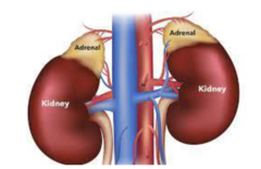
What does the endocrine gland do?
releases chemical messengers into the blood
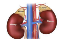
what are the parts of the adrenal gland?
1. cortex = controlled by the pituitary gland
2. medulla = controlled by sympathetic nerves (releases catecholamines (epinephrine and norepinephrine)

What are the functions of the parasympathetic nervous system?
1. rest and digest
2. decreased heart and respiratory rate
3. increased digestion
4. ganglia and located in or near the target organ
Parasympathetic ganglion are located ____________________________
in or near target organ
What are the characteristics of the preganglionic neuron in the motor pathway of the PSNS?
- long
- cell bodies in brain and cervical spinal cord
- synapses in ganglia near or in target organ
- neurotransmitter = Ach
- receptor = nicotinic

What are the characteristics of the postganglionic neuron in the motor pathway in the PSNS?
- short
- cell bodies in parasympathetic ganglia
- synapses on effector organ
- neurotransmitter = Ach
- receptor = muscarinic
The sensory pathways of the PSNS is similar to ____________________
cranial and spinal nerves
How does the sympathetic and parasympathetic nervous systems differ in how they regulate functioning organs?

What is the enteric nervous system?
a web-like series of neurons located in the wall of the intestine

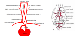
Circle of Willis
Formed by the basilar artery and internal carotid arteries. Major branches include:
Caudal Cerebellar Arteries: Arise directly off the basilar artery.
Circle of Willis Branches: Rostral cerebellar, caudal cerebral, middle cerebral, and rostral cerebral arteries.

Regional Distribution of arteries to the Telencephalon
Supplied by the rostral, middle, and caudal cerebral arteries.

Regional distribution of arteries to the diencephalon + mesencephalon
Supplied by perforating arteries.
Regional distribution of arteries to the rhombencephalon
Supplied by the vertebral, caudal, and rostral cerebral arteries.

Venous drainage of the brain
Small veins drain into venous sinuses located between folds of dura mater.
These sinuses drain into the internal jugular veins, which return blood to the heart via the cranial vena cava.

Spinal cord blood supply and drainage
Delivered by a midline ventral spinal artery and paired dorsal spinal arteries located within the dura mater (arising from aortic branches).
Drained by ventral and dorsal spinal veins.