Neuro E2 Hail Mary
 Call Kai
 Learn
 Practice Test
 Spaced Repetition
 Match
 Flashcards
 Knowt Play
1/55

Earn XP
Description and Tags
Godspeed
56 Terms
Ways Acetylcholine Agonists Exert Effects
1) Directly bind and activate ACh receptors (nicotine, muscarine)
2) Trigger massive presynaptic ACh release (black widow spider venom)
3) Inhibit acetylcholinesterase (AChE) from breaking down ACh (Sarin gas, VX nerve agent).
Ionotropic Receptors
Fast ligand-gated ion channels (made of 4 or 5 subunits forming a pore):
AMPA, NMDA, Kainate, GABA_A, Glycine, Nicotinic ACh (nACh), Serotonin (5-HT3), and Purines (P2X).
Metabotropic Receptors
Slow/modulatory G-protein-coupled receptors (GPCRs):
mGluRs (1–8), GABA_B, Dopamine (D1–D5), NE/Epi (alpha and beta), Histamine, Serotonin (all except 5-HT3), Muscarinic ACh (M1–M5), and Purines (A-type/P2Y).

Basic Structure of a GPCR
A single polypeptide with 7 membrane-spanning alpha-helices coupled to an intracellular 3-subunit (alpha, beta, gamma) G-protein.
Glutamate Receptors (Ionotropic)
AMPA (Na+ channel)
Kainate (Na+ channel)
NMDA (Na+ and Ca2+ channel that has 2 binding sites, is blocked by Mg2+ at resting potential, and requires membrane depolarization to open).
General Categories of Neurotransmitters
Amino acids (glutamate, GABA, glycine, aspartate)
Amines (catecholamines and indolamines)
Peptides (endorphins, enkephalins, substance P)
Esters (acetylcholine), and Others (ATP, NO, CO).

Filiform Papillae
Cone-shaped bumps located all over the tongue.

Fungiform Papillae
Mushroom-shaped bumps located on the sides and tip of the tongue.

Foliate Papillae
Fold-shaped ridges located on the back and sides of the tongue.

Circumvallate (Vallate) Papillae
Mound-shaped bumps arranged in a V at the back of the tongue.
Types of Taste Receptors
Saltiness (Na+ channel)
Sourness (H+/K+ channels)
Bitterness (T2R GPCRs)
Sweetness (T1R2+T1R3 GPCR)
Umami (T1R1+T1R3 GPCR).
Taste Perception Pathway
Tongue/Epiglottis ->
Cranial Nerves 7, 9, 10 ->
Medulla (gustatory nucleus / solitary tract) ->
VPM Nucleus of Thalamus ->
Primary Gustatory Cortex (Insular cortex: "What am I tasting?") & Parietal Cortex ("What texture is on my tongue?") (Ipsilateral).
Layout of the Retina
Divided into nasal and temporal halves;
features the optic disk (blind spot where blood vessels and optic nerve exit)
and the macula (center of retina containing the fovea pit).
Layers of the Retina (Inside-Out)
Light travels through: Ganglion cell layer -> Amacrine Cell (Inner plexiform layer) -> Inner nuclear layer → bipolar cell → horizontal (Outer plexiform layer) -> Outer nuclear layer -> Photoreceptor outer segments -> Pigmented epithelium.
Trichromatic Theory
Specific colors result from the relative activity of three cone types:
Short/Blue (~430 nm), Medium/Green (~530 nm), and Long/Red (~560 nm).
Targets of the Optic Tract
1) LGN of the Thalamus (conscious vision)
2) Hypothalamus (biological rhythms)
3) Tectum (pupil size and eye movement)
4) Superior colliculus (orients eyes/fovea to new stimuli).
Damage to Optic Nerve, Tract, and Chiasm
Left optic nerve cut = blindness in left eye (left hemifield)
Left optic tract cut = blindness in right visual field
Optic chiasm cut = tunnel vision (loss of peripheral vision).
Cells in Primary Visual Cortex (V1)
Simple cortical cells (monocular; respond to stationary bars/edges of light at a specific orientation with on/off areas),
Complex cells (binocular/monocular; respond to moving bars of light at a specific orientation)
Blobs (analyze color).
On-Center and Off-Center Cells
Retinal ganglion cells that detect illumination contrast/edges:
On-center cells fire action potentials when light hits the center (inhibited by light in surround)
Off-center cells fire when dark hits the center or light hits the surround (inhibited by light in center).

Structure of the Tympanic Membrane
Cone-shaped membrane (eardrum) at the end of the outer ear's auditory canal that vibrates in response to sound waves and moves the malleus.

Middle Ear and Its Components
Air-filled cavity with the Eustachian tube
two muscles (tensor tympani, stapedius), and three ossicles (Malleus, Incus, Stapes) that amplify vibrations from the tympanic membrane to the oval window.

Basilar Membrane Structure
Membrane in the cochlea supporting the Organ of Corti; narrow and stiff at the base (high frequencies, 20,000 Hz) and wider and less stiff at the apex (low frequencies, 20 Hz).
Flow of Fluid Through the Cochlea
Stapes pushes oval window ->
pushes perilymph in scala vestibuli to apex (helicotrema) ->
flows into scala tympani to round window ->
bends basilar membrane upward and bends hair cell stereocilia.

Structure of the Cochlea and Its Fluids
Divided into 3 fluid chambers by Reissner's membrane and the basilar membrane:
Scala vestibuli (top, perilymph),
cala media (middle, endolymph with Organ of Corti),
Scala tympani (bottom, perilymph).
Conduction deafness (earwax, tympanic membrane rupture, ossicle damage)
Nerve deafness (damage to the auditory nerve or hair cells).

Touch/vibration axons travel up the spinal cord ipsilaterally in the Dorsal Columns,
cross (decussate) in the Medulla (contralateral by the midbrain)
and travel up the Medial Lemniscus to the thalamus.
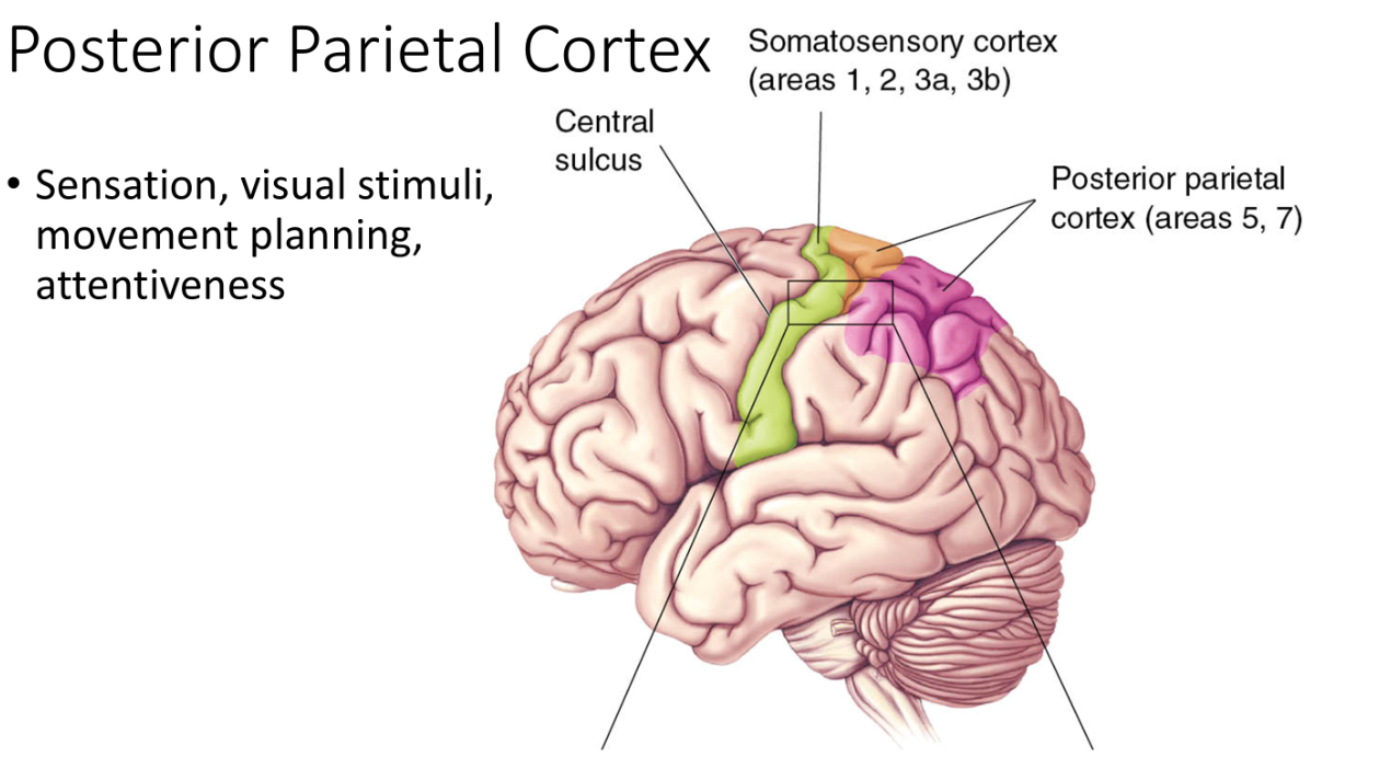
Somatosensory cortex (Areas 1, 2, 3a, 3b in the postcentral gyrus)
Posterior parietal cortex (Areas 5, 7).
30 total segments in 4 divisions:
Cervical (C1–C8), Thoracic (T1–T12), Lumbar (L1–L5), and Sacral (S1–S5).
