Chapter 7 - Bone Structure & Function
 Call Kai
 Learn
 Practice Test
 Spaced Repetition
 Match
 Flashcards
 Knowt Play
1/49

Earn XP
Description and Tags
Anatomy & Physiology (McGraw Hill)
50 Terms
regions of a long bone
diaphysis - shaft
provides leverage and support for long bone
compact bone on the outside, spongy bone on the inside
medullary cavity - hollow cylindrical space within diaphysis
contains red (children) or yellow (adult) bone marrow
epiphysis - expanded knobby region
thin layer of compact bone outside, extensive spongy bone inside
articular cartilage - hyaline cartilage that covers joint surface of epiphysis
reduce friction, absorbs shock
metaphysis - region of long bone where diaphysis widens into epiphysis
transfers force
epiphyseal plate (children) - thin layer of hyaline cartilage where bone length originates
epiphyseal line (adults) - thin, defined area of compact bone (remnants of epiphyseal plate)

periosteum
tough sheath that covers outer surface of bone (except areas covers by hyaline cartilage)
outer layer - fibrous, dense irregular connective tissue
protects bone
anchors blood vessels and nerves to bone surface
attachment site for ligaments and tendons
inner layer - osteoprogenitor cells and osteoblasts
bone width - both cells produce circumferential layers of bone matrix
perforating fibers - anchor periosteum to bone

endosteum
thin layer of connective tissue that lines internal surface of bone within medullary cavity
osteoprogenitor
osteoblasts
reticular fibers

diploe
region of spongy bone between two layers of compact bone
flat bones
no medullary cavity

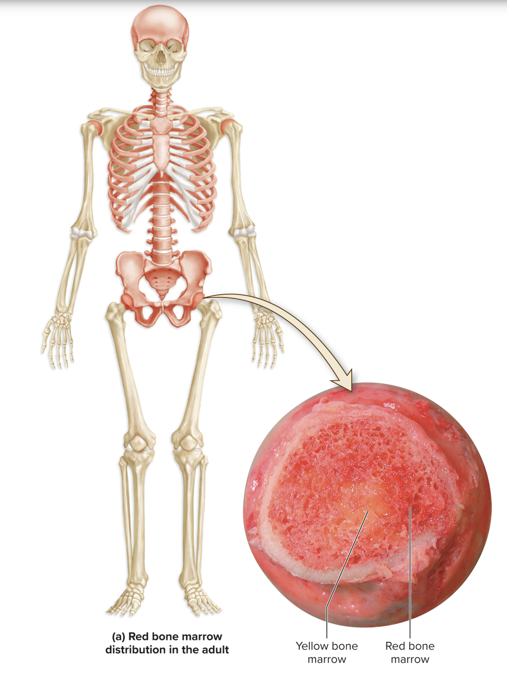
bone marrow
soft connective tissue of bone
structure
cells - developing red blood cells and/or adipocytes
reticular connective tissue
location - spongy bone in most bones, medullary cavity in long bones
red bone marrow - primarily developing red blood cells
widely distributed in children bones
selected portions of axial skeleton in adults
yellow bone marrow - primarily adipocytes
developing red blood cells decrease in adulthood, adipocytes increase
may convert back to rede bone marrow if red blood cells are needed
osteoprogenitor cells
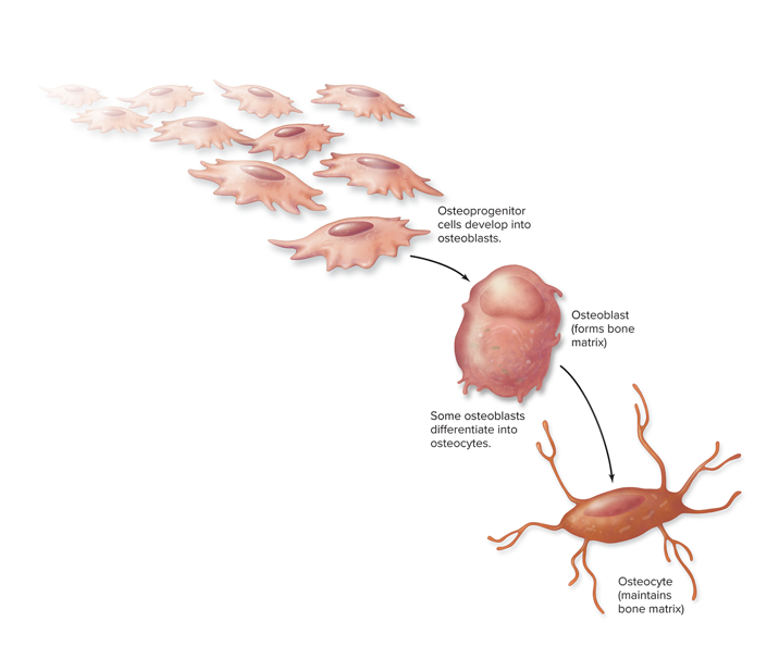
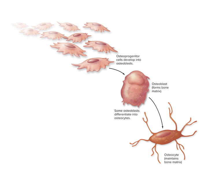
bone stem cells derived from mesenchyme
function - divide to produce mature osteoblasts
location - periosteum and endosteum
osteoblasts
matured osteoprogenitor cells
structure - cuboidal, abundant rough ERs and Golgi apparati
function - synthesize and secrete osteoid (bone matrix)
calcification - osteoid hardens due to calcium salt deposition
mature into osteocytes

osteocytes
matured bone cells derived osteoblasts
structure - contain cytoplasmic projections to maintain connection with neighboring osteocytes
function
maintain bone matrix
detect mechanical stress on bone
location - entrapped in calcified osteiod
lose ability to form new bone
osteoclasts
phagocytic cells derived from fused bone marrow cells
structure
large, multinuclear
ruffled border on bone surface increases surface area exposure to bone
function - bone resorption
location - resorption lacuna (pit on bone surface)

Osteitis deformans
disruption in balance between osteoclast and osteoblast function
excessive osteoclast activity - excessive bone resoprtion
oseoclasts are 5x larger than normal
contain 20+ nuclei (normal is 3-5 nuclei)
excessive osteoblast activity - excessive bone deposition
new bone is poorly formed and unstable
susceptible to deformation and fractures
extracellular matrix of bone connective tissue
osteoid (organic)
collagen proteins
semisolid ground substance - suspend and support collagen
contains proteoglycans, glycoproteins
crystal deposits (inorganic)
hydroxyapatite Ca10(PO4)6(OH)2
calcium phosphate crystals (CA3(PO4)2) + calcium hydroxide
other salts (i.e. calcium carbonate) and ions (i.e sodium, magnesium)
deposit around long axis of collagen fibers
calcification
bone formation; osteiod mineralizes/hardens
concentration of calcium and phosphate ions increase and precipitate out of solution
forms hydroxyapatite crystals that deposit around collagen fibers
vitamin D - enhances calcium absorption in GI tract
vitamin C - involved in collagen formation
bone resorption
bone matrix is destroyed by substances released by osteoclasts
proteolytic enzymes - chemically digest organic components (collagen, proteoglycans, glycoproteins)
hydrochloric acid - dissolves inorganic mineral (crystals)
osteolysis - release of calcium and phosphate from bone matrix
occurs during bone remodeling, low blood calcium levels
osteons
cylindrical structures in compact bone, basic structural unit
central canal - cylindrical channel in center of osteon
houses nerves and blood vessels
concentric lamellae - rings of bone connective tissue surrounding central canal
collagen fibers oriented in alternating patterns of direction, increases bone strength
osteocytes - mature bone cells in lacunae between lamellae
canaliculi - small interconnecting channels that connect adjacent lacunae
allow for communication between osteocytes
perforating canals - run perpendicular to central canals
connect multiple central canals within different osteons
contain nerves and blood vessels
circumferential lamellae - rings of bone that surround osteoblasts groups and extend entire circumference of bone
external - underneath periosteum
internal - over endosteum
interstitial lamellae - compact bone between osteons
leftover parts of osteons that have been partially resorbed
incomplete, no central canal

spongy bone anatomy
contais no osteons
trabeculae - open latticework of rods and plates of bone
space filled with bone marrow
parallel lamellae
lacunae - microscopic spaces between parallel lamellae
house osteocytes
canaliculi - microscopic channels that connects lacunae

cartilage growth
begins prior to birth during embryologic development
interstitial growth - grows in length
declines rapidly as cartilage matures
appositional growth - grows in width

intramembranous ossification
formation of bone tissue on thin layer of mesenchyme; flat bones, facial bones
ossification centers form in thickened regions of mesenchyme (8 weeks development)
mesenchymal cells differentiate into osteoprogenitor cells
osteoprogenitor cells become osteoblasts, begin to secrete osteoid
osteoid undergoes calcification
osteoblasts become entrapped, stop producing osteion and mature into osteocytes
woven bone (immature bone connective tissue) formed
mesenchyme surrounding woven bone thickens to form periosteum
mesenchymal cells produce additional osteoblasts
lamellar bone (mature bone) replaces woven bone
trabecular spaces filled with compact bone

endochondral ossification (gestation to childhood)
formation of bone in cartilage; most bones of skeleton
chondroblasts secrete cartilage matrix, forms hyaline cartilage (8-12 weeks development)
chondroblasts trapped within lacunae mature into chondrocytes
perichondrium surrounds cartilage
cartilage calcifies, periosteal bone collar forms
chondrocytes enlarge and resorb surrounding cartilage matrix, producing large holes in the matrix
cartilage matrix calcifies, chondrocytes die
blood vessels grow toward cartilage and penetrate perichondrium
stem cells in perichondrium develop into osteoblasts
periochiondrium becomes periosteum
osteoblasts under periosteum secrete osteiod, which hardens to form periosteal bone collar
primary ossification centers form in diaphysis (12 weeks development)
growth of capillaries and osteoblasts extends from periosteum into core of cartilage shaft
primary ossification center - osteoblasts secrete osteiod in center of cartilage shaft
extends in both directions toward epiphyses
healthy bone connective tissue replaces calcified, degenerating cartilage in shaft
secondary ossification centers form in epiphysises

endochondral ossification (childhood to adulthood)
bone replaces almost all cartilage, except for articular cartilage and epiphyseal plates
bones continue to grow in length until epiphyseal plates ossify into epiphyseal lines (adulthood)

interstitial bone growth
dependent on growth of cartilage in zones of epiphyseal plate
zone of resting cartilage
farthest away from medullary cavity of diaphysis, nearest to epiphysis
small chondrocytes in cartilage matrix (resembles hyaline cartilage)
secures epiphysis to epiphyseal plate
zone of proliferating cartilage
chondrocytes undergo rapid mitosis
align within lacunae into longitudinal columns parallel to diaphysis
zone of hypertrophic cartilage
chondrocytes stop dividing, enlarge greatly in size (hypertrophy)
walls of lacunae become thin as chondrocytes resorb matrix
zone of calcified cartilage
2-3 layers of chondrocytes
minerals deposited in matrix between columns of lacunae
calcification limits nutrients, causes chondrocyte destruction
chondrocyte death gives matrix opaque appearance
zone of ossification
walls between lacunae break down, forming longitudinal channels
channels invaded by capillaries and osteoprogenitor cells from medullary cavity
osteoprogenitor cells develop into osteoblasts, which deposit new bone matrix on remaining calcified cartilage

appositional bone growth
growth in bone width within periosteum
osteoblasts in inner cellular layer of periosteum produce/deposit bone matrix in external circumferential lamellae (tree rings)
as bone becomes wider, osteoclasts in medullary cavity resorb bone matrix

bone remodeling
continual addition of new bone tissue (bone deposition) and removal of old bone tissue (bone resorption)
occurs at different rates, depending on bone
compact bone remodels slower than spongy bone
achondroplasia dwarfism
abnormal conversion of hyaline cartilage to bone
long bones of limbs stop growing in childhood
bones of head and neck grow as expected
hormones that promote bone growth
growth hormone - released from anterior pituitary
stimulates liver to produce insulin-like growth factors (IGFs)
IGFs cause cartilage proliferation at epiphyseal plate
thyroid hormone - secreted from thyroid gland
stimulates metabolic rate of osteoblasts
calcitonin - promotes calcium deposition in bone, inhibits osteoclast activity
primarily in children, minimal effects in adulthood
sex hormones - stimulate osteoblasts
promote cartilage and bone growth at epiphyseal plate
also signals closure of epiphyseal plate
hormones that inhibit bone growth
parathyroid hormone - increases blood calcium levels
stimulate bone resorption by osteoclasts
calcitriol - increases blood calcium levels
stimulates bone resorption by osteoclasts
stimulates absorption of calcium ions from small intestine into blood
glucocorticoids - steroid hormones released from adrenal cortex
increases bone loss in when present high amounts
impair growth at epiphyseal plate in children
serotonin - prevents osteoprogenitor cells from differentiating into osteoblasts
enzymatic pathway of calcitriol (vitamin D) activation
UV light converts precursor molecules (in keratinocytes of skin) to cholecalciferol (vitamin D3) OR cholecalciferol (vitamin D3) from meal is absorbed from small intestine
cholecalciferol (vitamin D3) enters bloodstream
cholecalciferol (Vitamin D3) passes through liver
converted to calcidiol
Calcidiol passes through kidneys
covered to calcitriol hormone (active vitamin D)
effects of parathyroid hormone and calcitriol on blood calcium levels
blood calcium levels decrease (stimulus)
receptors of parathyroid gland detect drop in calcium levels
parathyroid gland release parathyroid hormone (PTH)
increases conversion of rate of calcidiol to calcitriol (vitamin D hormone)
PTH and calcitriol (vitamin D hormone) effect major organs
bone - increase osteoclast activity (bone resorption) to release of calcium from bone into blood
kidneys - triggers retention of calcium
small intestine - increases absorption of calcium into blood (calcitriol only)
net effect - more calcium in blood
removal of calcium from bone
increase of calcium resorption in kidneys
increase of calcium absorption from GI tract
Negative feedback - as blood calcium levels increase, PTH hormone secretion decreases

calcitonin
hormone that aids in regulating blood calcium in children
released from parafollicular cells in thyroid gland in response to high blood calcium levels or exercise stress
lows blood calcium levels
inhibits osteoclast activity
stimulates increase of calcium loss in urine
bone aging
bone strength decreases
osteoblasts produce less osteiod
bones become brittle
relative about of inorganic molecules (crystals salts) increases as protein decreases
bones become thinner
osteopenia - insufficient ossification as osteoblast activity declines
stress fracture
thin break in bone caused by physical activity

pathological fracture
fracture in bone that is weakened by disease
simple fracture
broken bone does not penetrate skin
compound fracture
one or both ends of broken bone pierce overlying skin

comminuted fracture
bone is splintered into several small pieces

transverse fracture
fracture is perpendicular to long axis of bone

spiral fracture
fracture spirals around axis of long bone, as a result of twisting stress
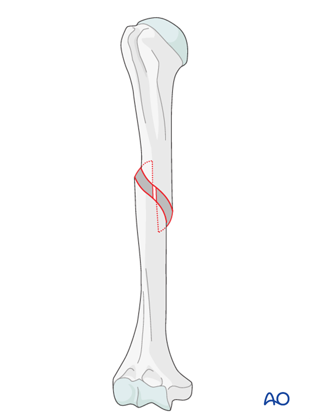
linear fracture
fracture is parallel to long axis of bone
avulsion fracture
bone fragment pulled off from rest of bone by soft tissue attachment
colles fracture
fracture of distal end of radius

complete fracture
bone is broken in 2 or more places
compression fracture
bone is squashed
depressed fracture
broken part of bone forms concavity (i.e. skull fracture)
displaced fracture
fractured bone parts are out of anatomical alignment
epiphyseal fracture
epiphysis is separated from diaphysis at epiphyseal plate
greenstick fracture
partial fracture
one side of bone breaks, other side is bent
more common in children

hairline fracture
fine crack in which sections for bone remain aligned
impacted fracture
one fragment of bone is driven into another
incomplete fracture
partial fracture extends only partway across bone
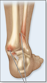
pott fracture
fracture at distal ends of fibula and fibula
bone repair
fracture hematoma forms as blood vessels inside bone are torn
fibrocartilaginous (soft) callus forms
blood capillaries infiltrate fracture hematoma
fracture hematoma reorganizes into growing connective tissue (procallus)
fibroblasts produce collagen fibers to connect broken ends of bones
chondroblasts form fibrocartilage and collagen fibers
procallus becomes soft callus
bony (hard) callus forms
osteoprogenitor cells in soft callus become osteoblasts
osteoblasts produce trabeculae of primary bone to replace soft callus
bony callus continues to grow and thicken for several months
bone is remodeled
osteoclasts remove excess bony material
compact bone replaced primary bone
