RTR.3 Clinical Principles
 Call Kai
 Learn
 Practice Test
 Spaced Repetition
 Match
 Flashcards
 Knowt Play
1/59

There's no tags or description
Looks like no tags are added yet.
60 Terms
US Breast
An alternative method for very dense breasts that may hide cancerous lesions that are difficult to see radiographically
High ability to distinguish between a cyst and a solid lesion
Screening Mammo Age Range
40- to 49-year age
Breast Tissue Types and Visualization
Glandular Tissue = denser, harder to visualize
Fatty Tissue = easier to visualize
Location of Highest Incidence of Breast Cancer
upper lateral quadrant

Mammo Technique
23-28 kVp; low kVp used to decrease Compton scatter and increase PE effect
Compression in Mammo
More uniform thickness
Improved spatial resolution
Reduces absorption blur and scatter → increased contrast resolution
Less breast thickness → less dose
Magnification in Mammo
Should not be used routinely
To investigate small, suspicious lesions or microcalcifications seen on standard mammograms
Image Intensifier
used in non-digital/conventional fluoro
Signal amplification device; converts photons that passed the patient into a visible light picture of high intensity
II Components
input phosphor: photon energy → visible light
photocathode: visible light → electrons
output phosphor: the site where accelerated electrons interact and produce visible light
Pulsed Fluoro Benefits
Reduces pt dose
Extends tube life
Misregistration Artifact in DSA
when the same anatomy is not registered in the same pixel of the image matrix due to motion
Can be fixed through registration = shifting the mask by one or more pixels so that superimposition of images is again obtained
Energy Subtraction
uses two different X-ray beams alternately to provide a subtraction image that results from differences in photoelectric interaction
Low kVp mask
High kVp mask w/ or w/out Contrast
Process to eliminate ST
Bone mask
Process to eliminate Bone
Hybrid Iodine Image
Arterial Access Methods
Seldinger: 18g inserted through the entire femoral artery; needle is withdrawn until flashback is seen → guidewire
Modified Seldinger: needle is pierced through only the anterior vessel wall
Trocar: non-vascular, for tubes and drains, used US to puncture the skin into ST
AP Axial Sacrum Beam Angle
15 ceph
AP Axial Coccyx Beam Angle
10 caud
AP Axial Calcaneous Beam Angle
40 ceph
Axiolateral Elbow Beam Angle
45 ceph
How does the scaphoid naturally sit
tilted anteriorly 20 degrees → the ulnar deviation helps brings it more // to the IR
LAT elbow radial tuberosity location
follows direction of 5th digit and palm surface
lateral: SI
supine: ant
prone: post
AP Femur Rotation
proximal
distal
hip: 5-10 int.rot’n
knee: 15-20 int.rot’n
What to do when unable to sep shoulders for Swimmers
3-5 caud CR
Additive Respiratory Pathologies -
atelectasis
bronchiectasis
edema
empyema
malignancy
miliary TB
pleural effusion
pneumonia
cardiomegaly
Destructive Respiratory Pathologies
emphysema
pneumothorax
Inlet Pelvis Positioning
40 caud
CP: ASIS

Outlet Pelvis Positioning
CP: 2’ distal to GT
male : 20-35 ceph
female: 30-45 ceph

Internal OBL Judet Best Views
posterior acetabular rim
anterior column / iliopubic

External OBL Judet best views
anterior acetabular rim
posterior column / ilioischial

AP Axial SIJ Positioning
2.5’ inf to ASIS
male = 30 ceph
female = 35 ceph

AP OBL SIJ Positioning
pt obl 25-30 away from IR, exact obl = ASIS → PSIS
1” med to raised ASIS
Bumped Up vs. Inferosuperior Shoulder
Bumped Up
15-20 angle
arms bent, elevated, and across chest
Inferosuperior
15-30 angle
arms abducted 90, ext rot’n
General Skull Positioning
PA = OML perp, petrous at supraorbital
PA Axial Caldwells = OML perp, 15 caud, petrous at lower 1/3
PA Axial Orbits = OML raised 30, petrous below infraorbital
AP Axial Townes = OML perp, 30 caud, dorsum sellae and clinoids visualized in the foramen magnum
PA Waters = MML perp to IR (37 from IR), petrous just below max sinus
PA Waters Orbits = OML 50-55 from IR, petrous mid max sinus
LAT = IOML perp to floor
AP Axial Modified Townes Positioning
CP = midway between vertex and supraorbital rims
Angle = 25 caud
Structures = vertex → supraorbital rims exclude eyes
Trauma Imaging Priority
Chest
CSP
T/LSP
Pelvis
Extremities
CTA Levels
Level 1: immediate physician attention; caridac/resp event, major trauma, shock
Level 2: within 15mins; altered mental state, substantial head inj, severe trauma, neonates
Level 3: within 30mins; modertate trauma, GI bleed, acute pain
Level 4: within 60mins; headache, nausea/vomitting
Level 5: within 120mins; minor trauma, mild ABD pain, vomitting/diarrhea
Vertebral Level - Xiphoid Process
T9/10
Vertebral Level - Coastal Angle
L1
Vertebral Level - Iliac Crest
L4
Vertebral Level - PSIS
L5/S1
Vertebral Level - ASIS
S2
Cervical Spine IVF Anatomy
45° anterior and 15° inferior from coronal
Lumbar Spine Z-joint Anatomy
45° posterior from coronal
Biliary Tree
Right and Left Hepatic Ducts join to form the Common Hepatic Duct
The CHD joins with the Cystic Duct to form the Common Bile Duct
The CBD joins with the Pancreatic Duct
The intestinal opening of the combined ducts is the Ampulla of Vater
Head to SVC - Venous System
vertebral vein
IJV, EJV
subclavian vein
brachiocephalic vein
SVC
Bifurcations from the Aortic Arch
Brachiocephalic (RT)
RT subclavian → RT vertebral
RT common carotid → RT ICA and ECA
LT common carotid → LT ICA and ECA
LT subclavian → LT vertebral
Branches of the Ascending Aorta
R/L Coronary Arteries as it forms the aortic arch

Pulmonary Arteries
R/L PAs arise from the pulmonary trunk
inf to the AA, between asc/desc aorta

Pulmonary Veins
R/L PVs begins at the hilum and carry oxygenated blood
inf to the PAs

Anterior and Middle Vasculature Head of the is supplied by
ICA

The Posterior brain vasculature is supplied by the
Basilar Artery

External Carotid Artery Supplies the
face, scalp, neck, tongue
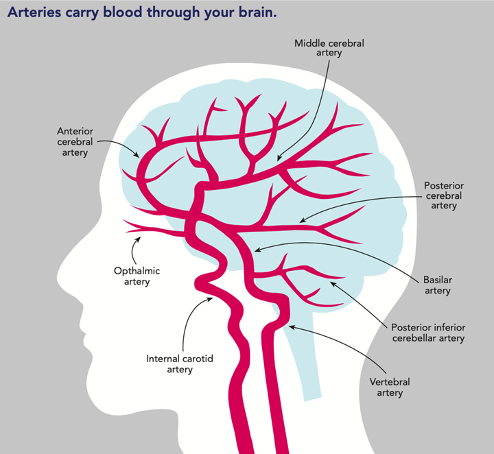
Circle of Willis Structure
ant/post CAs, ant/post communicating arteries, and ICA
important collateral pathway allowing blood flow between the cerebral hems in the event of arterial blockage

Dual Sinuses Structure
superior sagittal
inferior sagittal
straight
transverse
sigmoid
cavernous
petrosal sinus (sup, inf)

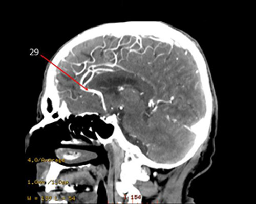
Anterior Cerebral Artery
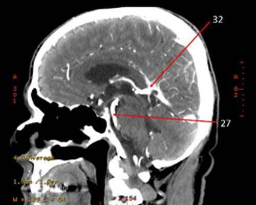
32?
Posterior Cerebral Artery

MIddle Cerebral Artery (largest)
Pharynx Divisions
Nasopharynx = nasal cav → hard palate
Oropharynx = soft palate → hyoid bone
Laryngopharynx = hyoid bone → esophagus

lateral → medial
EJV → SCM → IJV → CCA → Thyroid Gland
Heart Chambers Anatomy
RA = to the right
RV = most anterior
LA = most posterior
LV = to the left
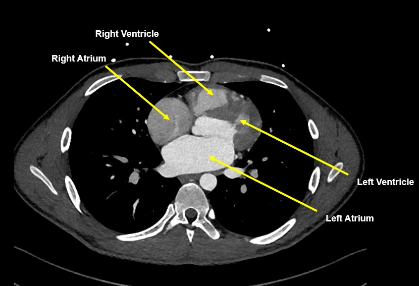
Female Pelvic Organs
Anterior → Posterior
Bladder
Uterus
Rectum
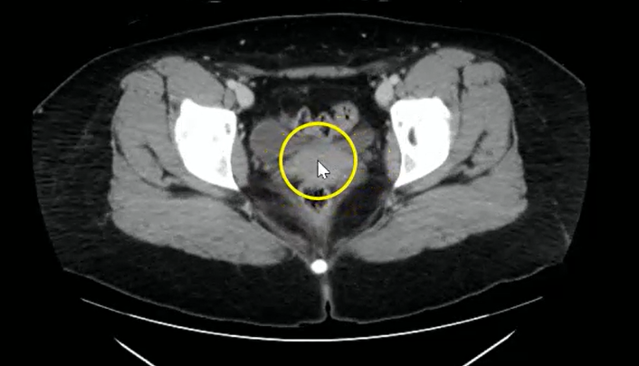
Male Pelvic Organs
Anterior → Posterior
Prostate gland (located inf to bladder)
Rectum
