Internal Environment- Cardiac Function
 Call Kai
 Learn
Learn Practice Test
 Spaced Repetition
 Match
Match Flashcards
 Knowt Play
1/25

There's no tags or description
Looks like no tags are added yet.
Name | Mastery | Learn | Test | Matching | Spaced | Call with Kai | Chat |
|---|
No analytics yet
Send a link to your students to track their progress
26 Terms
Draw, in correct temporal relationship, the pressure, volume, heart sounds, and ECG changes in the cardiac cycle. Identify the intervals of isovolumetric contraction, rapid ejection, reduced ejection, isovolumetric relaxation, rapid ventricle filling, reduced ventricular filling and atrial contraction
Wigger’s diagram → tied to CO
how much blood leaves the heart every minute (HR * SV = CO)
SV is how much blood leaves during a single contraction
when talking about CO, talking about left sided CO
right sides CO is going to the lungs, still the same (not lower)
if right side of heart didn’t pump as much blood out as left…
get more blood to body than lungs (bad things happen)
right sides gets more…
blood goes to lungs, not body
left-sided heart failure
end up with fluid in the lungs → pneumonia
left-side isn’t taking blood from lungs and transitioning it to body
right and left side balanced in a healthy individual
Describe the role of Starling’s Law of the Heart in keeping the output of the left and right ventricles equal
What keeps the right and left side balanced
Define ejection fraction and be able to calculate it from end diastolic volume, end systolic volume, and/or stroke volume
commonly used by cardiologists to do prognostication of how healthy a person is
ours is ~55%
every time heart contracts, ~55% of the blood in that ventricle goes out
when a person goes into heart failure, that ejection fraction begins to go down
right when you get at ~20% ejection fraction is the break point → heart can’t maintain flow (decompensation)
patient goes downhill, intervene medically and pharmacologically
Which of the following would not increase cardiac output? CO = HR * SV
Increased heart rate
Increased stroke volume
Increased resistance
Increased blood volume in the ventricle
Increased venous return
ICQ1 explanation
if either HR or SV go up, CO will go up
B1 stimulation inc. both HR & SV
inc. resistance → dec. CO
how much pressure is required to send blood into circulation
value we use is diastolic BP
point that blood begins to leave the heart & go into circulation
pressure required to open aortic valve
if that pressure goes up, the aortic valve doesn’t open at low pressure, it takes a higher pressure… the amount of time for blood to leave the heart gets cut down, less blood leaves the heart & CO drops
one of problems with hypertension with a high diastolic pressure
don’t get as much blood out of the heart for every beat
heart has to inc. rate to maintain CO… only choice is to beat faster
inc. venous return → inc. blood in the ventricles
the more blood we have in the ventricles, the harder the strength of contraction → Starling’s Law (mechanical example)
right now, most of blood is in our veins & lower extremities… when we stand up, blood is going to fall down bc of gravity… if our system doesn’t compensate, going to lay there… EMS/pass out
as you stand, the muscles squeeze the blood & more blood goes to the heart
the heart expands, physically stretches larger & contracts harder → Starling’s Law (automatic. adjustment based on varying the load)
50 mL of IV fluids directly into heart → heart would expand even more & pump out more than 50 mL
adjusts automatically
patient in heart failure → Starling’s Law is not working
The mitral or left AV valve opens when:
The atria contract
The ventricles contract
Ventricular pressure exceeds atrial pressure
Atrial pressure exceeds ventricular pressure
None of these are correct
ICQ2 explanation
the atria contract: valve was already open during atrial systole/contraction
the ventricles contract: closes valve → pressure is going to jump
ventricular pressure exceeds atrial pressure: valve closes
valve is sitting on the left side of the heart b/w the atria and ventricle
opens when pressure in the ventricle is lower than pressure in the atria
Inputs
Endocrine
Epinephrine
Stimulates B1 & B2 receptors of the heart
T3/T4 (excess thyroid hormone → higher rate & higher strength of contraction & higher BP)
Induces B1 receptor expression on the heart
GH/IGF-1
Required for normal cardiac function
High insulin
Disrupts B1 activity (type 2 diabetics that are high in insulin have impaired cardiac function/cardiac response)
CNS
Sympathetic
Increase rate & strength
Parasympathetic
Decrease rate
Inflammatory mediators
IL-5
TNF-a
when we look at coronary circulation, we have inflammation
damage coronary arteries & cause endothelial cell dysfunction
blame for everything
cause coronary arteries to not respond correctly
instead of relaxing, they constrict sometimes → can cause impaired blood flow
Cardiac Cycle
Pressure, volume, electrical activity, mechanical activity
Wigger’s Diagram
Independent variable
ECG
pacer → independent variable
causing everything else, are dependent on ECC
P wave, precedes atrial contraction
QRS, precedes ventricular contraction
Dependent variables
Aortic pressure
Ventricular pressure
Atrial pressure
Ventricular volume
Heart sounds (confirms what’s going on)
Wigger’s Diagram (cont’d)

Cardiac Output
Defined: the volume of blood pumped to the body per minute
Focuses on left side
Right-sided is equal to left
look at right side when looking at someone with pulmonary hypertension
total CO ≠ left + right
CO determines BP and blood flow to body
Determinants
Heart rate
Stroke volume
CO = HR x SV
Heart rate
Influences on pacing
Parasympathetic (muscarinic receptors → hyperpolarize pacemaker cells, action potential of the pacemaker cells, further from threshold takes them longer to reach it & get an AP)
Increases K+ permeability
makes cell more (-) → review
Decreases Na+f & Ca++t slope
positive, upward movement is slowed
Sympathetic
Increases Na+f & Ca++t slope
makes it happen quicker, B1 effect
put a patient on a B1 blocker → slowing that effect
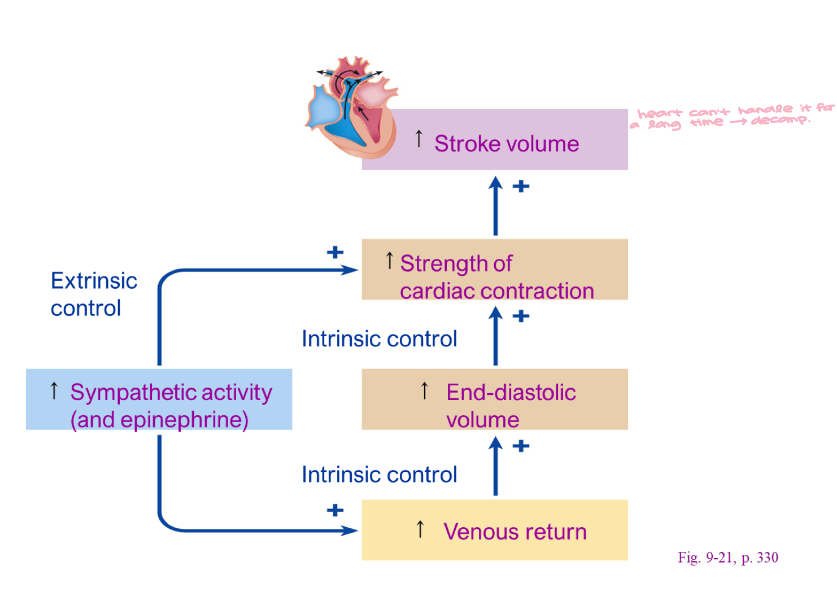
Stroke Volume (all about sympathetic, parasympathetic doesn’t impact directly)
Determinants
Sympathetic stimulation
Increased Ca++L influx
Venous return
Blood from the body to the heart
Greater volume of return = greater stroke volume

Stroke Volume (cont’d)
Determinants
Sympathetic stimulation
Increased Ca++L influx
Venous return
Blood from the body to the heart
Greater volume of return = greater stroke volume

Stroke Volume (cont’d 2)
Determinants
Sympathetic stimulation
Increased Ca++L influx
Venous return
Blood from the body to the heart
Greater volume of return = greater stroke volume

Stroke Volume (cont’d 3)
Determinants
Sympathetic stimulation
Increased Ca++L influx
Venous return
Blood from the body to the heart
Greater volume of return = greater stroke volume
Also known as Frank-Starling Law
Also known as preload
Afterload
Arterial pressure
occurs in striated muscle (cardiac & skeletal)
has a point where you get optimal alignment of the sarcomeres (A&I bands)
get muscle at right length, have a stronger contraction → everything is lined up
optimal alignment for heart is when it’s at its maximum fill of blood, going to contract its hardest
faint → elevate legs → more venous return → more blood goes back to the heart → heart stretches & contracts → brings BP up naturally bc of S.L.
problem with high BP is a lifetime of pushing harder at a faster rate causes S.L. to fail, goes past optimum

Stroke Volume (cont’d 4)
Determinants
Sympathetic stimulation
Increased Ca++L influx
Venous return
Blood from the body to the heart
Greater volume of return = greater stroke volume
Also known as Frank-Starling Law
Also known as preload
Afterload (pressure required to open aortic valve)
Arterial pressure
heart can handle a high preload, but not a high afterload
diastolic BP is high → isovolumetric ventricular contraction time got longer
ejection ends when T wave says it’s done
if we have less time to eject, we have less SV
someone who has hypertension has a higher HR bc they’re trying to maintain CO

figure 9-16, p. 322

Ejection Fraction
Percentage of End-diastolic volume that leaves the heart
Normal is roughly 55%
Formula
Ef= SV/EDV
SV → how much blood came out, EDV → how full was the heart
Ef declines in heart failure
SV & SL begin to fail
sympathetic will try to compensate

Overall Sympathetic Impact of Cardiac Output
Increased sympathetic activity in heart failure
Compensation
Failure of sympathetic to correct
Decompensation
dec. CO, pushing will cause more damage
Coronary Blood Flow
Coronary Artery Disease
Myocardial Perfusion
Myocardium is highly aerobic
Not constant
Greatest flow during diastole
want low HR
resting HR of 50 → diastole is long
muscle is happy bc it’s spending most of its time at rest instead of working (systole)
resting HR of 120 → diastole period is short, myocardium is not well perfused
Increased flow
Adenosine
healthy heart releases breakdown product of ATP, will cause coronary artery dilation in a healthy heart
B2 stimulation
both want to inc. blood flow to the heart
Decreased flow
Platelet activating factor
Endothelial cell dysfunction (causes vascular constriction of coronary arteries → bad)
Coronary artery disease

Coronary Artery Disease (blockage → flow drops)
Any impingement on a vessel negatively impacts flow
Poiseuille’s Law

Coronary Artery Disease (cont’d)
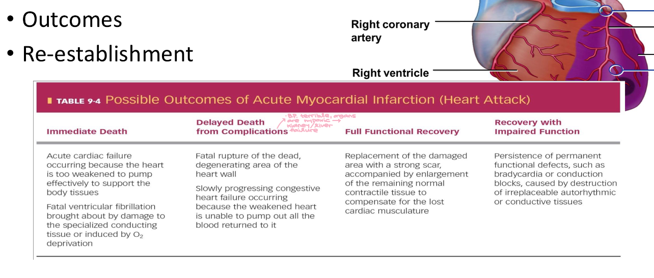
Blockage of a coronary artery is referred to as a Myocardial Infarct (MI)
usually due to a clot
get triangle shape piece of damage
Outcomes
Re-establishment

Coronary Artery Disease (cont’d 2)
Blockage of a coronary artery is referred to as a Myocardial Infarct (MI)
Outcomes
Re-establishment