MT 9
 Call Kai
 Learn
 Practice Test
 Spaced Repetition
 Match
 Flashcards
 Knowt Play
1/72

There's no tags or description
Looks like no tags are added yet.
73 Terms
⭐️ nervous system ⭐️
Cf for the cerebellum
Cerebell/o
It is located under the cerebrum and responsible for equilibrium.

Cerebellar, cerebellar hypoplasia
Decrease in the size of the cerebellum.
CF for the brain
Encephal/o
Ex. Encephalitis = Infl. Of the brain.
Herpes causes encephalitis (flames)
CF for the medulla oblongata
Modull/o
The part that connects the brain to the spinal cord and is responsible for breath, heart rate etc..
CF for the spinal cord
Myel/o
Myelitis
Infl. Of the spinal cord
Or can be inflammation of the bone marrow depending on context
CF for nerve
Neur/o
E.g: neuropathy
Alzheimer’s disease - AD
A disease characterized by diminished brain due to the protein accumulation leading to loss of mental ability.
(Alzheimer’s leads to dementia).

Concussion 🤕
A brain injury that can cause bruising, damage of the blood vessels and injury to nerves. Person may lose consciousness.

Epilepsy
الصرع
Chronic disorder with repeated seizures.
Brain neurons fire quickly leading to involuntary movements.
Glioblastoma
Gli/o = glial cells
Blast = immature
A malignant tumor of an increased production of immature (unspecified) glial cells.

What are the glial cells
They are cells that surround neurons and grow to have specific functions but they don’t carry signals.

Migraine
A severe headache usually on one side of the head. Caused by changes in the size of blood vessels and comes with vomitting, nausea and photophobia.

Multiple sclerosis- MS
The myelin sheath of the nerve cells is damaged and replaced by plaque leading to the neurons not firing well.

Syncope
Fainting because the brain is not getting enough oxygen
(During exercise for ex)
Cerebral angiography
X-ray imaging of the brain I cerebrum after a contrait material has been injected in the artery.

Cerebrospinal fluid (CSF) analysis
The use of a lumbar puncture - LP to remove the CSF and test sodium, chloride, protein, bacteria, cell counts, cultures to detect diseases of the brain and meninges.
Electroencephalography
(EEG)
Records the electrical activity in the brain.
Lumbar puncture
Process of putting a needle in the space around the spinal cord to take the CSF for analysis, and contrast material may be injected for myelography (spinal tap).
Magnetic resonance imaging - MRI
Magnetic waves and radiofrequency to make images for brain a spinal cord. in all three planes of the body

Positron emission tomography
(PET) scan
The brain takes up a radioactive material. This shows how the brain uses glucose and gives information about brain function.
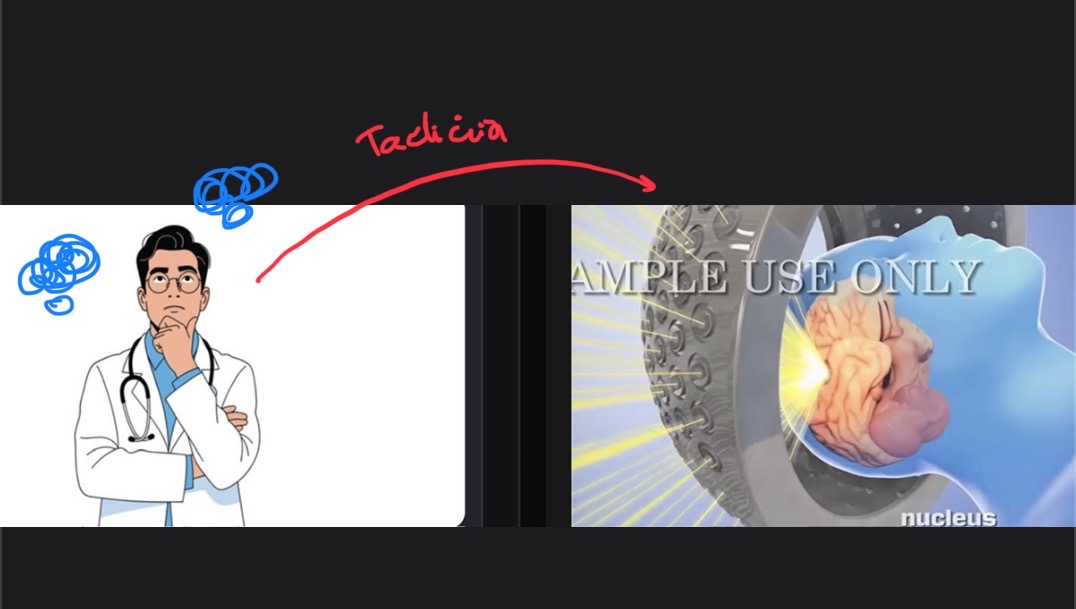
Stereotactic radiosurgery
Invasive surgery / radiation therapy. It treats abnormalities and smalltumors of the brain.
Also called CyberKnife or stereotactic radiotherapy.
It givesprecisely targeted radiation in fewer, high-dose treatments than traditional therapy.

Transcutaneous electrical nerve stimulation - TENS
A battery powered device is used to send stimulation signals to the nerves to relieve acute and chronic pain.
CNS
Central nervous system
CVA
Cerebrovancular accident
EEG
Electroencephalograply

TIA
Transient ischemic attack.
Temporary interference with blood flow to the brain. (Mini stroke).
⭐️SSS⭐️
Name the layers of the skin I what they contain
Epidermis = the outer layer of the skin
Dermis = hair root and follicle, the sebaceous gland, the sweat gland, nerve ending, blood vessels.
Subcutaneous tissue/ hypodermis = fat for thermal insulation.
CF for the skin
Derm/o ; epidermis
Dermat/o ; dermatology

CF for hair
Pil/o
Trich/o
Pilosebaceous
Pt. To the hair and the sebaceous gland.

Trichotillomania
Trich/o = hair
Till/o = trilling / حرث
Mania = obsessive preoccupation
Obsessive compulsive disorder (OCD) with the urge to pull one’s hair. (As a coping mechanism to stress etc)

CF for nail
Onych/o
Ungu/o
Onycholysis
Separation of the nail from the nail bed
Subungual (hematoma)
Pt. To under the nail , mans of blood under the nail.
CF for the eye
Ocul/o , ocular
Ophthalm/o , Ophthalmologist
What is the eye's pupil
The dark dot that expands depending on the concentration of light.
What is the Iris of the eye
It surrounds the pupil and gives the eye its color und helps dialate the pupil
What is the job of the cornea?
It bends the light.
What in the role of the conjunctiva
It covers the sclera (white area of the eye)

CF for the lens
Phak/o
Aphakia
Condition of the absence of the eye lens.
CF for the retina
Retin/o
Retinopathy
CF for the ear
Ot/o , otalgia
Aur/o , aural discharge

CF for the ear drum 💍
Myring/o
Tympan/o
Myringotomy
Insicion of the eardrum
Tympanoplasty
Surgical repair of the ear drum
Alopecia
Baldness


Cataract 💨☁
Clouding (opacity) of the lens of the eye causing poor vision/ blindness.

Conjunctivitis
Infl. Of the conjunctiva (the layer covering the sclera)
Also called pink eye.

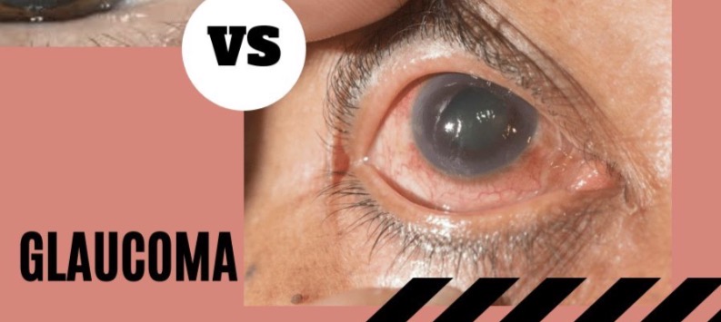
Glaucoma
Increased pressure due to fluid build up - aqueous humor- in the anterior chamber of the eye.

Melanoma
Malignant tumor of the pigment cells (melanocytes) and grows from the nevus in the skin
Nevus
A pigment lesion (benign) on the skin , mole.
Melan/o means:
Black

Stye / Sty
A purulent -pus filled - infection near the eye lid , caused by bacterial infection (hordeolum)

Tinnitus
An abnormal noise in the ears.
(Buzzing, ringing, roaring etc).
Allergy test
Allergy-causing substances are put on the skin and the reaction is noted.
1. In the patch test, a patch with a suspected allergen is placed on the skin. (No scratching the skin).
In the scratch test, several scratches are made and a small amount of allergen is put into the scratches.
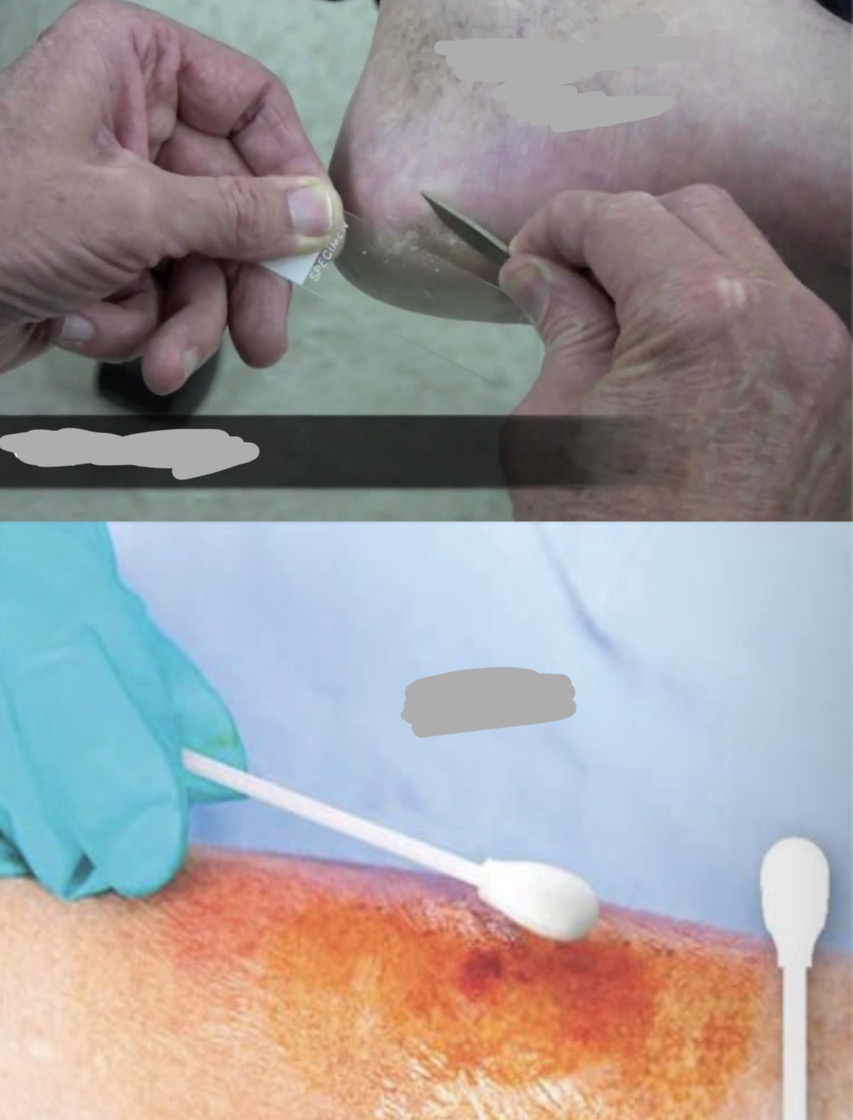
Bacterial and fungal tests
Samples from skin lesions are taken to find bacterial infection (they get a sample without scratching the skin) or fungal growth (scrape off the skin).
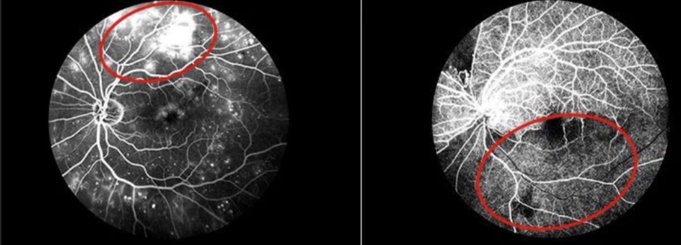
Fluorescein angiography
Fluorescein (a contrast substance) is injected into a vein.
The doctor uses ophthalmoscopy to watch the movement of blood in the back of the eye.
It is used to find diabetic or hypertensive retinopathy and degeneration of the macular (central) area of the retina.
Ophthalmoscopy ⬇
Visual examination of the inside of the eye.
Otoscopy ⬇
Visual examination of the inside of the ear (to the eardrum).
Skin biopsy
Sample of skin is removed and examined under a microscope (to check if the nevus is benign or malignant for ex)

Slit-lamp microscopy
Examination of the front of the eye like the cornea with a device shining intense light from a narrow opening.

Tuning fork test
A vibrating fork is held in front of the ear for air conduction (AC), then is placed on the mastoid bone to test bone conduction (BC).
Then they are compared (how well the hearing is) to detect potential hearing problems.
ENT
Ear
Nose
Throat
HEENT
Head, eyes, ears, nose, throat.
PERRLA
Pupils equal , round, reactive to light and accommodation.
Method of observation to identify “eye health”.
VA 🚶🥊
Visual activity (clarity of vision )
VF
Visual field (how much surrounded area can you see ).