Gastroenterology II: lower GI disorders
 Call Kai
 Learn
 Practice Test
 Spaced Repetition
 Match
 Flashcards
 Knowt Play
1/58

There's no tags or description
Looks like no tags are added yet.
59 Terms
Sudden, severe, persistent abdominal pain
Pain out of proportion to exam (mesenteric ischemia)
Nausea, vomiting, anorexia
Obstipation (no stool or flatus, obstruction)
symptoms associated with a acute/surgical abdomen
Cholecystitis, cholangitis, hepatitis, biliary colic, Fitz-Hugh-Curtis
DDx of RUQ pain
Splenic infarct/rupture, gastritis, pancreatitis, pneumonia
DDx of LUQ pain
Peptic ulcer disease, pancreatitis, MI, aortic aneurysm
DDX of epigastric pain
Early appendicitis, small bowel obstruction, mesenteric ischemia
DDx of periumbical pain
Appendicitis, Crohn's, ectopic pregnancy, ovarian torsion
DDx of RLQ pain
Diverticulitis, ovarian torsion, ectopic pregnancy
DDx of LLQ pain
Peritonitis, bowel obstruction, mesenteric ischemia, gastroenteritis
DDx of diffuse abdominal pain
peritonitis
Rebound + guarding +diffuse abdominal pain=
Cholecystitis/cholangitis
History: RUQ pain, worse after fatty meals, fever, nausea.
Charcot triad
RUQ pain, jaundice, fever/chills
associatedd with cholangitis
pancreatitis
History: Epigastric pain radiating to back, better leaning forward, N/V.
mesenteric ischemia
History: Severe pain out of proportion to exam, risk factors (AFib, atherosclerosis).
Labs: ↑ Lactate, metabolic acidosis.
perforated ulcer
History: Sudden severe epigastric pain.
Exam: Rigid abdomen, rebound, guarding.
Labs/Imaging: Leukocytosis, free air under diaphragm (upright CXR).
Bowel rest (clear liquid diet, advance as tolerated).
Oral antibiotics (covering Gram-negative + anaerobes, e.g., ciprofloxacin + metronidazole or amoxicillin-clavulanate).
Outpatient if stable.
management of acute uncomplicated diverticulitis
NPO, IV fluids, IV antibiotics.
Abscess drainage (percutaneous).
Surgery if perforation, fistula, or obstruction.
management of complicated diverticulitis (abscess, perforation, obstruction, peritonitis)
irritable bowel syndrome
Definition: Functional bowel disorder with chronic abdominal pain/discomfort + altered bowel habits (no structural cause).
Recurrent abdominal pain ≥1 day/week for 3 months, associated with ≥2 of: related to defecation, change in stool frequency, change in stool form
Rome IV criteria for diagnosis of IBS
infectious: Fever, chills, myalgias, nausea, vomiting
non-infectious: Bloating, weight loss, nocturnal diarrhea (IBD/malabsorption)
associated symptoms of infectious vs non-infectious diarrhea
infectious: Watery (viral, toxigenic bacteria) or bloody (invasive bacteria)
non-infectious: Fatty (steatorrhea in malabsorption) or mucus (IBS/IBD)
stool features of infectious vs non-infectious diarrhea
Sudan stain, 72-hour fecal fat collection
test for stool fat, indicating steatorrhea
D-xylose test
abnormal absorption suggests mucosal disease (celiac);
normal absorption if pancreatic insufficiency.

location: Colon only (always involves rectum), continuous
depth: mucosal and submucosal only
endoscopy: friable mucosa, continuous ulcerations
location, depth, and endoscopic findings of UC
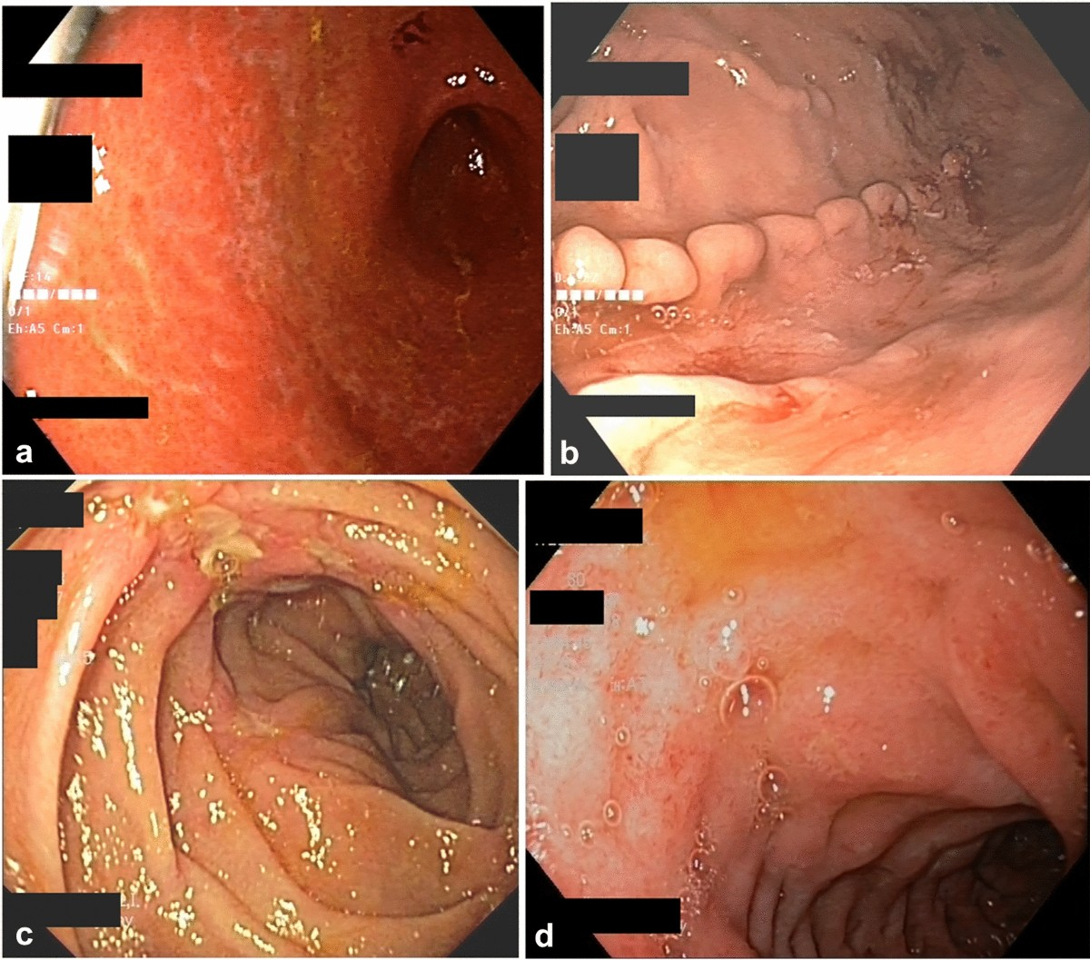
location: any part of GI tract, skip lesions
depth: transmurall
endosccopy: cobblestoning, strictures, fistula
location, depth, and endoscopic findings of crohn's disease

ulcerative colitis
histologgy: crypt abscesses, NO granulomas

crohn's disease
histology: noncaseating granulomas
Toxic megacolon, ↑ colorectal cancer risk
complications associated with UC
Fistulas, abscesses, strictures, malabsorption
complications associated with crohn's

Both can have arthritis, uveitis, erythema nodosum, pyoderma gangrenosum
extraintestinal effects associated with UC and crohn's
UC: smoking is protective
Crohns: smoking is a risk factor
the role of smoking in UC vs Crohn's
mild-moderate: 5-ASA (mesalamine, sulfasalazine)
severe: corticosteroids, bowel rest, biologics
treatment of acute exacerbation of IBD
Aminosalicylates (UC > CD).
Immunomodulators: azathioprine, 6-MP, methotrexate.
Biologics: anti-TNF (infliximab), anti-integrin (vedolizumab), anti-IL (ustekinumab).
maintenance therapy indicated for IBD
Chronic diarrhea, steatorrhea (bulky, greasy stools), bloating, flatulence, weight loss.
general symptoms of malabsorptive syndromes

Autoimmune deposition of IgA at the tips of dermal papillae
GI pathology (GERD, gastritis, IBD, hepatitis, pancreatitis) often causes inflammation and congestion
→ lymphatic drainage helps reduce edema and improve immune response.
rationale for lymphatic manipulative techniques in GI dysfunctions
>3mm thickness suggests pathology
define colonic wall thickening on radiographs
-IBD, infectious colitis, ischemic colitis
-colorectal carcinoma
-venous congestion
-toxic megacolon
DDx of colonic wall thickening on radiograph

thumbprinting GI
sign of mucosal edema/hemorrhage (ischemia, infection, IBD).
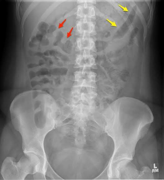

IBD, infectious colitis
symmetric long segment thickening on abdominal radiograph suggests
malignancy
asymmetric/focal irrregular thickening on colon radiograph inddicates
colonoscopy with biopsy
gold standard diagnostic test for IBD
CT enterography
specialized CT evaluation of the small bowel after the oral administration of low-density (0.1%) barium.
Outlines GI lumen → differentiates bowel from adjacent structures.
Used for suspected perforation, abscess, fistula, tumor.
indications for oral contrast in abddominal CT
Useful for distal colon/rectum evaluation.
Indications: suspected anastomotic leak, diverticulitis, distal obstruction.
indications for rectal contrast in abdominal CT
Highlights vascular structures & enhances solid organs.
Essential for ischemia, abscess, tumor, trauma, appendicitis, diverticulitis.
indications for IV contrast for abdominal CT
Adhesions, hernia, tumor, strictures
etiologies of SBO
SBO: Colicky abdominal pain, vomiting, distension, obstipation
ileus: distension, obstipation
Diffuse, mild abdominal discomfort, distension
symptoms of SBO vs ileus
High-pitched, hyperactive bowel sounds early → silent if late
exam findings associated with SBO

small bowel obstruction XR
Dilated small bowel loops (>3 cm), air-fluid levels, "step-ladder pattern," transition point
Post-op, meds (opioids), infection, electrolyte imbalance
etiologies of ileus
Hypoactive or absent bowel sounds
exam findings of ileus

ileus on XR
Diffuse bowel dilation (small + large bowel), no clear transition point
Functional, usually resolves with supportive care
Treatment | Indication / Purpose |
|---|---|
NPO initially | Bowel rest if significant distension, nausea, or vomiting |
IV fluids | Correct dehydration and maintain hydration |
Electrolyte correction | Correct hypokalemia and hypomagnesemia |
NG tube decompression | Significant vomiting or abdominal distension |
Stop opioids / anticholinergics | Medications that decrease bowel motility |
Early ambulation | Stimulates GI motility |
Early oral feeding as tolerated | Promotes recovery in uncomplicated postoperative ileus |
Treat underlying cause | Infection, inflammation, metabolic abnormalities |
clinical course of ileus
Clear liquid diet 24 hrs prior.
Bowel prep with laxatives/enema the night before.
NPO after midnight.
patient prep for a barium enema
-arterial emobolus (AFib, MI, valvular disease)
-Arterial thrombosis
-venous thrombosis
-hypotension, vasopressors
etiologies of ischemic bowel
acute ischemic bowel
Sudden severe abdominal pain out of proportion to exam.
Nausea, vomiting, diarrhea (may be bloody
Leukocytosis.
↑ Lactate & metabolic acidosis (late finding).
Possible elevated D-dimer.
lab findings associated with ischemic bowel
CT angiography (CTA)
diagnostic test of choice for ischemic bowel
ischemic bowel on XR
Bowel wall thickening, pneumatosis intestinalis (air in wall).
Portal venous gas.
Thumbprinting sign (submucosal edema/hemorrhage).