osborn- abdominal wall peritoneum
 Call Kai
 Learn
 Practice Test
 Spaced Repetition
 Match
 Flashcards
 Knowt Play
1/67

There's no tags or description
Looks like no tags are added yet.
68 Terms
abdominal cavity
muscle and bone bounded cavity lined internally by fascia (surrounds the peritoneal cavity)
borders of the abdominal cavity
cranial- diaphragm, caudal- pelvic inlet (linea terminalis), ventral- abdominal wall, dorsal-sublumbar mm.
abdomen
the part of the trunk that extends from the diaphragm to the pelvis
bony landmarks of the abdomen
costal arch, lumbar vertebral bodies, tuber coxae, promontory of the sacrum, iliopubic eminence, pecten of pubic bones

in this view of the equine left abdomen, name structures A-E
esophagus, descending aorta, diaphragm, external abdominal oblique, tunica flava (yellow tunic)
list the layers of the abdominal wall (superficial to deep)
skin (epidermis, dermis, subcutis), superficial layer of external fascia, cutaneous trunci, deep layer of external fascia/yellow tunic, external abdominal oblique, internal abdominal oblique, transversus abdominus, rectus abdominis, internal fascia of the trunk/transversalis, parietal peritoneum
what are the natural openings in the abdominal wall?
external and internal inguinal rings, umbiliicus (opening in linea alba)

what muscle is labeled ‘B’ in the image?
external abdominal oblique

what part of the muscle is indicated by the ‘A’ in the image?
aponeurosis
what is aponeurosis?
broad flat sheet of dense irregular collagen that functions as a tendon

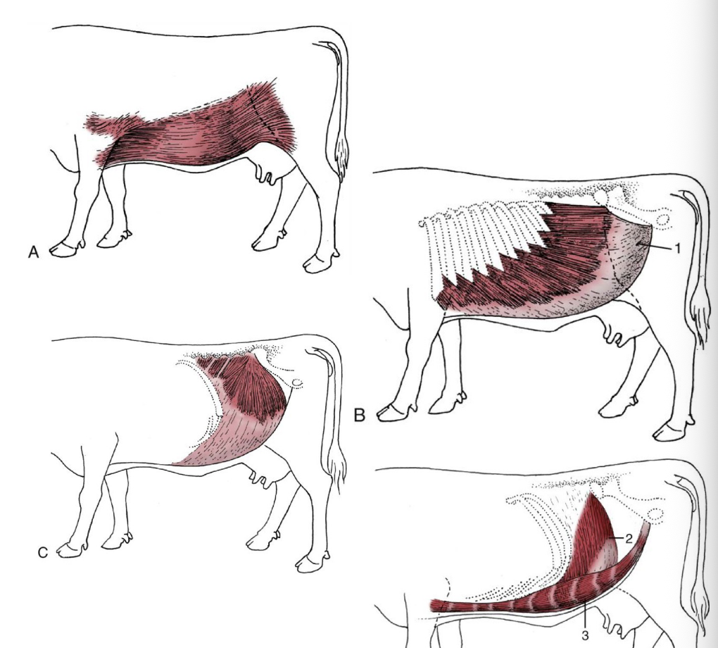
what is the origin of the external abdominal oblique muscle belly?
lateral ribs and lumbar fascia

what is the insertion of the external abdominal oblique aponeurosis?
linea alba and prepubic tendon
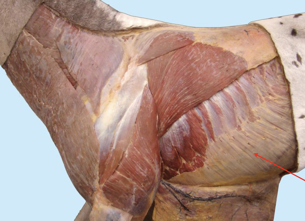
what structure is indicated by the red arrow? hint: unique to ungulates
tunica flava abdominis
what is the tunica flava abdominis?
found in ungulates, a deeper fascia consisting largly of elastic tissue and is yellow in appearance. well adapted to the passive support of the viscera and is the thickest ventrally where the burden is the greatest

what is created by COPD?
heave line

what is a heave line?
caudoventral boder of the muscle belly of the external abdominal oblique is prominent due to over-exercise

what are the origins of the muscle belly of the internal abdominal oblique?
tuber coxae and lumbar fascia

what are the insertions of the aponeurosis of the internal abdominal oblique?
costal arch, linea alba, and prepubic tendon

what part of the IAO muscle belly forms the cranial border of the deep inguinal ring?
caudal margin (5’ in the image)

what muscle (and part) is indicated by the letter ‘A’ in the image?
internal abdominal oblique, costal part

what muscle (and part) is indicated by the letter ‘B’ in the image?
internal abdominal oblique, abdominal part

in this image, what muscle is indicated by the letter ‘A’?
internal abdominal oblique

in this image, what lymph node is indicated by the B?
subiliac LN
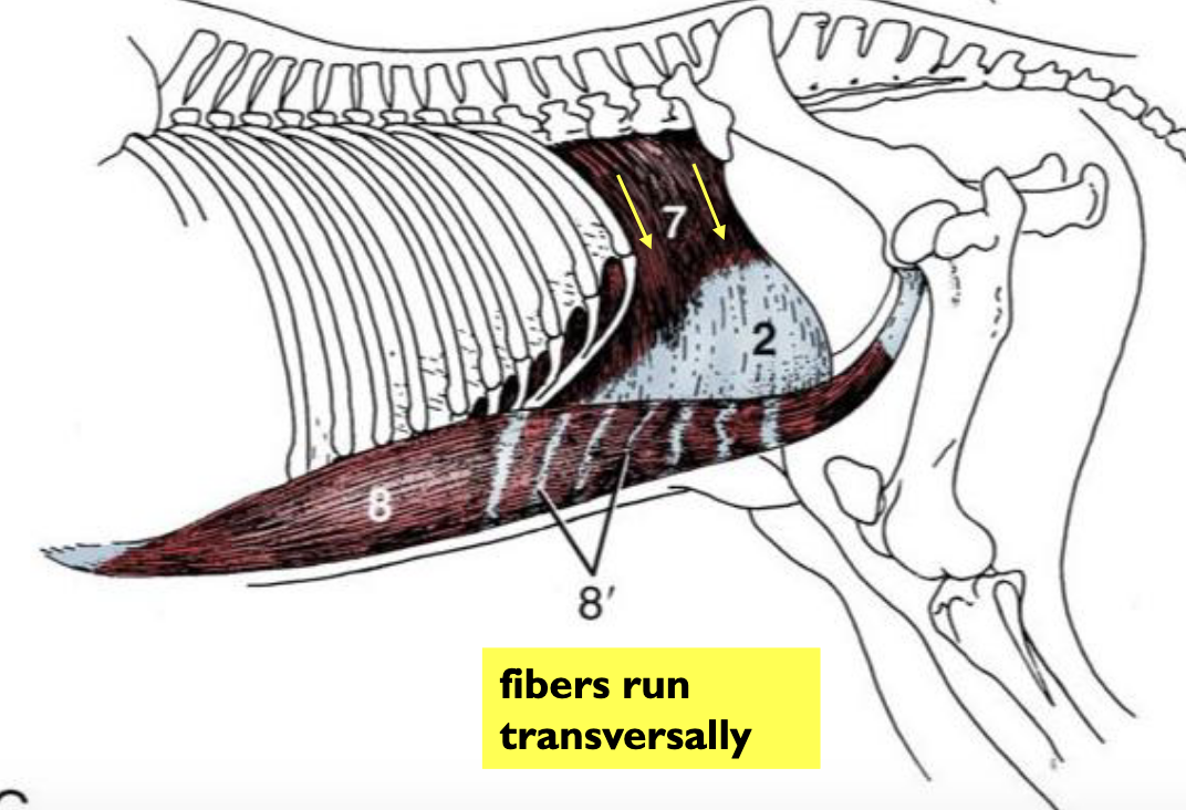
what are the origins of the muscle belly of the transversus abdominis oblique muscle?
costal arch, caudal ribs, transverse processes of lumbar vertebrae, lumbar fascia, and tuber coxae

what are the insertions of the transversus abdominis oblique aponeurosis?
linea alba and prepubic tendon

what runs within the fibers of the transversus abdominis oblique that distinguish it from the other abdominal oblique muscles?
ventral branches of the spinal nerves

what muscle is indicated by A?
transversus abdominus
what encloses the rectus abdominis oblique?
rectus sheath
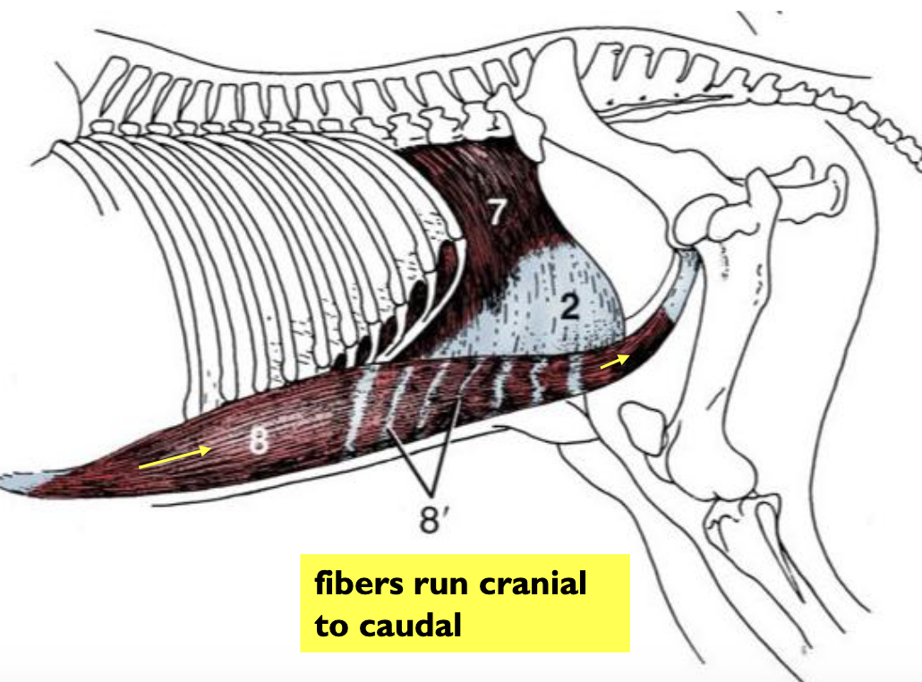
what are the origins of the muscle bellies of the rectus abdominis oblique?
outside sternum and sternal ribs/cartilages

what is the insertion of the aponeurosis of the rectus abdominis oblique? what is the name for the numerus white lines (8’)?
prepubic tendon, tendinous inscriptions

what muscle is indicated by the A in this image?
rectus abdominus
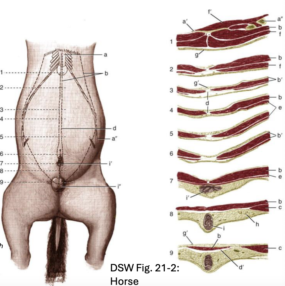
what muscle aponeuroses interweave and insert on the linea alba?
EAO, IAO, TA

where does the linea alba extend to?
from xiphoid process to cranial edge of pelvic symphysis
entire length of the linea alba: aponeurosis of the ______ _______ ______ muscle is external to the rectus abdominis muscle.
external abdominal oblique
cranial to umbilicus: aponeurosis of the ____ ____ ____ muscle splits and completely encloses the rectus abdominis muscle.
internal abdominal oblique

cranial to umbilicus: aponeurosis of the ______ ______ muscle is internal to the rectus abdominis muscle.
transversus abdominis

caudal to umbilicus: aponeurosis of _____ ______, and ______ abdominal muscles are external to the rectus.
external, internal, and transverse

clinical relevance: to close a midline incision, you need to incorporate full-thickenss bites of either one of what 2 structures in the sutures?
linea alba or external rectus sheath


what is the name of this opening in the lateral abdominal wall?
inguinal ring (external and superfical)
why are there openings/weaknesses in the abdominal wall?
fetal development
during development, what is the rotation of th emidgut (the twist of fate)?
in the embryo, the physical herniation of the midgut into the umbilicus. must happen in order to fit th eintestines into the abdominal cavity
what is the peritoneum?
thin, transparent sheet of mesothelium; serous membrane
what is the peritoneal cavity?
space between the parietal and visceral peritoneum; usually contains small amount od lubricating serous fluid to assist movements of organs

is the pelvic cavity ontinuous with the pelvic cavity?
no, makes several u-turns called ‘reflections’ that create extensions of the peritoneal cavity
name 4 extraabdominal extensions of the peritoneal cavity
pararectal fossa, rectogenital pouch, vesicogenital pouch, pubovesival pouch
define parietal peritoneum
single layer of peritoneum that lines the body wall

define visceral peritoneum
single layer of peritoneum that lines organ surfaces

define connecting peritoneum
double layer of peritoneum that extends vetween the body wall and viscera. extends between the viscera (ligament or fold)


what peritoneum is the yellow arrow pointing to?
parietal peritoneum

what peritoneum is lining the organs?
visceral peritoneum
3 examples of connecting peritoneum (2 layers)
mesenteries, ligaments and folds, omenta
define mesenteries
double layers of peritoneum that suspend the digestive tube from the body wall; blood and lymphatic vessels and nerves travel between the 2 mesothelial layers
dorsal mesentery covers which structures?
dorsal mesogastrium (becomes greater omentum), mesoduodenum, mesojejunum, mesoileum, mesocolon,mesorectum
ventral mesentery becomes which structures?
ventral mesogastrium (becomes lesser omentum), liver associated ligaments (coronary, triangular, falciform), medial ligament of the urinary bladder
describe the rotation of the midgut in development
intestinal loop rotates approximately 270 degrees counterclockwise, bringing the transverse colon cranial to the cranial mesenteric artery
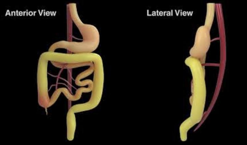
describe the ultimate outcome of the rotation of the midgut in all domestic species
transverse colon passes form right to left cranial to the cranial mesenteric artery

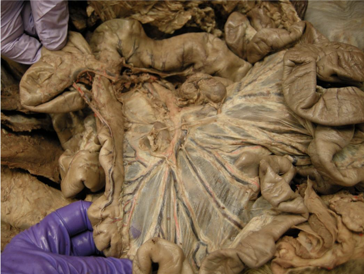
the mesentery (mesojejunoileum)
attatches to the dorsal abdominal wall by a short peritoneal attatchment known as “the root of the mesentery”

the root of the mesentery
surrounds the cranial mesenteric artery
what are peritoneal folds and ligaments?
double layers of peritoneum that connect viscera to viscera or viscera to the abdominal wall

describe omenta
double layers of peritoneum that develop from the dorsal and ventral mesenteries that suspend the stomach
which mesogastricum becomes the greater omentum and connects the greater curvature of the stomach to the dorsal body wall?
dorsal
which mesogastricum becomes the lesser omentum and connects the lesser curvature of the stomach and the liver?
ventral
what is the omental bursa?
potential space between the superficial leaf and deep leaf of the greater omentum

where is the superficial leaf of the greater omentum/mesogastrium?
(red) extends from the left longitudinal groove of the rumen to duodenum
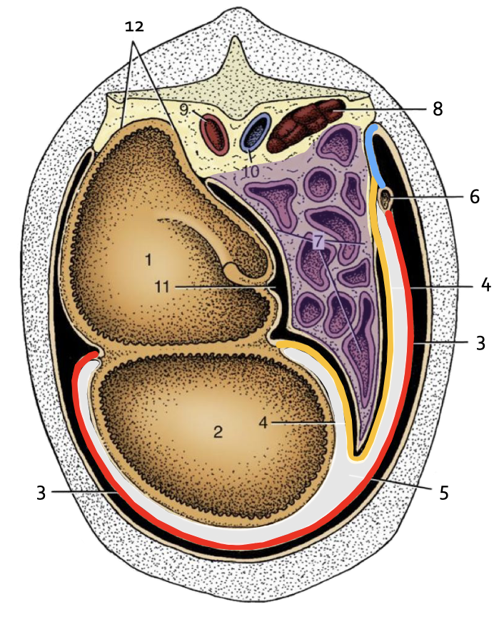
where is the deep leaf of the greater omentum/mesogastricum?
extends from right longitudinal groove of the rumen to mesoduodenum

in the ox, which leaf forms a sling for the intestinal mass called the supraomental recess?
deep leaf
what structure in ruminant is contained in the omental bursa?
ventral sac of the rumen
what is the epicloic foramen?
natural opening between the omental located caudal to the liver bordered by the vena cava dorsally and the portal vein ventrally
