Lt 12 Developmental Bone Disease
 Call Kai
 Learn
 Practice Test
 Spaced Repetition
 Match
 Flashcards
 Knowt Play
1/64

There's no tags or description
Looks like no tags are added yet.
65 Terms
Although radiographic signs vary by etiology, most are _____ in radiographic appearance
non-aggressive
Many degenerative diseases cause ______ lesions. Therefore, the other limb should always be radiographed for comparison.
bilaterally symmetric
Many developmental diseases result in _____
Degenerative joint disease
______ is failure of normal endochondral ossification caused by necrosis of epiphyseal cartilage
Osteochondrosis
Osteochondrosis leads to _____
divots in subchondral bone, progressive chondromalacia, and eventual defects in the articular surface
What factors can contribute to Osteochondrosis?
Anatomic conformation,
Rapid growth,
Nutrition,
Trauma/ischemia,
Hormonal influences
Clinical presentation for Osteochondrosis
Young, rapidly growing, large breed dogs;
Male >>> Female;
6-9 months old
What is the most common weight-bearing articular surface that gets Osteochondrosis?
Caudal aspect of the humeral head

Radiographic findings associated with Osteochondrosis?
Flattened or concave lucent defect in the subchondral bone.
Surrounded by bony sclerosis.
Widening of the joint space.
Synovial effusion/hypertrophy.

What is being highlighted by this image?
Osteochondrosis
Osteochondrosis Dissecans means that _____
Cartilage flap is present; "Joint mouse"

What is being highlighted in this image?
- Specifically the long arrow
Joint mouse
Prognosis for Osteochondrosis Dissecans
Caudal humeral head has excellent prognosis w/ treatment.
Fair to guarded prognosis for all other sites.

What is the most obvious/striking abnormality of this tarsus?
Degenerative changes and intracapsular soft tissue swelling

Is this a primary abnormality or is it secondary to some other process?
Secondary

What is the diagnosis for this image?
Osteochondrosis Dissecans

What is the next step for this patient?
Radiograph the other tarsus

Elbow dysplasia is a non-specific term for what 4 diseases that can affect the elbow?
1. Osteochondrosis (Distomedial aspect of humeral condyle/trochlea)
2. Ununited anconeal process.
3. Fragmented medial coronoid process.
4. Joint incongruity.
Ununited anconeal process is a common cause of Elbow dysplasia for _____
Large-breed dogs; Male >>> Female

Radiographic findings for Ununited anconeal process
Lucent line separating the anconeal process from the olecranon beyond 150 days
An ununited anconeal process can NOT be diagnosed until ______
5 months of age
What radiograph view needs to be taken to diagnose an ununited anconeal process?
Flexed lateral
Prognosis of an ununited anconeal process is ____
depends on degree of osteoarthritis and response to surgery
What is the MOST COMMON developmental disease of the cubital joint?
Fragmented Medial Coronoid Process
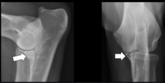
What part of the bone is being highlighted in this image?
- Normal presentation for this structure
Medial coronoid process

Radiographic findings for a
Fragmented Medial Coronoid Process
- Osteophyte formation on anconeal process and radial head.
- Indistinction or abnormal contour of cranial margin.
- Sclerosis of subchondral bone of Trochlear notch.
- Joint incongruity.

What is the BEST diagnostic tool for identifying a Fragmented Medial Coronoid Process?
Arthroscopy > CT > Radiographs

What is being highlighted in this image?
Elbow dysplasia; Fragmented medial coronoid process and Ununited anconeal process
Aseptic Necrosis of the Femoral Head is also called _____
Legg-Calve-Perthes disease
Aseptic Necrosis of the Femoral Head is common for _____
Young, SMALL-breed dogs; NO sex predisposition
Aseptic Necrosis of the Femoral Head is caused by _____
Compromised blood supply to the femoral capital epiphysis
Aseptic Necrosis of the Femoral Head progresses to _____
Subchondral bone necrosis and remodeling of the femoral head
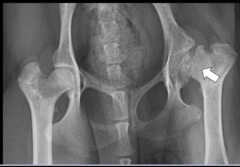
Radiographic findings for Aseptic Necrosis of the Femoral Head
- Lucent areas within the epiphysis and metaphysis.
- Flattening of the femoral head.
- Linear lucent areas within the subchondral bone.
What is the treatment of choice for Aseptic Necrosis of the Femoral Head ?
Femoral head and neck ostectomy; Tx is palliative.
What diagnostic needs to be done to confirm Aseptic Necrosis of the Femoral Head?
Histopathology
_______ is a condition that causes delayed closure of the capital physis and multiple other growth plates in cats.
Contains abnormal clusters of chondrocytes and excessive connective tissue.
Physeal Dysplasia
The capital physis normally closes around ______
7.5 to 11 months old

What condition is being highlighted in this image?
Physeal Dysplasia
Clinical presentation for Physeal Dysplasia?
Spontaneous fracture of the capital physis NOT associated with trauma;
Male cats >>> Female cats;
Obese;
VERY often bilateral condition

What are the radiographic signs of Physeal Dysplasia?
- Femoral head and neck lysis with sclerosis and remodeling.
- Capital Physeal fracture.
-Metaphyseal resorption with chronicity.

Treatment for Physeal Dysplasia
Femoral head and neck ostectomy; Tx is palliative.
9 month old, MN, Golden retriever with right forelimb lameness and pain on shoulder extension
Osteochondrosis
1.5 year old, FS, Golden retriever with a hopping gait, pain on hip extension, decreased thigh musculature, and a positive Ortolani sign
Hip dysplasia
1 year old, MI, German shepherd with bilateral thoracic limb lameness with pain on elbow flexion
Elbow dysplasia; Fragmented medial coronoid process and Ununited anconeal process
7 month old, MN, Rottweiler with right forelimb lameness and pain on elbow flexion
Elbow dysplasia; Fragmented medial coronoid process and Ununited anconeal process
1 year old, FS, Toy poodle with a sudden non-weight bearing lameness of the left pelvic limb, decreased left thigh musculature, and pain on left hip extension
Aseptic Necrosis of the Femoral Head and Neck
_______ is a self-limiting idiopathic disease of the long bones.
Increased osteoblastic and fibroblastic activity in the medullary cavity, endosteum, and periosteum.
Panosteitis
Does inflammation play a role in Osteitis?
No, its a misnomer
Clinical presentation for Panosteitis
large breed dogs;
Males >>> Females;
5-12 months old up to 7 years old
Clinical presentation for Osteitis
Pain on palpation of the diaphysis of the long bones

Radiographic findings of Osteitis
increased opacity of the diaphyseal medullary cavity of long bones; Often near the nutrient foramen

Is this an early or late presentation of Osteitis? Why?
Early presentation; Patchy and heterogenous lesion
What would a late presentation of Osteoitis look like?
Diffuse and homogenous lesion
Prognosis for Osteitis is ____
Excellent and often self-limiting
Treatment for Osteitis
Rest,
Analgesics,
Weight management

What is being highlighted in this image?
Osteitis of the radius and ulna
Causes of Hypertrophic Osteodystrophy
Oversupplementation of vitamins and minerals,
Hypovitaminosis C,
Sterile inflammation,
Canine distemper virus
What of Hypertrophic Osteodystrophy?
Neutrophilic inflammation of the metaphysis causing necrosis, hemorrhage, and increased osteoclasts —> Collapse of the metaphysis.
Congestion and edema in surrounding tissues.
Clinical presentation for Hypertrophic Osteodystrophy?
Large breed dogs; 2-8 months old
Swollen, firm, and painful metaphyseal region of long bones.
Fever, lethargy, anorexia.

Radiographic findings for Hypertrophic Osteodystrophy
Lucent zones int he metaphysis that are parallel to the physis -> "Double Physeal line sign".
Subchondral bony sclerosis.
Irregular widened physis.
Which bones is Hypertrophic Osteodystrophy most common in?
Distal radius and ulna
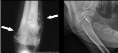
What is being highlighted in this image?
Hypertrophic Osteodystrophy; "Double Physeal sign"
Prognosis for Hypertrophic Osteodystrophy
Good with treatment and usually self-limiting.
Premature Physeal closure or permanent bony changes.
Treatment for Hypertrophic Osteodystrophy
Analgesics,
IV fluids,
Decreased caloric intake in fast-growing dogs

8 month old, MI, German shepherd with right thoracic limb lameness, sometimes switches to left thoracic limb lameness
Panosteitis; Multifocal, ill-defined, ovoid to irregularly shaped sclerotic lesions of the humeral, radial, ulnar diaphyses.