Lab Exam 1
 Call Kai
 Learn
Learn Practice Test
 Spaced Repetition
 Match
Match Flashcards
 Knowt Play
1/73

There's no tags or description
Looks like no tags are added yet.
Name | Mastery | Learn | Test | Matching | Spaced | Call with Kai | Chat |
|---|
No analytics yet
Send a link to your students to track their progress
74 Terms

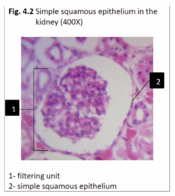
epithelial tissue that is most suited to
Diffusion of gases across lung air sacs
Simple Squamous epithelium
Located: Alveoli of lungs, glomerular
capsule of kidney (filtration
membrane), endothelium of blood
vessels

epithelial tissue that is most suited to
Mechanical protection & waterproofing
stratified squamous keratinized epithelium
Located: Skin, scalp


epithelial tissue that is most suited to
Secretion of mucus & movement of mucus
Pseudostratified ciliated columnar epithelium
Located: Trachea, bronchi, auditory canal of middle
ear


epithelial tissue that is most suited to
Stretchy quality that allows expansion as the lumen is filled
transitional epithelium
Located: Urinary bladder, ureters, calyces (funnels) of
kidney

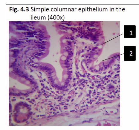
epithelial tissue that is most suited to
Secretion, absorption & chemical protection
Simple columnar epithelium
Located: Small intestine (duodenum, jejunum,
ileum), colon, stomach, gallbladder, uterus


epithelial tissue that is most suited to
Mechanical protection of a moist surface
Non-keratinized stratified squamous epithelium
Located: Esophagus, tongue, vagina, anus, pharynx
(throat)


A Pap smear involves collection & microscopic examination of epithelial cells from the nonkeratinized stratified epithelium of the vagina & cervix. Which surface of the cells is the sample scraped from?
scraped from the apical surface of the epithelial cells.

fill in the blanks
simple squamous epithelium
filtering unit
simple cuboidal epithelium
lumen

stratum corneum

transitional epithelium

simple cuboidal epithelium

pseudostratified ciliated columnar epithelium
trachea

stratified squamous epithelium
tongue

simple squamous epithelium

simple columnar epithelium
uterus

what kind of tissue?
location
Areolar connective tissue
lining the digestive, respiratory, urinary, and reproductive tracts
anchoring the epidermis and dermis to underlying muscles while allowing the skin to move freely


what kind of tissue?
location
adipose connective tissue
Directly beneath the skin (hypodermis) across the entire body


what kind of tissue
location
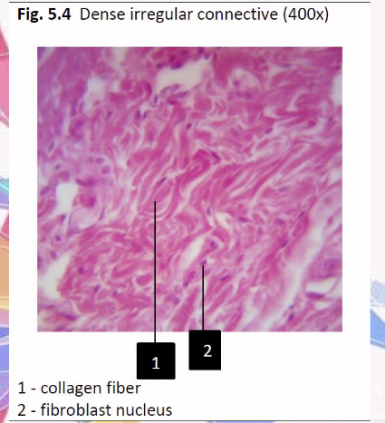
dense irregular connective
areas that need strength against pulling dif directions
skin, organ covering etc.

what kind of tissue
dense regular connective
tendons, ligaments, and aponeuroses


what kind of tissue
location
hyaline cartilage connective
joints, the respiratory tract, lines the ends of bones in movable joints


what kind of tissue
location
bone connective tissue
skeletal system
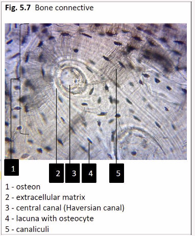

what kind of tissue
location
blood connective
throughout the body inside blood vessels cardiovascular system

elastic cartilage
location
flexible, shape-retaining cartilage
the external ear (pinna), and parts of the larynx.
fibrocartilage
Fibrocartilage is a tough, shock-absorbing tissue
high-stress areas of the human body
spine, knee joint, pelvis, jaw
Considering that a hydrophilic stain was used to stain the skin, explain why the fat vacuoles in
adipocytes are white
hydrophilic “like’s water”
fats repel water so the fat filled adipocytes stay white and wont absorb the hydrophilic stain.

How do chondrocytes differ from fibroblasts in appearance?
chondrocytes: inside round lacunae. often stacked or clustered. hyaline cartiladge
Fibroblasts: flattened or oval shaped between between collagen and elastic fibers in loose or dense connective tissues

tissue that
Resists pulling forces at attachment points
dense regular connective tissue

tissue that
Smooth, slipper surface that reduces friction
hyaline cartilage

tissue that
Resists pulling forces from many different directions
Dense Irregular Connective Tissue

tissue that
Stores lipids
adipose tissue


what kind of muscle
cardiac muscle tissue
involuntary
striated
1-2 center nuclei per cell
intercalated discs
branched muscle fibers
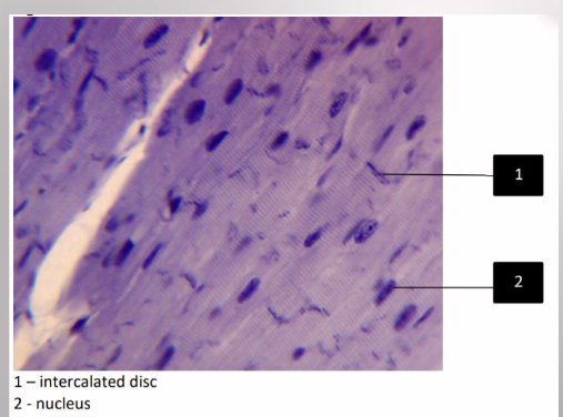

what kind of muscle
skeletal muscle tissue
voluntary
striated
long cylindrical
parallel fibers

what kind of muscle
smooth muscle
involuntary
non-striated
Cells are spindle shaped (pointed at both ends)
single central nucleus
location: walls of hollow organs, blood vessels,

what kind of tissue
Nervous Tissue

When one muscle cell dies, the tissue is usually replaced by dense irregular connective tissue. How do these tissues differ structurally & functionally?
Called fibrosis
forms a non-contractile scar
dense irregular connective tissue does not contract for movement
goes from elongated muscle fibers to thick interwoven bundles
Dense irregular connective tissue is sometimes confused with smooth muscle. How can you
distinguish between these two?
Dense:
thick collagen fibers between some cells
fibroblasts flattened between nuclei
Smooth:
tightly packed smooth muscle cells
nuclei in the center of cells

Write the parts of a multipolar neuron in the correct order of receiving & sending the impulse
Dendrites (receives the incoming signal)
Cell body (soma)
Axon
Synapse / Next Neuron

What is this tissue
name the parts
epidermis of thick skin
CLGSB - chat lets get sun burn
Stratum corneum
Stratum lucidum
Stratum granulosum
Stratum spinosum
stratum basale

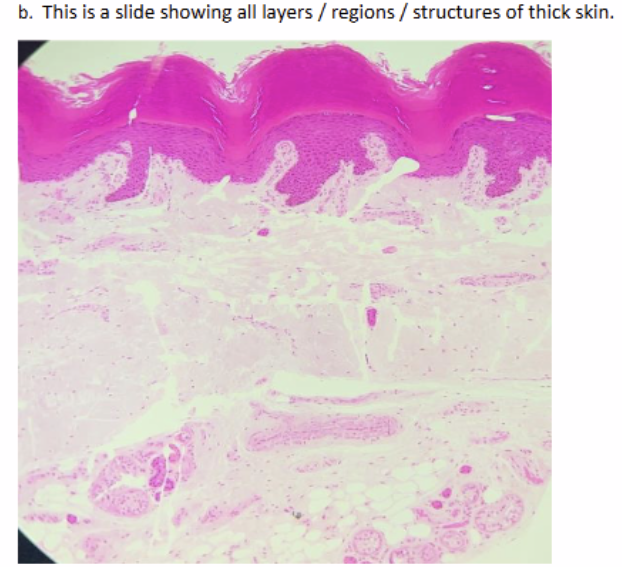
name of tissue
name parts
thick skin
epidermis: CLGSB
dermis: Papillary region, Reticular region
hypodermis


what tissue
what structures
thin skin
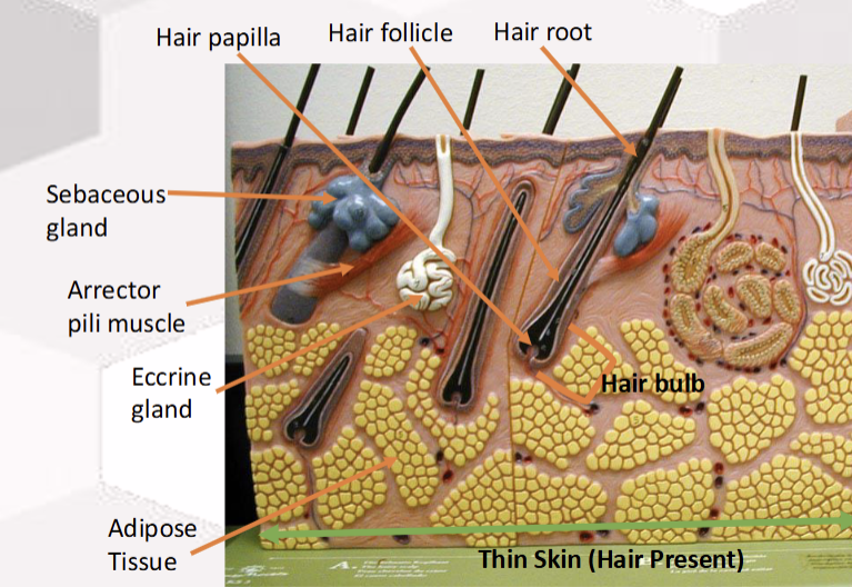
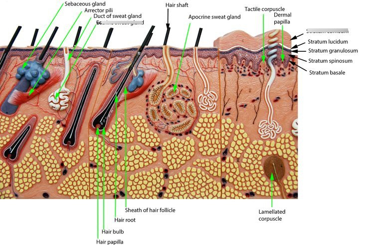
idk read these
The epidermal layers of the skin in which these skin cancers arise:
Basal cell carcinoma
basal cell layer of the skin
deepest layer of the skin
Ultraviolet (UV) radiation damages the DNA inside these basal cells, leading to uncontrolled growth
The epidermal layers of the skin in which these skin cancers arise:
melanoma
basal cell layer
deepest layer
malignant transformation of melanocytes,
The epidermal layers of the skin in which these skin cancers arise:
Squamous cell carcinoma
stratum spinosum
keratin producing squamous cells produced here
Which part of the skin peels off with a minor sunburn?
epidermis
are permanent tattoos made by injecting pigment into the dermis or epidermis? Explain your
answer
dermis
Epidermal cells constantly die and flake off, renewing themselves every few weeks.
Which sub-layer of the epidermis has the oldest cells?
stratum corneum
topmost layer of the skin
cells get pushed up as they die
Explain why a superficial scrape doesn’t bleed
it only scratches the epidermis
epidermis has no blood capillaries of its own
IPMATC
i passed my anatomy test calmly
mnemonic for cell division
Interphase, (Mitosis:) prophase, metaphase, anaphase and telophase), and cytokinesis
difference between mitosis and meiosis
Mitosis makes two identical body cells for growth and repair, while meiosis makes four unique sex cells (gametes) for reproduction.
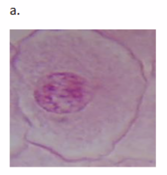
What cell cycle stage is this
Name the structures
interphase
spaghetti like chromatin
the nuclear envelope and a distinct nucleolus are clearly visible inside the nucleus

What cell cycle stage is this
Name the structures
Left cell: Prophase — Chromosomes are condensing and becoming visible.
Right cell: Interphase — Chromatin hasn’t condensed into visible chromosomes yet

What cell cycle stage is this
Name the structures
metaphase - chromosomes have lined up
spindle fibers are visible

What cell cycle stage is this
Name the structures
Anaphase - spindle fibers pull the chromosomes/sister chromatids apart

What cell cycle stage is this
Name the structures
Telophase - chromosomes pulled to opposite poles
guided by spindle fibers
cleavage furrow begins to split them during cytokinesis
how to interpret/calculate cell cycle phases of normal tissues and cancerous tissues
Mitotic Index: # of cells in mitosis (P, M, A, T)
Total number of cells counted
Normal tissues: have a mitotic index range of 1% to 5%
Cancerous tissues: Significantly higher sometimes exceeding 10%
Why is cell division important for both single-celled and multicellular organisms?
it allows single-celled organisms to reproduce and multicellular organisms to grow, develop, and repair damaged tissues
Why does cell division remain important to an adult organism even after it is fully developed?
to repair damaged tissue, heal wounds, and replace old or dead cells through continuous cell turnover
Cells divide, differentiate, or die. What is differentiation?
Differentiation is when unspecialized or immature cells (such as a stem cell) changes into a specialized cell type with a distinct structure and function (like a muscle cell, nerve cell, or red blood cell
What is apoptosis? What is its purpose?
the body's natural, programmed process of controlled cell death
Keeps the right balance of cells in your body by getting rid of old cells
kills cells before they become cancerous
Shapes tissues and organs during embryonic development
What are cell cycle regulators?
special proteins and molecular signals
Stimulating proteins drive the cycle forward
Inhibitory proteins stop the cycle
What happens if cell cycle regulators don’t function properly?
the body produces too many or too few cells
can lead to uncontrolled growth (cancer), tissue damage, or genetic disorders
Cells go through periods of growth and division. Cell division occurs during ___________________
mitosis

G1: gap 1 - The cell grows and makes new proteins and organelles.
S: synthesis - The cell replicates its DNA.
G2: gap 2 - The cell grows more and gets ready to divide.
In general, what is the purpose of a checkpoint in the cell cycle?
checkpoint - checks if previous stage is complete, cell size, nutrients, growth factors, and DNA damage.
If conditions fail, the cell may enter G0 a resting state or undergo programmed cell death (apoptosis)
What is the G0 phase of the cell cycle
G0 cells remain active but do not divide usually (after G1 phase)
causes: differentiated cells lost the need or capacity to divide, nutrient-deprived or energy-limited environment, Absence of Growth Factors, DNA damage or cellular stress
some cells enter G0 temporarily
What are cell cycle regulators
Stimulating proteins are encoded by proto-oncogenes
Inhibitory proteins are encoded by tumor suppressor genes
Examples include: cyclin-dependent kinases (CDKs)
Cancer is the result of an improperly regulated cell cycle. Describe two reasons why cells can form tumor
uncontrolled cell division and a failure of programmed cell death (apoptosis).
In some types of colon cancer, stem cells have a mutation in the APC gene. What happens if the APC gene is mutated?
APC gene produces a tumor suppressor protein that destroys beta-catenin when it is not needed
When APC is mutated or shortened, it fails to break down beta-catenin, which triggers uncontrolled cell growth and leads to colorectal cancer
Normally, proto-oncogenes stimulate the cell cycle. What are oncogenes and how do they affect the cell cycle?
mutated proto-oncogenes are oncogenes
they drive cells to grow and divide out of control.
To cause cancer, proto-oncogenes require _________ allele(s) to be mutated and therefore are
considered ______________________ . The mutation results in a ________ of function
proto-oncogenes require ONE allele to be mutated
Considered dominant because a single altered copy is enough to promote abnormal growth, even if the second allele is normal.
mutation results in a gain of function that pushes the cell through the cell cycle continuously.
Normally, tumor suppressor genes inhibit the cell cycle. How do mutated tumor suppressor genes affect the cell cycle?
Mutated tumor suppressor genes lose their ability to stop the cell cycle, leading to uncontrolled cell division and tumor growth
To cause cancer, tumor suppressor genes require __________ allele(s) to be mutated and therefore are considered ______________________ . The mutation results in a ________ of function.
tumor suppressor genes require both(or two) alleles to be mutated
are consideredrecessive (require both)
mutation results in a loss of function.