non-squamous malignancies
 Call Kai
 Learn
 Practice Test
 Spaced Repetition
 Match
 Flashcards
 Knowt Play
1/59

Earn XP
Description and Tags
lecture given 11/16/2026
60 Terms
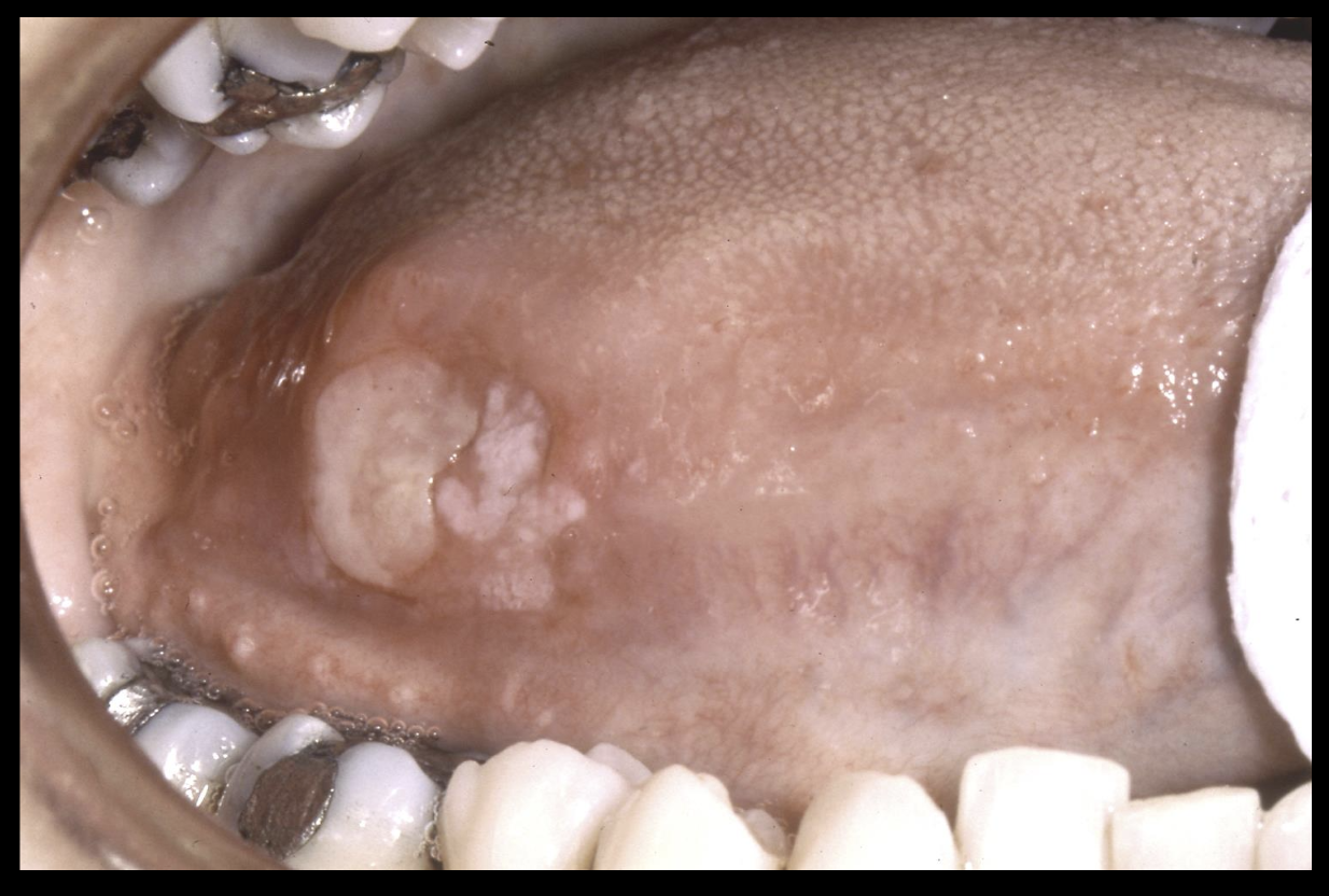
high risk sites for oral squamous carcinoma
lateral tongue
ventral tongue
floor of mouth
retromolar trigone
soft palate
squamous carcinoma
leukemia
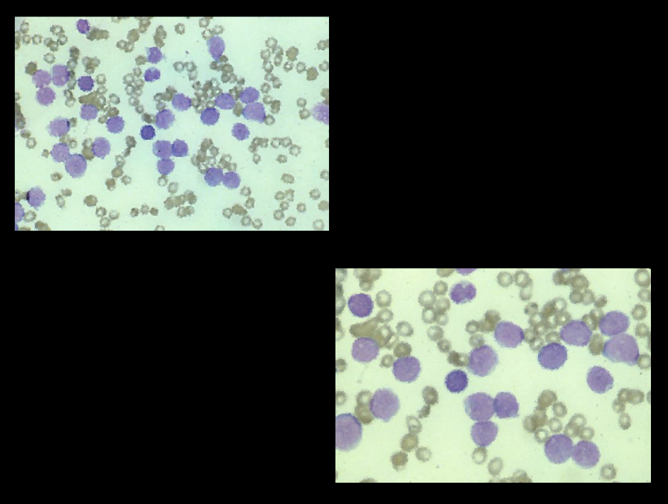
malignancy of hematopoietic stem cell derivation
malignant transformation of one of the stem cells
initially proliferates in the bone marrow and eventually is seen in the peripheral blood
leukemic cells crowd out the normal white and erythrocyte precursors
results from environmental and genetic factors
classification- acute myeloid, chronic myeloid, acute lymphocytic
signs and symptoms- myelophthisic anemia, splenomegaly, lymphadenopathy, hepatomegaly, thrombocytopenia, fever associated with infection
oral manifestations- oral ulceration, candidiasis, herpetic infections, diffuse gingival enlargement/tumor like growth, gingival hemorrhage, leukemic infiltration of periapical tissues
diganosis- confirmed via peripheral blood and bone-marrow analysis / immunohistochemical markers, cytogenetic, and molecular studies are done to classify the type
treatment- chemotherapy
histopathology- loss of normal tissue architecture to a monotonous proliferation of poorly differentiated cells with myelomonocytic or lymphoid features
leukemia

leukemia

leukemia

leukemia

leukemia
hodgkin lymphoma
malignant lymphoproliferative disease in lymph node
neoplastic cells make up only 0.1-2% of the cells in the enlarged lymph nodes
unknown cause but may be linked to EBV infection
most common sites- cervical and supraclavicular lymph nodes (75%), axillary and mediastinal lymph nodes (10%), inguinal and abdominal lymph nodes (<5%)
bimodal peak at 15-35 yrs and 50+
males > females
signs and symptoms- persistently enlarging non-tender mass in lymph node region, weight loss, fever, night sweats, generalized pruritis
diagnosis- history and physical exam, microscopic examination, chest radiographs, CT scans, PET scans, MRI studies, hematological studies
treatment- depends on stage of involvement, stage 1&2 local radiation therapy, stage 3&4 chemo and radiation
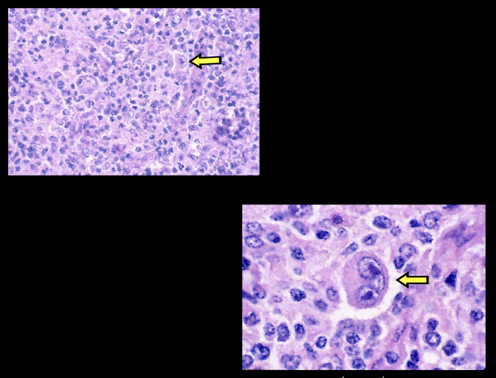
histopathology- normal nodal architecture is replaced by a diffuse mixed inflammatory cell infiltrate, interspersed in btwn these cells are neoplastic large atypical lymphoid cells, atypical cells are called reed sternberg cells, typically binucleated, several histopathologic subtypes are recognized
hodgkin lymphoma

hodgkin lymphoma
non-hodgkin lymphoma
diverse group of malignant lymphoproliferative disease
initially arise within lymph nodes and slowly enlarge as solid masses
complex classification scheme- modified REAL/WHO classification
adults > children
most commonly develops in lymph nodes, slowly enlarging non tender mass, cervical/axillary/inguinal nodes, gradually other lymph node groups are affected, invasion of adjoining normal tissues
20-40% of cases are extranodal lymphomas, oral lesions most commonly B cell in origin
soft tissues- non tender/diffuse swelling, buccal vestibule, gingiva, posterior hard palate, boggy consistency
central bone lesions- vague pain/toothache, paresthesia, ill-defined radiolucency, bony expansion with cortical perforation
diagnosis- history and physical exam, microscopic/immunohistochemical/cytogenetic studies, chest radiographs, CT scans, PET scans, MRI studies, hematologic studies
treatment- depends on the stage and grade, chemotherapy/radiation therapy
histopathology- infiltrative monotonous proliferation of lymphocytic appearing cells destroying normal tissue architecture showing varying degrees of differentiation; lesional tissue necrosis and vague germinal center formation may be seen / patterns- nodular/follicular, diffuse
non-hodgkin lymphoma

non-hodgkin lymphoma

non-hodgkin lymphoma

burkitt lymphoma
malignancy of B lymphocyte origin that represents undifferentiated B cell lymphoma
unique geographic differences in clinical presentation
largely associated with EBV; represents the first time virus identified in human tumor pathogenesis
cytogenic chromosomal translocations- most common translocation places oncogene c-myc on chromosome 8 under control of IgH enhancer on chromosome 14, remainder exhibit translocation btwn c-myc and either kappa or lambda light chain
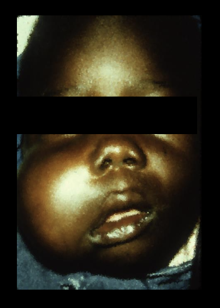
endemic (african) form- central africa, rare in US, 50-70% present in jaws, maxilla>mandible, posterior>anteriorn, affects children, male to female 2:1, 90% of the cases are associated with EBV infection
sporadic (american) form- lesions typically present in abdomen (rarely jaws), affects pts over wide age, 15-30% EBV associated
signs and symptoms- facial swelling and proptosis, marked tooth mobility, radiolucent destructive lesions with ragged, ill-defined borders and patchy loss of lamina dura
histopathology- sheets of small hyperchromatic tumor cells with round nuclei and minimal cytoplasm, macrophages in lesional tissue give starry sky appearance
burkitt lymphoma
burkitt lymphoma

burkitt lymphoma

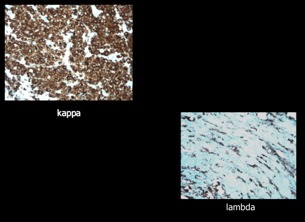
multiple myeloma
uncommon maligancy of plasma cell origin, multi-centric within bone, unknown cause
1% of all malignancies, 10-15% of all hematologic malignancies
monoclonal proliferation of plasma cells
signs and symptoms result from uncontrolled proliferation of the tumor cells and the uncontrolled manufacture of their protein products
presents in older men, males>females, blacks>whites
signs and symptoms- bone pain/pathologic fracture, anemia/petechial hemorrhage, fever, radiolucent punched out lesions of bone, kidney failure (accumulation of bence jones proteins), amyloidosis- deposition of amyloid in various soft tissues of the body, predilection for tongue/periorbital skin
histopathology- diffuse monotonous proliferation of neoplastic plasmacytoid cells, mitotic activity
multiple myeloma

multiple myeloma

multiple myeloma

multiple myeloma

multiple myeloma

multiple myeloma

multiple myeloma

multiple myeloma
multiple myeloma

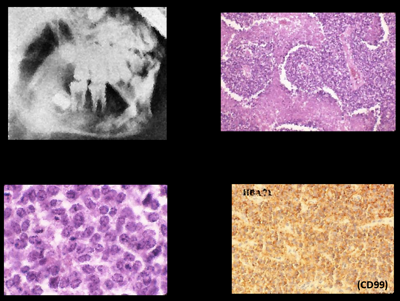
ewing sarcoma
primary malignant tumor of bone, composed of small undifferentiated round cells
less than 3% originate in the jaws, uncertain histogenesis; likely neuroectodermal origin
80% occur in pts <20 yrs, white males, mandible > maxilla
signs and symptoms- pain associated with swelling, fever, leukocytosis, elevated erythrocyte sedimentation rate, cortical perforation, paresthesia, loosening of teeth, ill-defined radiolucency
histology: small round blue cell tumor, sheets or lobules of tumor cells, glycogen granules often found in cytoplasm, fish/RT-PCR/immunohistochemistry confirms diagnosis
pleomorphic adenoma

ewing sarcoma
rhabdomyosarcoma
malignant neoplasm that is characterized by skeletal muscle differentiation
more common in children, head and neck is most common site
several microscopic patterns noted- embryonal & alveolar subtypes common in the head and neck area
rapidly growing painless infiltrative mass
face and orbits are the most common location
palate is the most common intraoral site
local surgical excision followed by chemotherapy, 5 year survival rate 55-7%
rhabdomyosarcoma

rhabdomyosarcoma

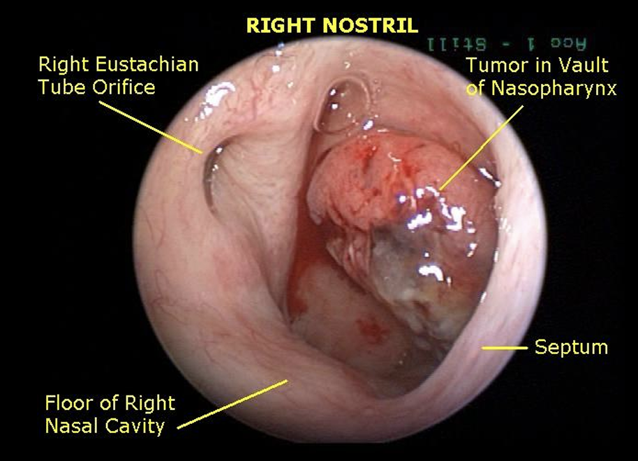
nasopharyngeal carcinoma
arise from the lining epithelium of the nasopharynx which is rich in lymphoid tissue
rare in US, increased incidence in southern china, southeast asia, inuits of alaska, and greenland
contributory factors- EBV infection, diets deficient in vitamin C, consumption of salt fish containing carcinogenic N-nitrosamines, tobacco, HPV
40-60 yrs, male>female
arises in the lateral nasopharyngeal wall, primary lesion may be small and difficult to detect
enlarged cervical lymph node may be the first sign of the disease
epistaxis, nasal obstruction, pharyngeal pain
radiation therapy/chemotherapy, prognosis fair
nasopharyngeal carcinoma

nasopharyngeal carcinoma
nasopharyngeal carcinoma

osteosarcoma
malignancy of the mesenchymal cells that have the ability to produce osteoid or immature bone
etiology unknown, 6% of all osteosarcomas occur in the jaws
occurs in pts 20-40 yrs of age, males > females, maxilla = mandible
signs and symptoms: swelling, pain, loosening of teeth, paresthesia, nasal obstruction, some osteosarcomas may exhibit slow growth
radiographic features: radiopaque or mixed radiolucent-radiopaque or entirely radiolucent lesion with ill-defined borders, cortical distruction, cortical expansion and periosteal reactions may be seen, classic sun burst pattern, symmetrical widening of the PDL space, triangular elevation of the periosteum, spiking root resorption
treatment and prognosis: radical surgical excision, radiation, chemotherapy, 60-70% 5 yr survival rate, metastatic lesions from jaw rare (metastatic sites lymph nodes, lung, brain)
osteosarcoma

osteosarcoma

osteosarcoma

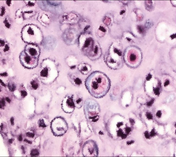
chondrosarcoma
malignant neoplasm, tumor cells form cartilage
1-12% occur in head in neck area
average age 51 yrs, males > females, maxilla = mandible
signs and symptoms: painless swelling, loosening of teeth, nasal obstruction/epistaxis
radiographic features: ill defined radiolucency with varying degrees of radiopacity, occasionally multilocular radiolucency, cortical penetration may yield sun burst pattern, extensive infiltration btwn bony trabeculae, root resorption and symmetric widening of PDL space
histopathology: cartilage showing varying degrees of maturation and cellularity round or spindle shaped cells, often has a lobular pattern with fibrous connective tissue septae, calcification of chondroid matrix may occur
treatment and prognosis: radical surgical excision, poorer prognosis than osteosarcoma, head and neck chondrosarcomas are locally aggressive with low metastatic potential
chondrosarcoma

chondrosarcoma
melanoma
malignant neoplasm of melanocytes
skin version more common in fair skin pts, oral version more common in dark skin pts
4 of 5 oral melanomas occur on the palate or maxillary alveolus
more common in older pts
although rare in the oral cavity it has a terrible prognosis
ABCDE features
melanoma

melanoma

melanoma

salivary gland malignancy
uncommon but not rare, more common in older pts
risk factors are not identifiable in most cases
common tumors- mucoepidermoid carcinoma, adenoid cystic carcinoma, PLGA
common intraoral locations- palate, upper lip, buccal mucosa, floor of mouth, tongue
usually present as a painless slow growing swelling
prognosis depends on tumor type and clinical stage at diagnosis
adenoid cystic carcinoma

metastases to the oral cavity
uncommon
metastasis can occur to the jaw bones or to the oral soft tissues
management is usually palliative in nature
overall prognosis is poor
metastatic tumors to the jaws
common form of cancer involving bone metastasis from breast, lung, thyroid, prostate, and kidney carcinomas
older pt predilection for posterior mandible
pain, swelling, tooth mobility, paresthesia, may also be asymptomatic
jaw lesion may be the first sign of the malignant disease
ill defined or moth eaten radiolucencies
microscopic appearance resembles the tumor of origin
metastatic tumors to the oral soft tissues
gingiva- most common soft tissue site
tongue- next most common site
nodular mass that resembles hyperplastic tissue like PG
adjacent teeth may be mobile
middle aged/older pts
lung cancer, renal carcinoma, melanoma, breast
oral lesion may be the first sign of the malignant disease
microscopic appearance resembles the tumor of origin
sources of oral metastasis
breast, lung, GI tract, thyroid, prostate, kidney
metastatic cancer

metastatic cancer

metastatic cancer

metastatic cancer
