Abdomen Ultrasound II- Discussion 9
 Call Kai
 Learn
 Practice Test
 Spaced Repetition
 Match
 Flashcards
 Knowt Play
1/79

Earn XP
Description and Tags
Abdominal Vasculature Pathology
80 Terms
What is the largest artery in the body?
aorta
Where does the aorta originate?
Left ventricle of the heart
Where is the aorta positioned?
Anterior to the spine, just left of midline
Arteries become thick and stiff restricting blood flow to your organs and tissue
arteriosclerosis
The buildup of plaque, which can restrict blood flow.
Atherosclerosis
Full occlusions of the abdominal aorta tend to occur in the __________ portion of the aorta and/or _____________________ with thrombus spreading upward.
lower; common iliac
Ultrasound features of atherosclerotic plaque:
Luminal irregularities – plaques
Aortic wall calcifications- sharp, hyperechoic, can display acoustic shadowing
Tortuosity (twisting)– mild or markedly
Causes of atherosclerosis:
HTN, high cholesterol, high triglycerides, smoking, insulin resistance or diabetes, inflammation from diseases or infections
Treatments for atheroscelrosis:
Lifestyle changes, such as eating a healthy diet and exercising, are often the most appropriate treatment for atherosclerosis. Medication or surgical procedures such as angioplasty and stent placement.
What is the most frequent cause of embolism?
plaque breaking loose and traveling distally until it lodges in a small vessel
Clinical findings of aneurysm:
Pulsatile abdominal mass, abdominal bruit, back pain, abdominal pain, lower extremity pain
What is the measurement for an AAA?
>3 cm
The _____________ the dilation, the more risk there is for potential rupture.
larger
Present when there are areas of dilation less than 3 cm or when there are irregular margins and a non-tapering profile
ectasia
Most aortic aneurysms are _____________.
fusiform
How is the aorta measured?
outer to outer
Risk factors for AAA:
Atherosclerosis, tobacco use, hypertension, trauma, chronic obstructive pulmonary disease and positive family history for AAA, Marfan syndrome, inflammation of media and adventitia
Inherited, degenerative disorder of the connective tissue, which gives shape and structure to tissues in the body and holds them in place
Marfan’s syndrome
Symmetric and gradual dilatation of vessel wall with proximal and distal tapering. Due to chronic degenerative change in arterial wall (makes it slow growing)
Fusiform aneurysm
Focal, eccentric dilatation of vessel wall. Suggests possible inflammatory or infectious etiology
Saccular aneurysm
Not full thickness of vessel wall. Usually related to prior instrumentation including prior aneurysm repair
Pseudoaneurysm
Blood enters the media of the aorta and splits the aortic wall
Dissecting aneurysm

Fusiform aneurysm
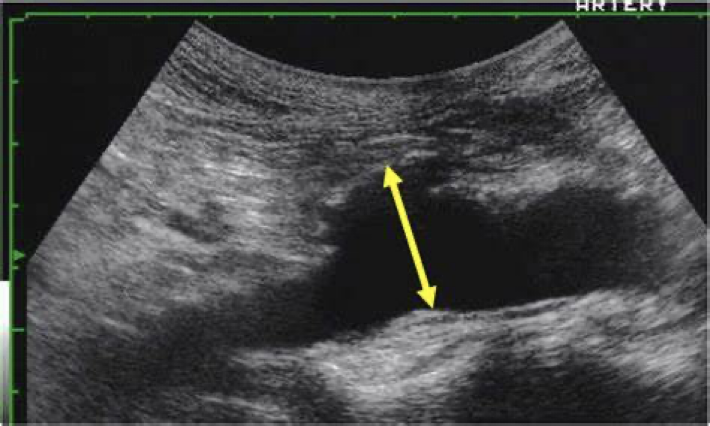
Saccular aneurysm

Pseudoaneurysm
Pseudoaneurysm sonographic findings:
A perivascular hematoma that contains swirling blood and has a neck connecting it to the vessel, color Doppler demonstrates turbulent flow within the mass, the spectral waveform obtained from the neck of pseudoaneurysm typically displays a high velocity, bidirectional or “to-and-fro” signal (ying-yang)
What is the most common type of splanchnic artery aneurysm?
splenic artery
Second most common type of splanchnic vessel aneurysms
encountered.
75% extrahepatic in origin.
25% occur intrahepatically.
Right hepatic arterial branch more often affected than the left.
Rare and tend to male preponderance.
Hepatic artery aneurysm
Rarest splanchnic artery aneurysms.
Branch superior mesenteric arterial aneurysms are also rare.
SMA aneurysm
aneurysm caused by infection are termed mycotic aneurysms
Mycotic aneurysm
small saccular aneurysm commonly located in the cerebrum (brain vessels)
brain aneurysm
More commonly seen in very large aneurysms. Usually eccentric in location. Causes narrowing of vessel lumen.
Fresh clots (acute) are hard to see, less echogenic; while chronic clots more echogenic.
Thrombi-blood clots
The most serious complication of AAA is ________ of the aorta into the retroperitoneum
rupture
Risk of rupture increases with aneurysms larger than ____.
7 cm
Mortality rate for untreated rupture is virtually _______.
100%
With surgery, mortality rate for rupture ranges from
40-60%
Ruptures aortic aneurysm symptoms:
severe abdominal pain, hypotension, shock, decreased hematocrit, and an expanding abdominal mass
Sonographic appearance of ruptures aortic aneurysm:
Varies
Appears anywhere from completely cystic to complex collection in the abdomen
It is difficult to identify the exact location of a rupture with ultrasound
CT is the imaging modality of choice with suspected AAA rupture
Surgical intervention is considered when the aneurysm reaches _____ or greater.
5 cm
Correction of pseudo aneurysm may be accomplished by:
Surgical repair, prolonged compression repair, ultrasound guided thrombin injection repair
Aortic dissection almost always begin in the _______ aorta and extend down into the abdominal aorta.
thoracic
Aortic dissection sonographic appearance:
Identify 2 channels of flow, using 2D or Color flow Doppler
A linear intimal flap, may be noted within the aortic lumen. This flap may be visualized swaying in the current of the passing blood
Determine extent of dissection in either direction
Determine the velocity of flow and existence of turbulent flow
Type A aortic dissection:
Tear in the ascending aorta, which may extend into the abdominal aorta -- More common and dangerous type
Type B aortic dissection
Tear in the descending aorta which may also extend into the abdominal aorta
Aortic dissection causes and risk factors:
Occurs in a weakened area of the aortic wall
Chronic HTN may stress the aortic wall making it more susceptible to tearing
Conditions associated with a weakened and enlarged aorta, such as Marfan syndrome can also increase the chances of occurrence
Aortic dissection most commonly occurs in:
Men during the 6th-7th decade of life (>40 years)
Treatments for Type A aortic dissection:
Requires open heart surgery
Treatments for Type B aortic dissection:
Can be treated with graft repair
Type A aortic dissection symptoms:
Sudden, sharp, stabbing pain in the chestType B aortic dissection symptoms: sudden, sharp,
Type B aortic dissection symptoms:
sudden, sharp, stabbing pain in the back

Aortic dissection
caused by obstruction of the terminal aorta. It usually occurs in males
Leriche syndrome
Leriche syndrome symptoms:
fatigue in the hips, thighs, or calves on exercising
absence of pulsation in the femoral arteries
Impotence
coldness/pallor of the lower limbs
Characterized by compression of the third (transverse) portion of the duodenum against the aorta by the superior mesenteric artery
SMA syndrome
The superior mesenteric artery usually forms an angle of approximately ___ with the abdominal aorta.
45°
Median Arcuate Ligament Syndrome (MALS)
In some people, the MAL passes anteriorly to the celiac artery and may cause celiac artery compression during expiration due to upward motion of the diaphragm.
Increased velocities are noted in the celiac artery during compression by the median arcuate artery
There is an immediate decrease in PSV with Deep Inspiration
What is the largest veining the body?
IVC
IVC measurement
Measure less than 2.5 cm and should be measured during suspended respiration
What is the normal response of the IVC when blood flow is obstructed?
To increase in diameter behind the point of obstruction
What is the most common cause of IVC obstruction that leads to enlargement?
Right-sided heart failure
Metastatic tumors of the IVC most often occur from __________________.
renal cell carcinoma
How can you tell the difference between a blood clot and a tumor?
A tumor will demonstrate blood flow
IVC thrombosis/DVT clinical signs:
Leg edema, low back pain, pelvic pain, gastrointestinal complaints
Gelatinous clump composed of fibrin and blood cells due to blood coagulation
Blood clot
What is fibrin?
A fibrous mesh that forms during the clotting of blood
T/F An IVC thrombus, or any DVT, is life threatening.
true
A DVT that dislodges from the vessel wall becomes:
An embolus
An embolus that travels to the lungs is referred to as a:
pulmonary embolism (PE)
What is the most common vascular problem affecting the IVC?
thrombosis
The most common findings of IVC thrombosis are:
IVC enlargement, absence of flow, and thrombus noted within the IVC lumen
Acute thrombus may be completely ___________ or ___________ and a free-floating thrombus tail may be seen in the very acute phase
Anechoic; hypoechoic
With time, thrombus will become more ___________ and may even calcify and produce acoustic shadowing
echogenic
Thrombosis treatment:
Focus on preventing a new clot and reducing risk of embolization, medications, IVC filter
Budd-Chiari Syndrome most common cause:
Blood clots that completely or partially blood flow from the liver
Compression or invasion of the hepatic veins and/or the inferior vena cava by a lesion that originates outside of the vein (for example, a malignancy –hepatocellular carcinoma or adrenal or renal carcinoma)
Secondary Budd-Chiari syndrome
When 2 hepatic veins get occluded, it causes _____________________ that stretches the liver capsule. The fluid starts passing through the liver capsule leading to ascites, portal hypertension and decreased flow into the liver from the portal vein
venous congestion
A vascular compression disorder when the aorta and SMA compress or pinch the left renal vein
Nutcracker syndrome
Nutcracker syndrome symptoms:
Flank pain, hematuria, oliguria, typically asymptomatic
An abnormal connection between an artery and a vein…essentially bypassing capillary beds and diminishing blood supply to surrounding tissues in the affected area
Arteriovenous fistulas (AVF)
Acquired AVF is normally due to:
injury