Kap. 2 - Cognitive Neuroscience
 Call Kai
 Learn
 Practice Test
 Spaced Repetition
 Match
 Flashcards
 Knowt Play
1/13

There's no tags or description
Looks like no tags are added yet.
14 Terms
What is the difference between dualism and materialism?
Dualism
The idea of a separate, nonphysical mind
R. Descartes
That the mind receives information from the brain and directs the body via the brain
.
Materialism
The mind as entirely a product of the brain
Cognition has a neural basis!
no brain = no mind
Modern psychology and neuroscience take this position!
What is cognitive neuroscience?
How is it similar to and different from cognitive psychology?
How does neuroscience inform the understanding of the mind?
Cognitive neuroscience
Interdisciplinary study of the neural mechanisms of cognition and behavior
.
Cognitive neuroscience compared with cognitive psychology
Similarities
Goal:
To understand how we perceive, think, learn, communicate and control action
Differes
Focus in trying to achieve the goal:
Cognitive neuroscience: On the role of brain mechanisms
.
Neuroscience inform/advances the understanding of the mind/cognition
Linking physiological and computational complexity to behavior and the mind
E.g.
How people respond to visual signals
The nature of consciousness
What is functional specialization? - Give some examples
Functional specialization
The principle that different brain areas serve different perceptual and cognitive skills
BUT: all the modules of the brain work together
E.g:
Fusiform gyrus: face recognition
Lesion —> prosopagnosia
Hippocampus: memory
Lesion —> memory loss / afasia
Broca’s area: produce spoken language
Lesion —> inability to speak
Wernicke’s area: understand spoken language
Lesion —> inability to comprehend language
What are the three major components of neurons and their functions?
Components of neurons
Dendrites
Receives input from sensory receptors or other neurons
Soma (cell body)
Receives input
Provides the metabolic machinery for the neuron
Axon
Transmits signals to other neurons
Which aspects of neural activity is electrical and which is chemical?
Electrical neural activity
Action potential:
If input from dendrites and cell body exceeds a firing threshold, electrical signal is generated down the axon
All-or-nothing
Triggers release of neurotransmitters at the axon’s end
.
Chemical neural activity
Neurotransmitter release
Release of chemicals triggered by the action potential
Neurotransmitters = the “messages” from presynaptic neuron to other networked neurons (postsynaptic) - across the synapses
Neurotransmitter - depending on the type - either facilitate or inhibit the firing of the postsynaptic neuron
.
Plasticity
Connections between neurons are plastic
Their strength can change with learning / experience
Foundation of memory
Learning makes it more/less likely that signals will transmit from one neuron to another
What are the main functions of glial cells?
Glial Cells
More numerous than neurons
Provide structural and functional support to neurons
Where are the four lobes and cerebellum located?
Summarize each of their specialized functions!
Cerebral cortex
A sheet of neurons - the outer layer of the brain
Has grooves (sulci) and bumps (gyri)
Split in two hemispheres
Divided into 4 lobes!
Frontal
Thinking
Planning
Decision making
+ With parietal:
Complex cognitive operations:
E.g. memory, attention, decision making
.
Parietal
Controlling action
+ With frontal:
Complex cognitive operations:
E.g. memory, attention, decision making
.
Temporal
Complex perception
Memory
Language
.
Occipital
Visual perception
.
Cerebellum
Control of fine movements
Coordination of complex thought

In comparing the strengths and weaknesses of each cognitive neuroscience research method, what are three dimensions to consider?
Spatial resolution
Ability to pinpoint where neural activity occurs
Variety:
From neuron level to large brain areas
.
Temporal resolution
Ability to pinpoint when neural activity occurs
Variety:
From milliseconds to seconds (or longer)
.
Invasiveness
Degree of impact on the individual whose brain is being studied
Determines how practicality and broad it can be used
Variety:
From implanting electrodes to observing a person
What are single dissociation studies and double dissociation studies?
Explain why double dissociation studies can provide stronger evidence for specialization of the brain than single dissociation studies can
Single dissociation
A demonstration that lesion to brain region A - but not B - disrupts function X but not function Y
.
Double dissociation
A demonstration that lesion to brain region A impairs function X but not function Y, and that lesion to brain region B impairs function Y but not function X
.
Double dissociation gives stronger evidence
Provide stronger evidence that function X and function Y are supported by independent mechanisms
e.g. Brocas (impacts spoken language) vs. Wernicke (impacts understanding language) afasi and lesion!
You now can infer causality!
BUT: with methodological restrictions
Plasticity
Activity in lesion brains may just show how the brain now compensates, and not the original activity of the lost brain area
Anatomical overlap
Lesion from natural causes are seldom area specific
Complex networks
Functions of the brain usually works in complex networks, so lesions may only indicate the areas importance on some level (not all levels)
A single dissociation could however potentially be explained by other factors
e.g. task difficulty differences
What is neuropsychology?
What is its primary advantage over other methods, and what are its limitations?
Neuropsychology
Studies the behavioral consequences of brain damage (i.e. lesions)
Either from natural or unnatural causes
Identifying overlap of behavior and damaged area across patients
Possible because of functional specialization
.
Advantage over other methods
Allows functional brain mapping
Identification of brain regions required for specific functions
Can help clinicians in treating patients
E.g. where a tumor may be located and what may happen if removed
Establishing ROIs (regions of interest) (for further study)
OBS! Important to avoid reverse inference!
i.e. illogically infering mental process from the activation of a brain region
.
Limitations
Low chance of consistency across patients
Lesions are imprecise and often affect multiple regions
Comparing with control groups is hard
Lesions usually impairs several functions
Prioritization of treatment over testing
Ethics over scientific gain
What is the subtraction method in cognitive psychology and in neuroscience?
AI answer:
Function:
An experimental design used to isolate the brain activity or time required for a specific mental process
Method:
Researchers compare two very similar tasks—one with the target process and one baseline task without it—and subtract the baseline measurements from the experimental measurements
.
Subtraction method in cognitive psychology experiments
e.g. - F.C. Donders - early work in cognitive psychology
Baseline: Simple reaction time
Experiment: Choice reaction time

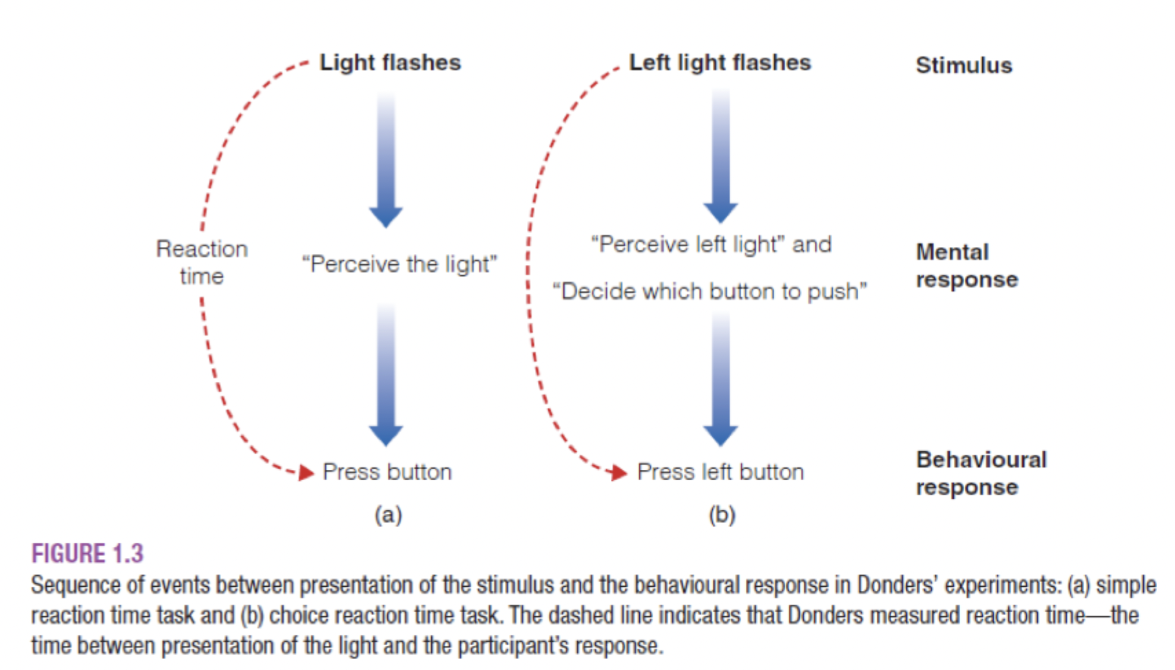
.
e.g. The Posner Task
Tested reaction time based on type of mental representation:
Results - infered by subtraction method (comparing the reaction times based on type of mental representation):
The brain activates different mental representations (visual, phonetic, semantic) in specific time order:
Visual (AA): fastest
Phonetic (Aa): second fastest
Semantic categorization (AU/SC): slowest
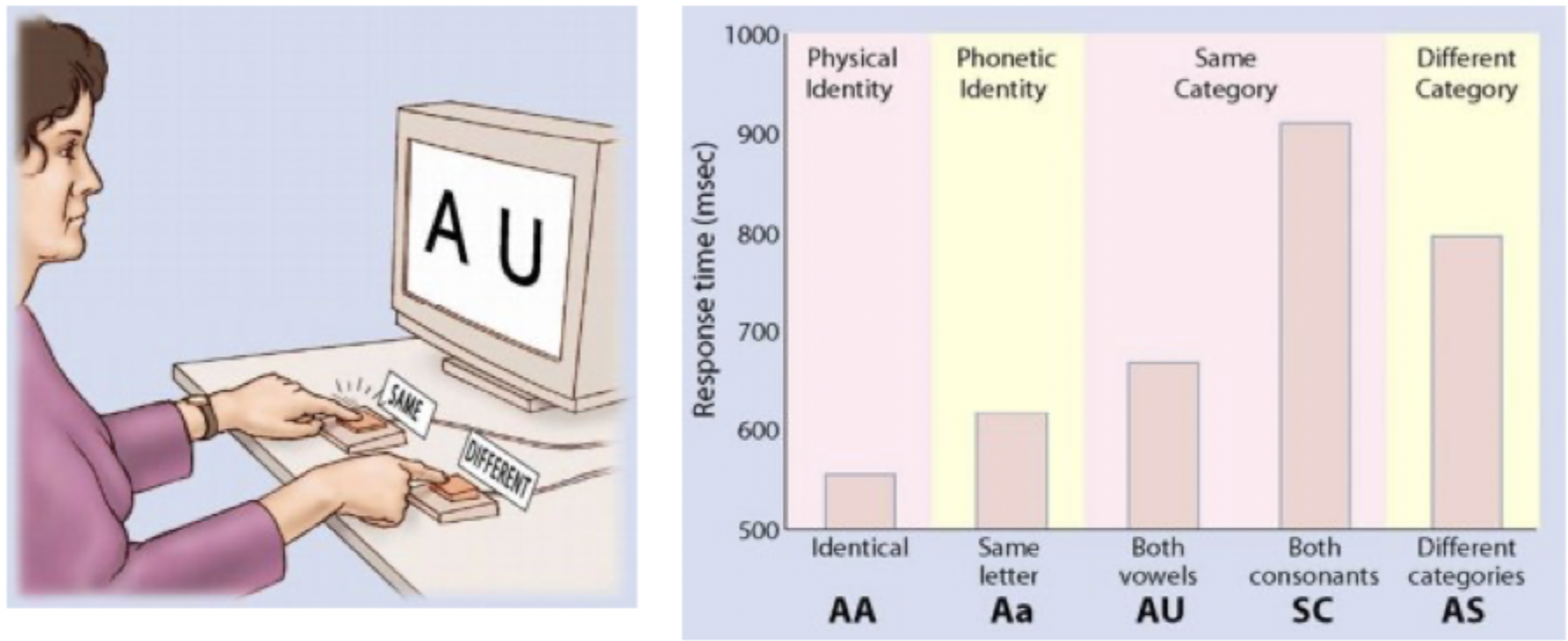
.
e.g. Spreading Activation Theory
Experimental condition: being shown new words that are somehow related to the practiced list
e.g. being shown “sleep” after coding words in the list, like “bed" or “dream”
Control condition: being shown new words not related to the practiced list
e.g. being shown “tractor”
Test: determining if a word shown was a part of the practiced list.
—> more false “rememberances” for related words than for random new words
Subtraction method:
Number of false alarms for related words - number of false alarms for unrelated words

.
Subtraction method and brain activity (neuroscience)
e.g. The visual system: isolation of color (V4) and movement (v5)

Problem:
When you look at a colorful pattern or a flickering/moving checkerboward, V1 and V2 will light up - since both tasks involve visual stimuli
Solution: subtraction method:
Color condition - motion condition:
The general activity in V1 and V2 are subtracted/cancelled out - only leaving significant activity change in V4 remaining
Proving that V4 is specialized for color
Motion condition - static control (fixation point):
Removed baseline noise, isolating activity in V5, specialized for motion
Representative image by averaging the differences of the individual participants
Revealing activity in V1, V2 and V5 for the movement control
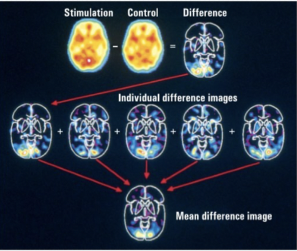
.
The brain is always active!
The factor underlying the subtraction method
You cannot assume that the way a persons brain is active during an experimental condition directly shows a significant activity pattern for that condition - since the brain would also show activity in similar areas during a control condition
.
e.g. The Peterson studies
Goal: finding where the brain performs deeper, semantic evaluations—such as finding a verb that matches a noun
(e.g., seeing the word "cake" and generating the verb "eat").
Step-by-step hierarchical subtraction:
1. Hearing = Hearing – Fixation Point:
Isolates pure, passive auditory processing in the auditory cortex.
2. Seeing = Seeing – Fixation Point:
Isolates pure, passive visual processing in the visual cortex.
3. Speaking (repeating words) = Speaking – Hearing/Seeing:
Strips away the sensory inputs and isolates the motor areas controlling mouth movements and articulation.
4. Generating Verbs = Generating Verbs – Speaking:
This is the crucial step. Both just repeating a word and coming up with a new verb require speaking. By subtracting "just speaking" from "generating," the researchers are left with only the purely cognitive process: semantic association and selection for action.


What is neuronal firing rate, and why do we study its change?
How do you demonstrate the selectivity of a neuron, such as in response to faces?
Neural firing rate
Frequency of a neuron reaching action potential / per second
Action potentials are brief and binary (all-or-nothing)
Higher frequency = more activity
.
Why study change in firing rate?
To figure out the neuron’s function (i.e. its selectivity)
.
Demonstrating neuron’s selectivity
In electrophysiology, finding experimental manipulations that change the firing rate of the neuron
Manipulation by measuring when the neuron is responsive to a stimulus being presented / a task being done:
The neuron’s firing rate will change in comparison to its baseline firing rate
.
Different methods!
Single-cell electrode:
Good/bad?
High spatial + temporal precision
Invasive
ECoG (electrocorticography)
Intracranial recordings (used for clinical purpose in seizures, but can also give scientific benefits)
Good/bad?
High spatial + temporal precision
Invasive
EEG (electroencephalography)
Electrodes on scalp to detect and amplify global electrical activity - recording graded potentials from thousands of cells
ERP (event-related potential)
EEG pattern in response to a stimulus/task
ERP is a time-locked segment of EEG data extracted from a specific interval following a stimulus.
Rather than being a single snippet, it is the average of many identical segments - cancelling out random background noise
Different ERPs are associated with different mental operations
BUT: Control conditions and subtraction method are fundamental - since there is no direct map between ERP components and mental operation
Provides information about timing and activation of different stages of information processing
ERPs get names! (a letter and a number)
e.g. face processing and the n170 effect
Good/bad?
Limited spatial precision
High temporal precision + non-invasive (+ convenient)
.
E.g. in response to faces (in animals):
1. Placing electrode in brain region hypothesized to be important for visual processing
2. Measuring baseline
(when subject is looking at nothing/something neutral)
3. Presenting various stimuli
To see which type of stimulus (in this case faces vs non-faces/controls) makes the neuron fire differently
“Claims for selectivity are only as good as the control stimuli tested“
Subtraction method: measured with stimuli - measured baseline = ev. significant activity
What is Functional Brain Imaging? What is fMRI?
Compare fMRI with EEG: What are the advantages and disadvantages of fMRI and EEG relative to each other?
Functional Brain Imaging
What is it?
AI answer:
FBI measures active areas of the brain by tracking changes:
in blood flow, metabolism, or electrical activity
Types
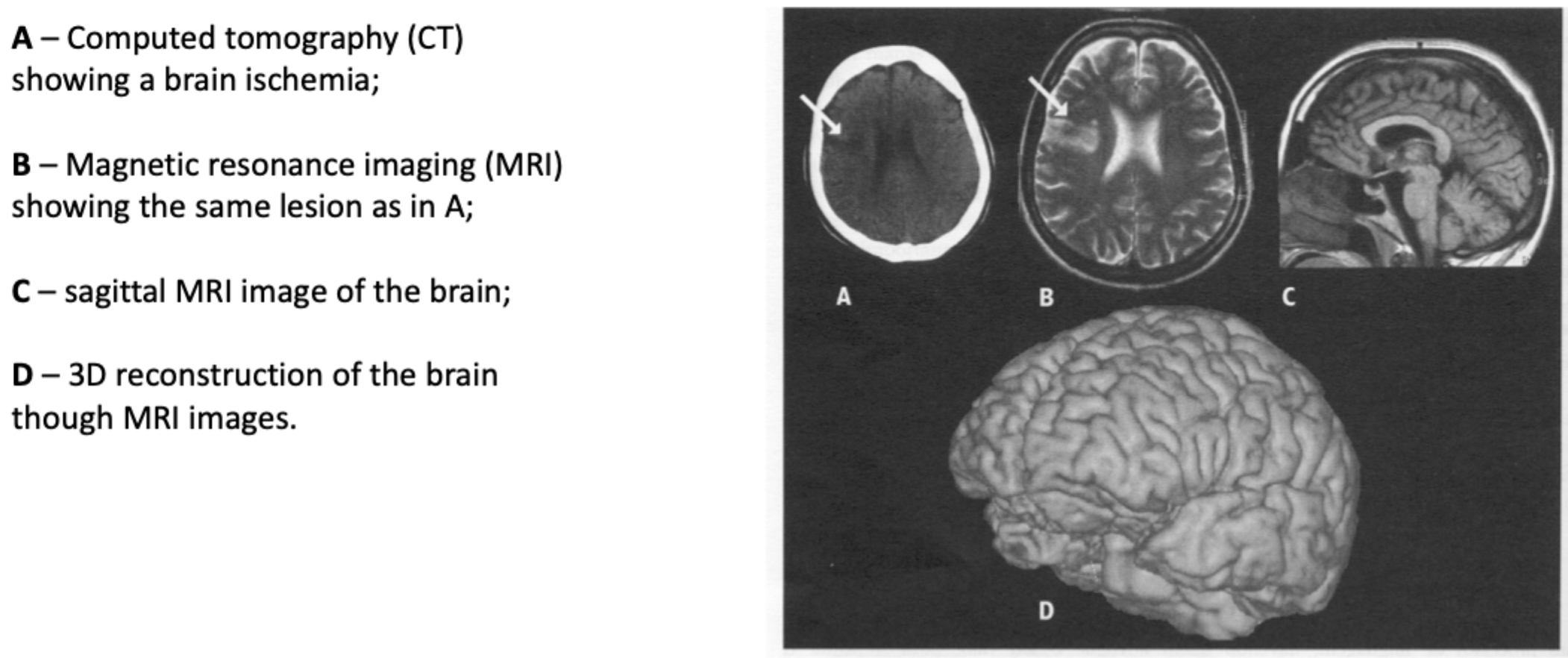
CT
PET
MRI
Detects differences in magnetic properties of different brain structures
Creates 2D images from numerical values of 3D volumes (brains)
e.g.

fMRI
Functional Magnetic Resonance Imaging
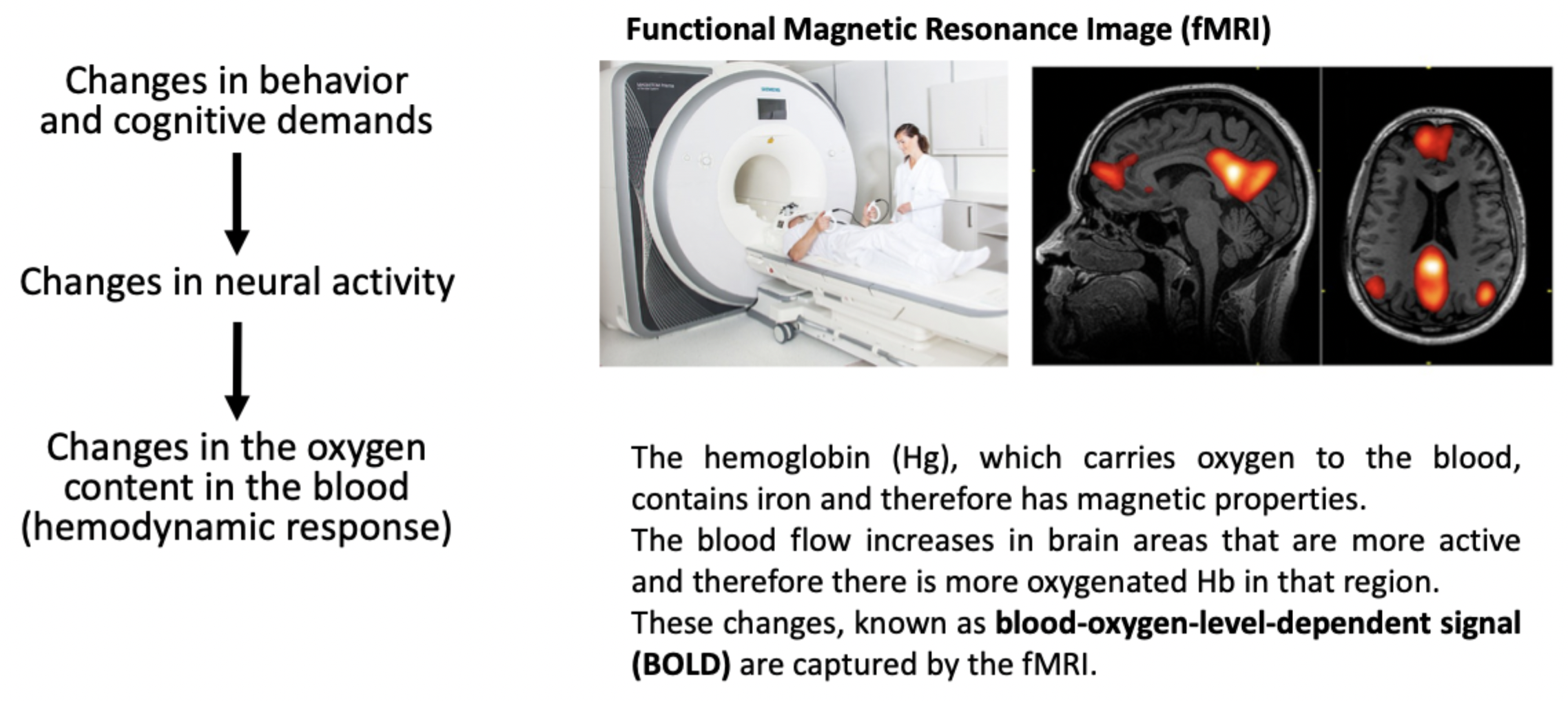

Measures and localizes blood oxygen levels throughout the brain
Infering brain activity —> Active brain areas have higher levels of blood oxygen
(Since it consumes more energy + oxygen supplied through the bloodstream)
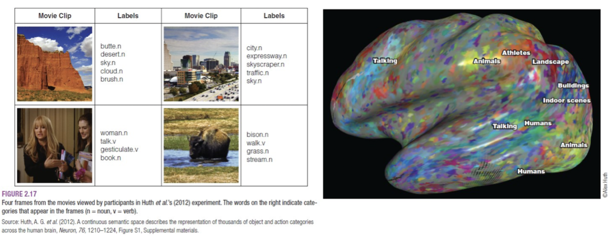
fMRI subtraction method:
Allows comparison of activity patterns for two categories of stimuli
.
fMRI - compared to EEG
Advantages
Higher spatial resolution
Functional brain mapping + locating ROIs
Less invasive
(doesn’t even touch the patient)
Disadvantages
Lower temporal resolution
More expensive
Less convenient
More safety measures + claustrophobia + noisy
Risk of reverse inference
.
EEG - compared to fMRI
Advantages
Higher temporal resolution
Cheaper
More convenient - portable
Possible to use on babies
Disadvantages
Lower spatial resolution
What is the correlation and causation problem in neuroscience, and what methods are available to overcome it?
Correlation and causation problem
Confusing correlation as a cause and effect
e. g. Reverse inference
.
Methods to overcome the problem
TMS (transcranial magnetic stimulation)
Temporarily disrupt brain activity - “virtual lesions“ - using focal magnetic pulses targeted over different areas of the scalp
Advantages
Generally safe method
Determine causality
Whether a brain region is important for a particular function
Temporal precision
Limitations
Spatial precision
Reach - only within ca 3 cm of the scalp
Can cause discomfort an small risk of seizures
.
Double disassociation in lesion studies
.
tDCS (transcranial direct current stimulation)
Applying weak electrical current across the skull to modify brain activity
Increasing/decreasing likelihood of neurons firing
Advantages
Safe + non-invasive