L14: Skeletal Muscle
 Call Kai
 Learn
 Practice Test
 Spaced Repetition
 Match
 Flashcards
 Knowt Play
1/96

There's no tags or description
Looks like no tags are added yet.
97 Terms
What is a muscle?
Whole organ that contains bundles of muscle fibers
What is a fascicle?
Bundle of muscle fibers (cells) wrapped together by connective tissue (perimysium)
What is a muscle fiber?
A single long muscle cell that can contract
What is a myofibril?
Long, rod-like contractile structure inside muscle fiber
What are myofilaments?
Thread-like protein strands
What is the organization of skeletal muscle tissue from smallest to largest?
Myofilament
Myofibril
Muscle fiber
Fascicle
Muscle
What connective tissue layer surrounds the entire muscle?
Epimysium
What connective tissue layer surrounds fascicles?
Perimysium
What connective tissue layer surrounds individual muscle fibers?
Endomysium
What surrounds and separates groups of muscles?
Deep fascia
What is in the presynaptic terminal of NMJ?
ACh synaptic vessicles
What is the motor end plate in the NMJ?
specialized, folded sarcolemma that contains nicotinic ACh receptors
What is sarcolemma?
Specialized cell membrane that surrounds muscle cells
Which structure within the synaptic cleft breaks down ACh?
Basal lamina
What travels down the motor neuron’s axon to presynaptic terminal?
Action potential
What opens to allow Ca2+ to enter the terminal?
Voltage-gated Ca2+ channels
What triggers synaptic vesicles to fuse with membrane?
Ca2+
What binds to nicotinic receptors?
ACh
What produces an end-plate potential?
ACh binding to nicotinic receptors
What does end-plate potential do?
Triggers muscle action potential
What binding happens at the NMJ that elicits action potential?
ACh binds to nicotinic receptors on motor end plate, opening ligand-gated channels
What happens with the ions after ACh binds?
Influx of Na+ and K+ efflux
What causes depolarization (EPP)?
Entry of positive charge
What happens in sarcolemma if threshold is reached?
Depolarization opens voltage-gates Na+ channels in adjacent sarcolemma
Where does action potential propagate?
Muscle fiber
What is excitation-contraction coupling?
Process where an action potential in sarcolemma leads to Ca2+ release from sarcoplasmic reticulum—resulting in initiation of muscle contraction
What is the resting membrane potential of muscle fibers?
-85mV
Does muscle tension last longer than electrical signal?
Yes
Where does AP propagate?
Sarcolemma and T-tubules
What enables muscle contraction?
Ca2+ released from sarcoplasmic reticulum
What causes contraction summation?
AP triggers additional force before muscle fully relaxes (multiple action potentials)
Why can contraction summation happen?
Skeletal muscle refractory period ends before contraction ends
What is sarcoplasm?
Cytoplasm of muscle fiber
What is T-tubule?
Inward fold of sarcolemma that extends into muscle fiber

What does T-tubule allow?
Rapid transmission of action potentials from surface to interior
What is sarcoplasmic reticulum?
Smooth ER that stores and releases Ca2+ for muscle contraction and takes it back up for relaxation
What is terminal cisternae?
Enlarged regions of SR beside the t-tubules
What does terminal cisternae store/release?
Ca2+
What is triad made of?
T-tubule + 2 terminal cisternae
What are the 3 structures that link excitation to contraction?
NMJ
triad
sarcomere
How does NMJ link excitation to contraction?
Initiates muscle action potential
How does triad link excitation to contraction?
Couples action potential in T-tubule to Ca2+ release from SR
How does SR link excitation to contraction?
Generates contractile force
Where does electrical signal travel along for calcium release?
Sarcolemma
Where does action potential travel down for calcium release?
T-tubules
What does depolarization activate in calcium release?
DHP receptors
What does activation of DHP receptors do?
Triggers opening of ryanodine receptors in SR, which releases Ca2+ in sarcoplasm
What does Ca2+ enable?
Actin-myosin interaction
What does actin-myosin interaction lead to?
Muscle contraction
What are actin filaments made of?
Globular actin (G-actin) units arranged in 2 intertwined strands
What does each actin unit have?
Myosin-binding site covered by tropomyosin at rest
What is troponin?
Small protein complex attached to tropomyosin that has 3 subunits
What are the 3 subunits of troponin?
Tn-C (Troponin C)
Tn-I (Troponin I)
Tn-T (Troponin T)
What does Tn-C do?
Binds Ca2+, which triggers contraction
What does Tn-I do?
Inhibits—blocks actin-myosin interaction at rest
What does Tn-T do?
Anchors troponin to tropomyosin
Tropomyosin function?
Prevents contraction when muscle is relaxed
What does myosin look like?
Has a tail that forms the filament and two heads
Which filament is thin/thick?
Actin= thin
Myosin= thick
What does myosin head do (3)?
Binds to actin
Binds + hydrolyzes ATP
Power stroke (pulls for force generation)
What are light chains on myosin?
Small proteins attached to the neck that help stabilize and regulate movement
What moves during power stroke?
Myosin neck
What does ATP binding do to myosin head?
Causes myosin to detach from actin (new ATP forces the head to let go of actin)
What does ATP hydrolosis do to myosin head?
Breaks ATP molecule to provide energy to pull myosin head back into position
What allows myosin heads to bind and pull?
Ca2+ exposes myosin-binding sites on actin
Where does Ca2+ bind?
Troponin C
What happens when Tn-C binds to Ca2+?
Tropomyosin shifts, which exposes the binding site
What happens after myosin binds actin?
Myosin head pulls and there is contraction
What happens to myosin at rest?
Can’t bind b/c Tropomyosin blocks myosin-binding sites on actin
When is cross-bridge formed?
Cross-bridge is formed when myosin binds to actin
How is myosin head energized?
ADP + Pi bond
When does binding occur?
When Ca2+ exposes binding sites on actin
What promotes power stroke?
Pi is released
What pulls actin toward center of sarcomere?
Myosin head pivots
When is ADP released?
When myosin head pivots
What binds to myosin head after ADP is released?
New ATP
What causes myosin head to let go of actin?
ATP binding to myosin head
What hydrolyzes ATP?
Myosin head
When does myosin detach?
ATP binding
When does myosin re-cock?
ATP hydrolosis
What do myofibrils contain?
Repeated sarcomeres arranged end-to-end
What is the smallest unit of muscle contraction?
Sarcomere
Where does sarcomere extend from?
One Z disc to next Z disc
What is the Z disc line?
Protein structure that anchors actin and marks boundary of sarcomere
What is M line?
Structural proteins that hold and align thick filaments at sarcomere center

What is Titin?
Keeps thick filaments centered and supports elastic recoil
What is H zone?
Central part of A band that contains thick filaments only
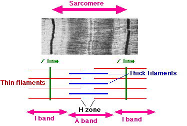
What is I band (light)?
Contains thin filaments but no thick filaments
When does muscle contraction occur?
When thin actin filaments slide past thick myosin filaments
Why do muscle fibers shorten?
Because sarcomeres shorten
What remains the same when sarcomere shortens?
Filament length remains unchanged and A band remains the same
What changes when sarcomere shortens?
I band shortens and H zone decreases
What is dystrophin?
Structural protein located under sarcolemma
What is function of dystrophin?
Protects muscle fiber
What links cytoskeletal actin to extracellular matrix?
Dystrophin
What does connection between cytoskeletal actin and ECM do?
Stabilizes sarcolemma and protects it from damage during contraction
What is duchenne muscular dystrophy caused by?
Repeated sarcolemma damage