Pathology
 Call Kai
 Learn
 Practice Test
 Spaced Repetition
 Match
 Flashcards
 Knowt Play
1/195

There's no tags or description
Looks like no tags are added yet.
196 Terms
ATP depletion, membrane damage, metabolic damage, genetic damage
What are the 4 mechanisms of cell injury?
cellular injury
Oxygen deficiency, physical damage, infectious agents, nutritional imbalance, genetic damage, workload imbalance, toxic damage, immune dysfunction, and aging are all causes of _______________
hypoxia
An oxygen deficiency due to decreased circulating oxygen
ischemia
An oxygen deficiency due to decreased flow of oxygen and nutrients to and from tissues
physical damage
Trauma, temperature extremes, radiation, and electricity are all forms of _______________ that can cause cellular injury
reversible
Hydropic degeneration is a form of ______________ cellular injury in which the cell swells due to water accumulation
hydropic degeneration
reversible cell injury in which the cell swells due to water accumulation resulting in an increased cell size and volume and cytoplasmic vacuolation
hypoxia (less oxidative phosphorylation so less ATP for Na/K pump)
What causes hydropic degeneration?
sodium
Hydropic degeneration starts with hypoxia to the tissue preventing oxidative phosphorylation. This means that less ATP is produced in the cell so the Na/K pump cannot move sodium out of the cell and K into the cell. The accumulation of ______ increases the osmotic pressure of the cell, causing water to follow which distends the cisternae of the organelles leading to rupture and vacuolation
irreversible
Necrosis is a form of ____________ cellular injury
necrosis
Cell death after injury by hypoxia, ischemia, or cell membrane damage
necrosis
Acute cell swelling can lead to ___________ after it passes a “point of no return”
cell membrane
Chemicals and free radicals can cause chemical injury to the _________________ resulting in necrosis of the cell
anti-oxidants
Superoxide dismutase, glutathione peroxidase, and vitamins E and C are ______________ that cells use as protective mechanisms to avoid free radical injury
ischemia-reperfusion
________________ injury follows restoration of blood supply to ischemic tissue causing the generation of Reactive Oxygen Species (ROS) following restored oxygen supply and a Ca influx due to impaired ion pumps that can lead to necrosis of tissue
pyknosis
Shrunken, dark, round spots representative of nuclear changes caused by tissue necrosis
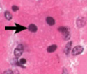
hypereosinophilia
Cytoplasmic ___________________ is a histologic change of necrosis in which the cytoplasm becomes bright red/pink when stained with Eosin
karyorrhexis
Nuclear fragments representative of nuclear changes associated with tissue necrosis

karyolysis
A pale nucleus due to the dissolution of chromatin which is representative of nuclear changes associated with tissue necrosis

pyknosis, karyorrhexis, karyolysis
List the three nuclear changes associated with necrosis as they would progress through the necrotic process: karyorrhexis, pyknosis, karyolysis
coagulation
______________ necrosis is a morphologic change of necrosis in which the cells die but the overall structure remains

vascular
Sharp lines of demarkation and geometric shapes are indicative of lesions of ___________ origin
caseous
______________ necrosis is a morphologic change of necrosis in which the cells have a “cheesy” appearance

liquefactive
______________ necrosis is a morphologic change of necrosis which is associated with fluid changes in the necrotic tissue

gangrenous
______________ necrosis is a morphologic change of necrosis which occurs when you have ischemic necrosis where tissues meet, caused by putrification or dessication of the cells
wet
Putrification caused by bacteria leads to ______ (wet, dry) gangrene
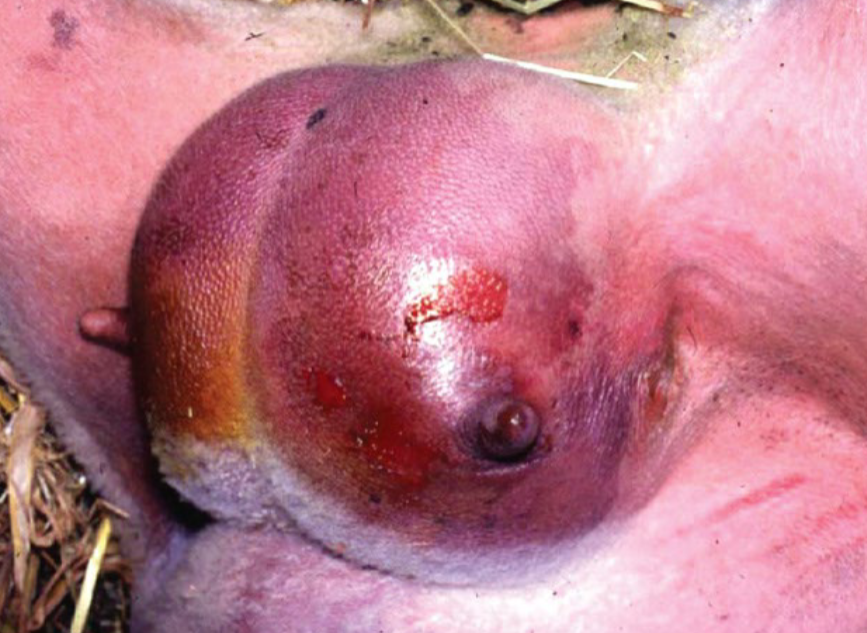
dry
Dessication of cells leads to __________ (wet, dry) gangrene

fat
______________ necrosis is a morphologic change of necrosis in which saponification (“soap making”) of fat cells occurs due to released lytic enzymes

apoptosis
programmed cell death caused by:
Physiologic processes - involution after hormone withdrawal, elimination of self-reactive lymphocytes, age-related thymic involution
Pathologic processes - viral infections, gland involution following duct blockages, accumulation of misfolded proteins, immune-mediated
extrinsic
The ____________ pathway as a mechanism of apoptosis occurs with the binding of cell surface death receptors (TNF family receptors)
FAS
What triggers the caspase cascade that initiates apoptosis for the EXTRINSIC pathway?
intrinsic
The ____________ pathway as a mechanism of apoptosis occurs with increased mitochondrial permeability causing a release of pro-apoptotic molecules into the cytoplasm
cytochrome c
What triggers the caspase cascade that initiates apoptosis for the INTRINSIC pathway?
caspase
Both the extrinsic and intrinsic pathways of apoptosis lead to the activation of the ___________ cascade which culminates in DNA degradation and apoptosis
shrinkage, without
Morphologic features of apoptosis include:
Cell _______________ (swelling, shrinkage)
Condensed chromatin
Cytoplasmic buds (forms apoptotic bodies for phagocytosis)
____________ (with, without) inflammation
apoptotic bodies
With the process of apoptosis, the cell shrinks and forms cytoplasmic buds off called __________________ which can be phagocytized by other cells in the body for more controlled cell death
cell shrinks with apopotosis but swells with necrosis, inflammation with necrosis but not apoptosis
What are the two primary differences between apoptosis and necrosis?
dystrophic
____________ calcification occurs in LOCALIZED areas of necrosis where calcium accumulates in the mitochondria of cells
metastatic
_____________ calcification occurs SYSTEMICALLY in normal tissue secondary to hypercalcemia due to diseases such as renal failure, Vitamin D toxicosis, elevated parathyroid hormone or parathyroid hormone related peptide, or neoplastic bone destruction
PTHrp (parathyroid hormone related peptide)
Complex that mirrors the function of parathyroid hormone to mobilize Ca which is produced by neoplastics
lymphoma, AGASACA
What two types of neoplasm produce parathyroid hormone related peptide (PTHrp) which acts like parathyroid hormone to induce primary hyperparathyroidism that can cause metastatic calcification?
cell function/viability, cellular replication
Cellular aging causes a decrease in:
____________________ - progressive exposure to exogenous influences and accumulation of cellular damage, reduced oxidative phosphorylation, and reduced protein/nucleic acid synnthesis
____________________ - progressive telomere shortening
intracellular accumulations
_____________________ result from dysregulated cellular metabolism such as:
Normal production with inadequate removal
Normal production but defective enzymatic removal
Abnormal production due to gene mutation
Abnormal exogenous substances being present in the cell
hepatic lipidosis
An intracellular inclusion of lipids caused by the accumulation of intracellular triglycerides due to increased supply of free fatty acids or decreased metabolism or excretion of lipoproteins which can be seen in early lactation dairy cows and obese animals causing distinct, clear, smooth walled cytoplasmic vacuoles on histology and pale tan, friable/greasy lesions grossly
glycogen
Intracellular accumulations of _____________ often occur in hepatocytes due to corticosteroids, diabetes mellitus, or glycogen storage diseases
exogenous
Carbon (such as from smoking) and tattoo ink are examples of ____________ pigments that can cause intracellular accumulations
endogenous
Melanin, lipofuscin, and hematogenous (iron based) pigments including hemosiderin are examples of ____________ pigments that can cause intracellular accumulations
inflammation
A well-organized cascade of fluid and cellular changes within vascularized tissue which is a host response to remove damaged and necrotic tissue or foreign invaders
fluid
What is the first change in the well-organized cascade of inflammation?
heat, redness, swelling, pain, loss of function
What are the five cardinal signs of inflammation?
repair, healing
Inflammation is part of the process of ________ and ___________, designed to destroy, dilute, or wall of injurious agents and initiate healing and tissue repair
protective
Fundamentally, inflammation is a ______________ response to get rid of pathogens, remove necrotic debris, and repair damage to return normal structure and function, but it can be harmful, particularly in excessive amounts
rapid, short, edema, neutrophils
Acute inflammation is characterized by:
___________ (rapid, slow) development
___________ (long, short) duration
___________ - exudation of fluid and plasma protein
Emigration of leukocytes, primarily ____________
slow, long, macrophages, lymphocytes
Chronic inflammation is characterized by:
___________ (rapid, slow) development
___________ (long, short) duration
Emigration of leukocytes, primarily ____________ and _____________
Proliferation of blood vessels and connective tissue (fibrosis)
increased (arteriole dilation, venule dilation)
The first stage of Acute Inflammation is vascular alteration leading to ____________ blood flow
permeability
The second stage of Acute Inflammation is an increase in microvasculature ____________ of the epithelium to allow leakage of fluid and plasma proteins that induce edema and movement of leukocytes
emigration
The third stage of Acute Inflammation is the ___________ of leukocytes, primarily neutrophils, into the perivascular area
acute
Infections, trauma, physical and chemical agents, tissue necrosis, foreign bodies, and immune reactions are all potential stimuli for _______ inflammation
exudate
Inflammatory extravascular fluid with a HIGH protein concentration and HIGH cell count leading to a high specific gravity (over 1.020)
permeability
Exudate is formed when there is significant alteration in small blood vessel ____________ at the site of injury associated with vascular damage or inflammation
transudate
Clear/transparent extravascular fluid with a LOW protein concentration and LOW cell content leading to a low specific gravity (less than 1.012)
ultrafiltrate
Transudate is due to _____________ of blood such as increased hydrostatic pressure or decreased oncotic pressure due to hypoproteinemia (low plasma proteins/albumin in blood)
edema
The accumulation of excess interstitial fluid due to:
Increased vascular permeability (exudate)
Increased intravascular hydrostatic pressure (transudate)
Decreased intravascular osmotic pressure (transudate)
Decreased lymphatic drainage
hydrostatic, osmotic
Normal movement of fluid in and out of the blood vessel is controlled by a balance of ______________ pressure caused by blood pressure and colloid ___________ pressure caused by the presence of plasma proteins in the blood vessel
transudate
An increase in hydrostatic pressure due to obstruction or congestive heart failure could cause accumulation of ____________ fluid
transudate
A decrease in colloid osmotic pressure due to decreased protein synthesis from liver failure or increased protein loss from kidney disease could cause accumulation of ____________ fluid
exudate
Inflammation leading to vasodilation and increased endothelial permeability of the blood vessel allowing fluid AND protein leakage could lead to the accumulation of ___________ fluid
pus
A purulent exudate which is rich in leukocytes (mostly neutrophils) and cell debris
purulent
Adjective use of pus
arterioles
Vasodilation is the first step of inflammation, beginning in the _____________
histamine, nitrous oxide
When the arterioles dilate during the first vasodilation step of inflammation, ____________ and _____________ act as bioactive mediators to open new capillary beds by causing the smooth muscle to relax
viscosity (greater concentration of red cells = thicker = slower moving)
With increased permeability of the blood vessels, fluid pours out into the extravascular tissue, resulting in a loss of fluid in the blood vessel which increases the blood _____________ and induces blood stasis
stasis
Blood ________ created by the increased blood viscosity from the loss of fluid to extravascular tissue during inflammation allows leukocytes to marginate, in which they accumulate along the endothelium and stick to the wall for emigration
endothelial contraction
Mechanism for vascular leakage in acute inflammation mediated by histamine, bradykinins, and leukotrienes which stimulate the endothelial cells to contract, thereby increasing the interendothelial space
direct endothelial injury
Mechanism for vascular leakage in acute inflammation in which direct damage to the endothelium from burns or lytic bacteria causes necrosis and detachment, thereby increasing the microvascular permeability
leukocyte-dependent injury
Mechanism for vascular leakage in acute inflammation in which activated leukocytes secrete free radicals and proteolytic enzymes designed to kill invading pathogens, unintentionally leading to damage of the endothelial cells which increases microvascular permeability
transcytosis
Mechanism for vascular leakage in acute inflammation in which there is an increase the transport of fluid and protein through the endothelial cells from the blood vessel to the tissue
thrombosis (platelet clotting)
Unlike endothelial contraction, direct endothelial injury starts immediately and lasts until ____________ occurs or the endothelium is repaired
endothelial contraction
What mechanism for vascular leakage is rapid and short lived?
cytokines
The endothelium of blood vessels must be activated by ______________ released by macrophages stimulated by the presence of a pathogen or other phagocytized molecule to “catch” leukocytes because this does not occur in normal blood flow
selectins
The cytokines released by activated macrophages in tissue upregulate the presentation of ____________ on the surface of blood vessels which allow leukocytes to adhere transiently to the endothelium and roll until they become firmly attached
integrins
____________ on leukocytes adhere to immunoglobulin family receptors on the blood vessel endothelium surface firmly to allow transmigration into the tissue
immunoglobulin family receptors
Integrins on leukocytes adhere to ___________________ on the blood vessel endothelium surface firmly to allow transmigration into the tissue
weak (allows rolling)
Leukocytes bind with _________ (strong, weak) affinity to selectins
strong (allows transmigration)
Leukocytes bind with _________ (strong, weak) affinity to immunoglobulin family receptors
margination
The movement of leukocytes toward the endothelium of the blood vessel with blood stasis
rolling
The slow movement of leukocytes along the endothelium of blood vessels due to weak affinity binding with selectins
adhesion
The firm attachment of leukocytes to the endothelium of blood vessels due to strong affinity binding between integrins and immunoglobulin family receptors that allows transmigration into the tissue
transmigration (diapedesis)
Movement of leukocytes through the endothelium into tissue, primarily in venules
migration
Movement of leukocytes in interstitial tissue toward a chemotactic stimulus
genetic
___________ deficiencies in leukocyte adhesion are heritable deficiencies in type and amount of adhesion molecules which can inhibit leukocyte transmigration and acute inflammation
recurrent
Genetic deficiencies in leukocyte adhesion can cause a poor inflammatory response to pathogens and dead tissue, allowing ___________ infections
pseudopod
During transmigration, leukocytes insert a ___________ into the junction between endothelial cells and squeezes through
chemotaxis
The movement of leukocytes in tissue along a chemical gradient
exogenous
_____________ products from bacteria can create a chemical gradient for leukocytes to follow during chemotaxis
endogenous
Complement system components serve as ______________ products that act as a chemical gradient for leukocytes to follow during chemotaxis
C5a
What complement system component from the COMPLEMENT CASCADE serves as an endogenous product as a chemical gradient for leukocytes to follow during chemotaxis?
leukotriene B4
What complement system component from the ARACHADONIC CASCADE serves as an endogenous product as a chemical gradient for leukocytes to follow during chemotaxis?
TNF-alpha, IL-1
What two complement system CYTOKINES serves as an endogenous product as a chemical gradient for leukocytes to follow during chemotaxis?
filopodia
Leukocytes move through tissues by extending a __________ that pulls the back of the cell forward