Neuroscience Lab 2
 Call Kai
 Learn
Learn Practice Test
 Spaced Repetition
 Match
Match Flashcards
 Knowt Play
1/50

There's no tags or description
Looks like no tags are added yet.
Name | Mastery | Learn | Test | Matching | Spaced | Call with Kai | Chat |
|---|
No analytics yet
Send a link to your students to track their progress
51 Terms

Precentral Gyrus: Primary motor area
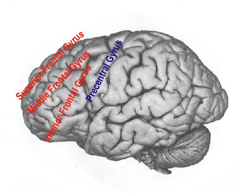
Structures of the frontal gyruses?
Superior Frontal Gyrus, Middle frontal gyrus, inferior frontal gyrus

Structure of the baby blue area?
Lateral premotor area
Lateral premotor area’s function
motor programming, sensory guidance of limb movements, initiation and sequencing of mvmts.
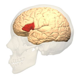
Broca’s area

Broca’s Area made up of which 2 components:
Pars opercularis + pars triangularis

Prefrontal area 2 portions
Dorsolateral prefrontal: task switching, working memory, inhibition
Vetrolateral prefrontal: risk/reward, motivation, threat detection


Postcentral gyrus - somatosensory area

Structure? - green selection probably expands slightly more
Superior parietal lobe
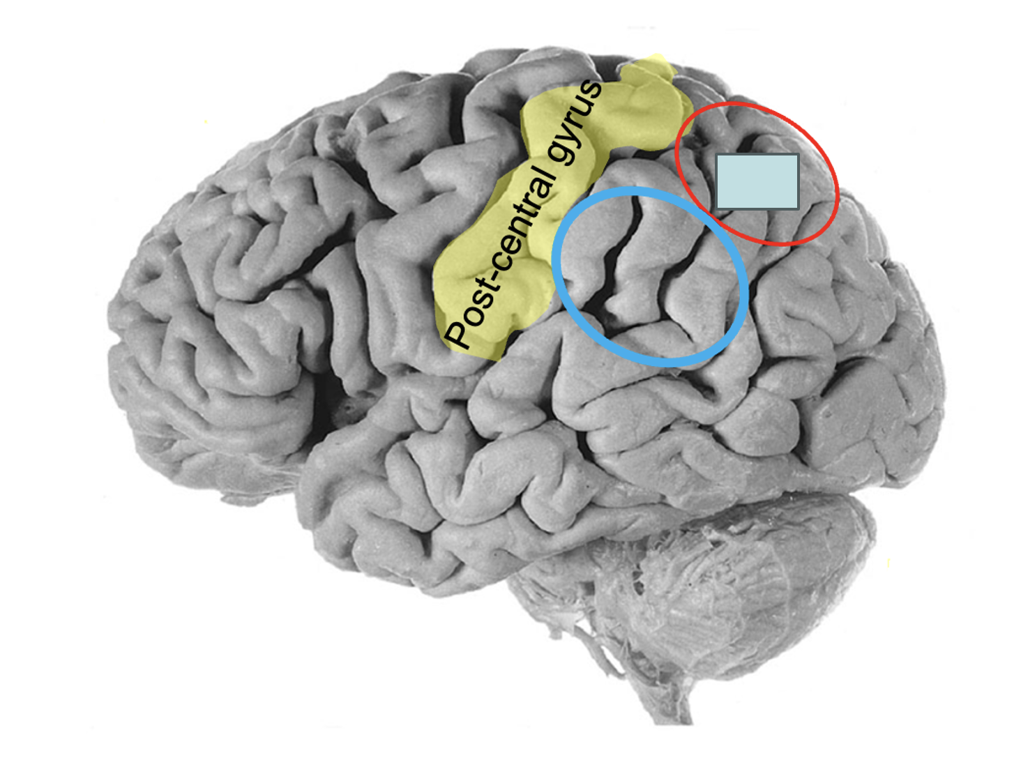
Blue circle
Inferior parietal lobe
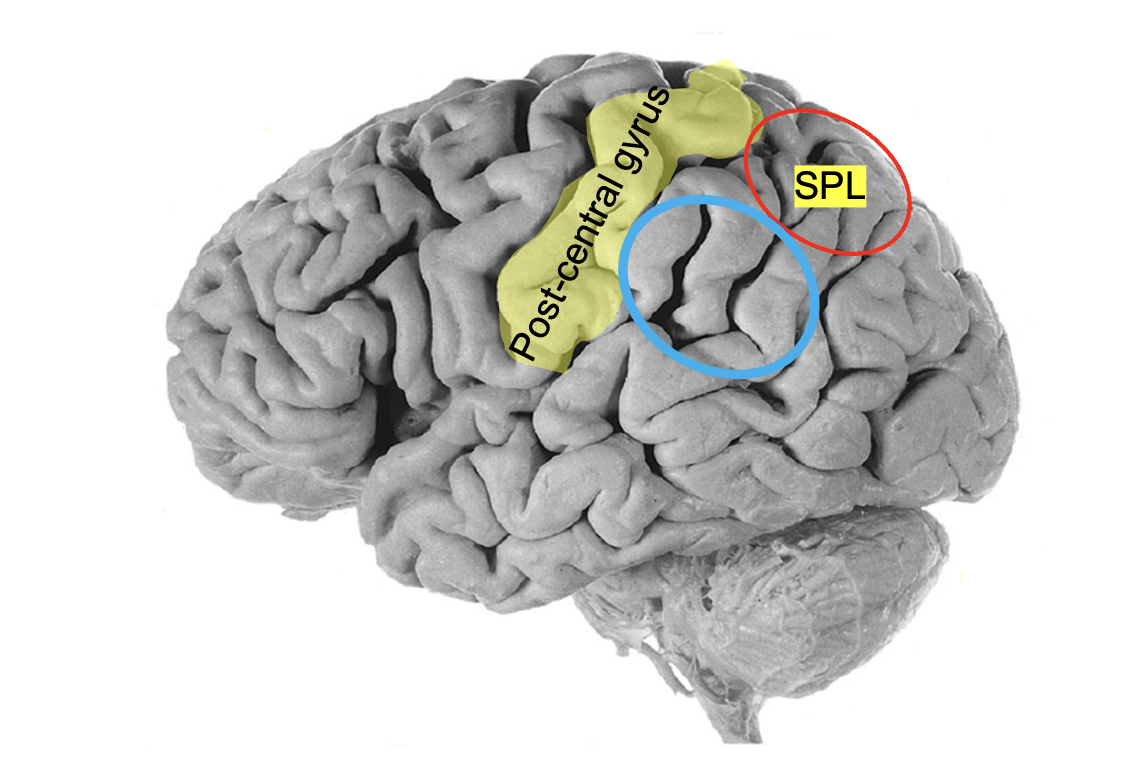

Inferior parietal lobe made up of:
Supramarginal gyrus (anterior), angular gyrus (posterior)


Orange line structure (medial view)
Cingulate sulcus

Larger area where arrow is pointing to?
Lateral occipital gyri (association areas for vision)

Solid Red region?
Primary vision

3 parts of the temporal lobe?
Superior, middle, inferior temporal gyrus,

Posterior region of superior temporal gyrus
Wernicke’s area (speech perception)

Transverse temporal gyri of (Heschl)

Red area structure (medial view)
Paracentral lobule

Precuneus - medial view of the parietal lobe

Cuneus - medial view of the occipital lobe

Green line structure
Calcarine sulcus - primary vision area 17

Blue line around the cingulate sulcus
Marginal Branch of cingulate sulcus - anterior border of precuneus

Structure of F?
Parieto-occipital sulcus - posterior border of precuneus
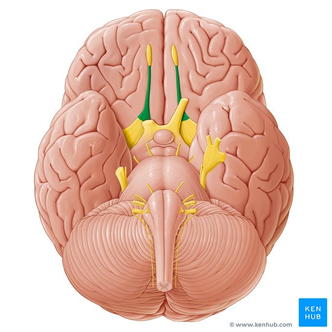
Inferior view, two yellow dots
Olfactory tract and bulb (CN1 - sense of smell)
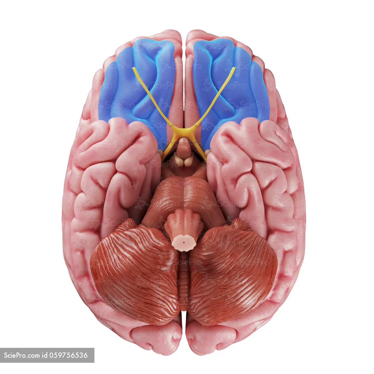
Orbital gyri

Parahippocampal gyrus
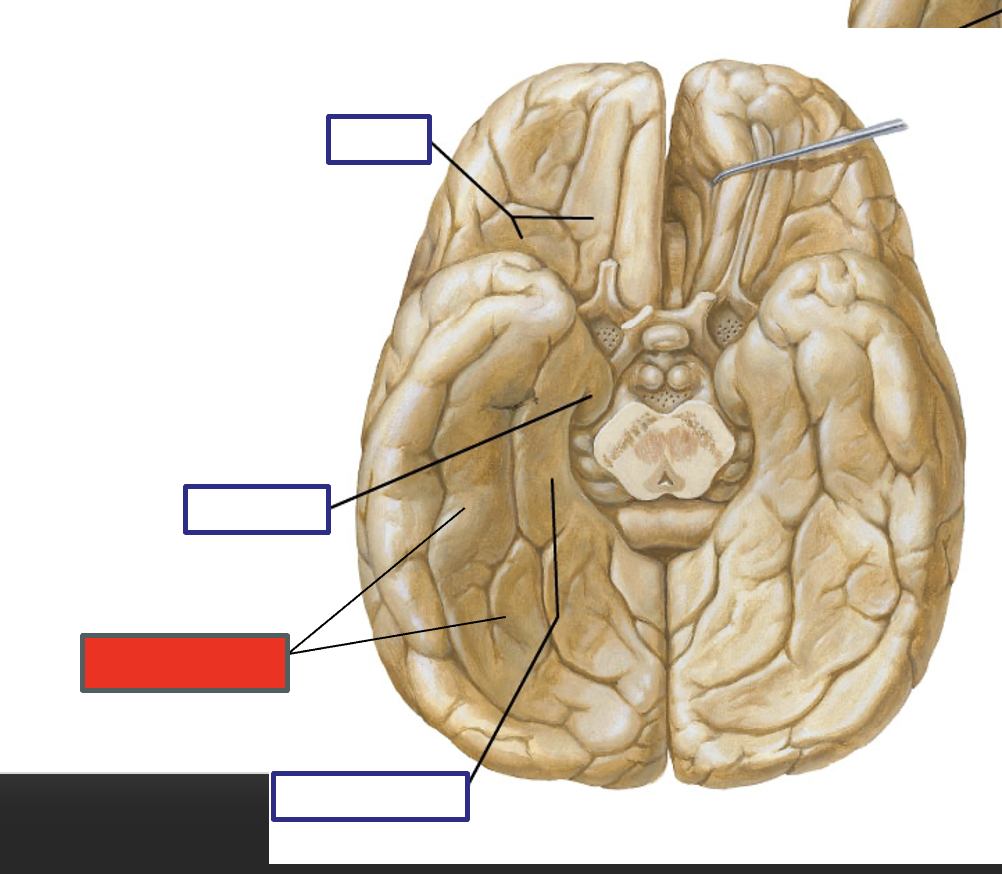
Structure of the Red label?
Occipitotemperal gyrus

Uncus - role in charged behaviors; amygdala lies deep inside this structure

Cingulate gyrus
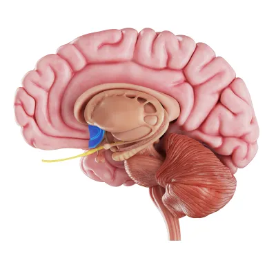
Blue area
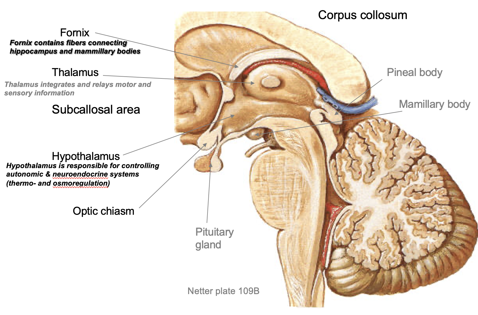
Subcallosal area - mood and affective behaviors (located beneath the anterior portion of the corpus callosum)
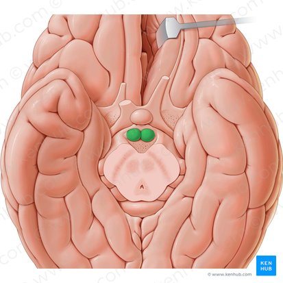
Green area
Mammillary body - aspects of memory

What is this structure?
Fornix - connects Mammillary body to hippocampus
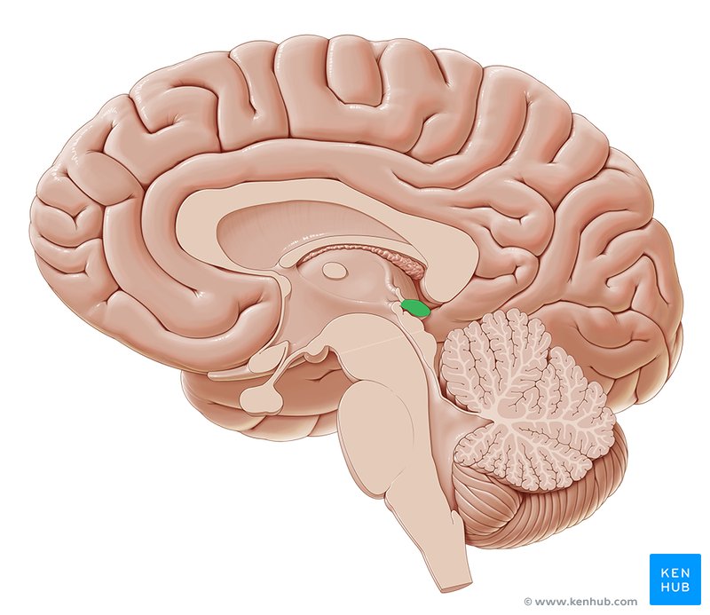
Pineal body - controls sleep
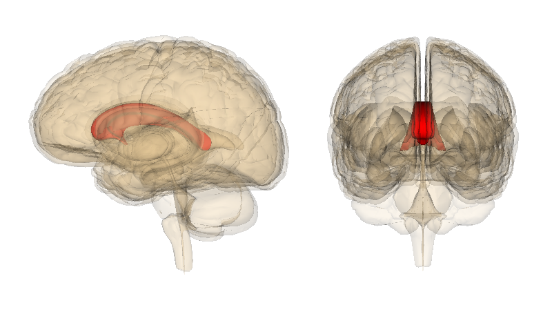
Corpus callosum

Tectum - roof of midbrain
Tectum consists of…
Superior & inferior colliculus

Tegmentum - floor of the midbrain

Purple arrow is pointing to…
Interpeduncular fossa - space between two cerebral penduncles
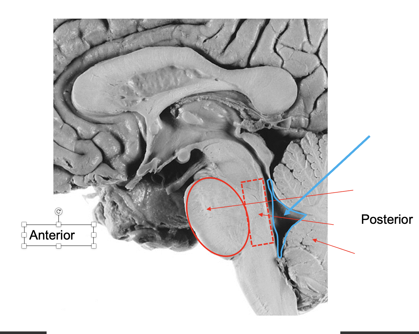
Name these structures from anterior to posterior
Basilar pons, pontine tegmentum, 4th ventricle, cerebellum

Name these two parts of the medulla
Ventral - Pyramids (descending path of motor signals), decussation (crossing of the descending motor fibers)

Sensory fibers - Dorsal side of medulla, ascending path for sensory signals

Ventral view, green region? purple region?
Diencephalon: optic nerve/chiasm/tract
Midbrain: Cerebral peduncles, Interpeduncular fossa

Ventral view, yellow region? Red region?
Pons: basilar aspect of pons
Medulla: olives
Cerebral peduncles function:
white matter pathway for descending from cerebral hemisphere to brainstem
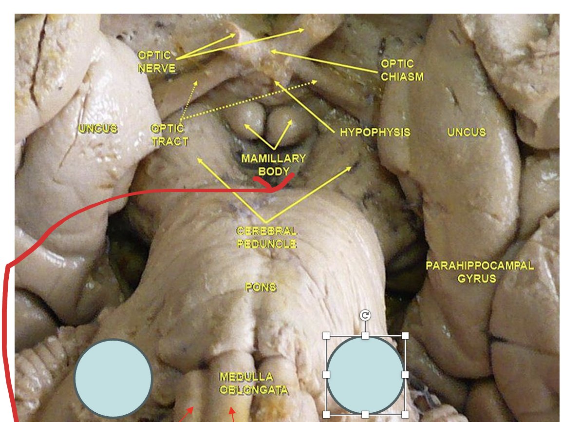
The gray circles are which structures in the metencephalon?
Middle cerebellar peduncle


Label these structures of a ventral view of brainstem
Answers

Superior colliculus associated with?
Inferior colliculus associated with?
Superior colliculus associated with VISION
Inferior colliculus associated with AUDITION
Transverse temporal gyri of Heschl’s function
Primary auditory + auditory associated area (speech & language comprehension)
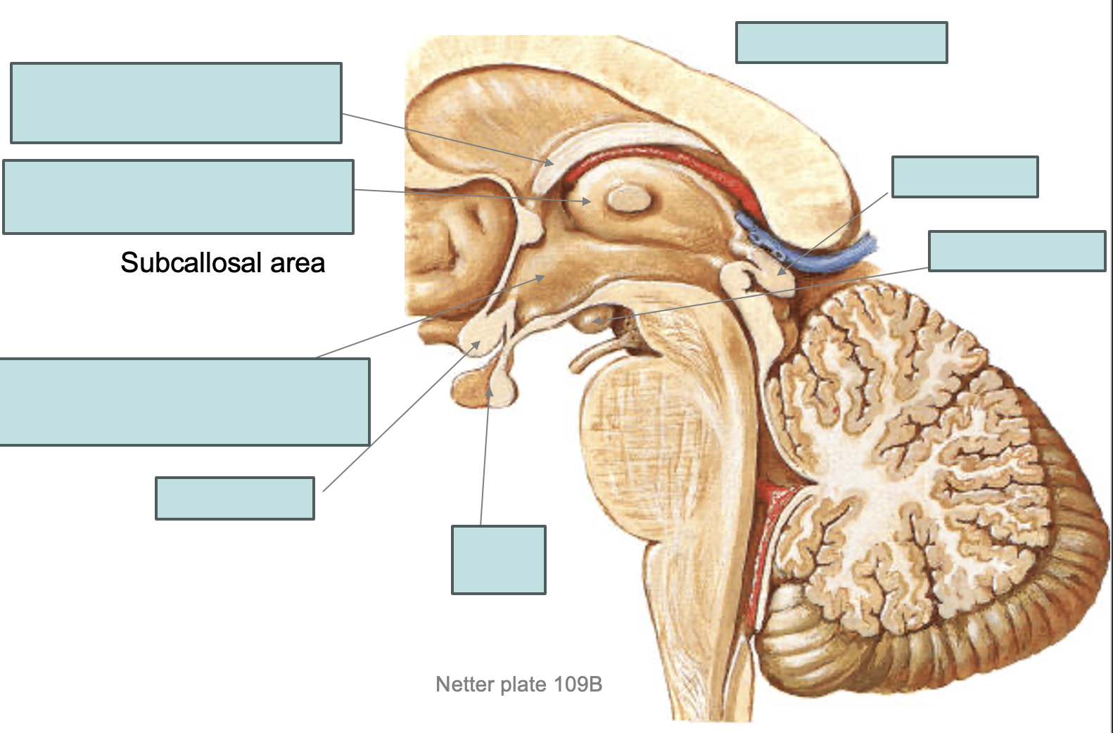
Fill in the blanks (medial view)
Paracentral lobule function:
Medial representations of sensory and motor cortices of the lower extremity
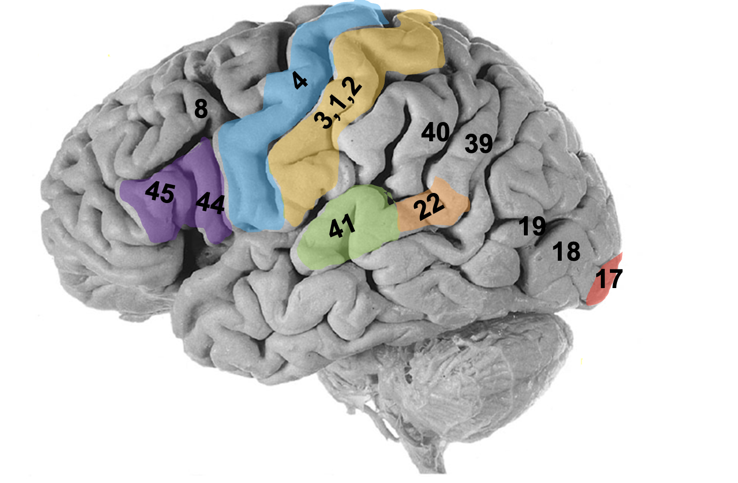
Bordmann’s numbers
44, 45: broca’s area
4: precentral gyrus
1,2,3: postcentral gyrus
17, 18, (19): primary vision and lateral occipital arrea
22: wernicke’s area
40, 39: supramarginal, angular gyrus
41: transverse temporal gyri of heschi
