Cellular Movement and Muscles
 Call Kai
 Learn
Learn Practice Test
 Spaced Repetition
 Match
Match Flashcards
 Knowt Play
1/19

There's no tags or description
Looks like no tags are added yet.
Name | Mastery | Learn | Test | Matching | Spaced | Call with Kai | Chat |
|---|
No analytics yet
Send a link to your students to track their progress
20 Terms
what physiological processes depend on movement?
all of them?

what r the 2 component of all cellular machinery?

what r the 2 elements of cytoskeleton and what 3 ways is it used for?
can change the shape of cell
analogy: muscle contraction

from the second compoent of cellular machinery, microtubules, what is it and wat r some characteristic of it
Are tubelike polymers of protein tubulin (similar protein in diverse animal groups, multiple isoforms)
Are anchored at both ends
microtubule organization center (MTOC) (-) near the nucleus (centrosome, near negative end)
Attached to integral proteins (+) in the plasma membrane
negative end of microtubule is close to nucleus, which has the centrosome.
fluorescent microscope of a cell: the cirlce is the nucleus of cell,

what are the functions of microtubules:
spindle formation if microtubules
in mitosis, meiosis
sperm
in some animals they cause rapid color change of skin


explain this
housed in a bathtub
we toook frogs out and ave them in clar plastic box. they were no longer black, they were light. They were resembling the environment they were in, to camouflage. this was due to the movement of pigments.
Melanophore: +MSH gormone causes a chemical rxn that stimulated kinasyn to bind to this pigments. the kynasin would walk along the microtubules to change the color
what is the conformation of microtubules:
Tubulin: alpha and beta tubulin
microtubule shave polarity (±)

explain the process of microtubule assembly
Activation of tubulin monomers by GTP
Monomers join to form a tubulin dimer
Dimers form a single-stranded protofilament
Many (13) protofilaments form a sheet
Sheet rolls up to form a tubule
Dimers can be added or removed from the ends of
the tubule: Asymmetrical growth and Growth is faster at + end
Cell regulates rates of growth and shrinkage
they r formed from alpha and beta tubulin
they can bind to GTP, have higher affinity to each and form dimers
beta tubuling can hydrolyze GTP into GDP
once they reach certain length, they become a sheet
the dime that ws at the positive side will end up moving to the negative side because the microtubule keeps growing.
13 of this protomers create a sheet
it can grow and shrink at both ends

what r the 5 factors affecting growth and shrinkage:

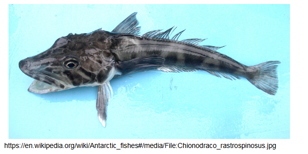
talk about the antarctic fish having stable microtubules:
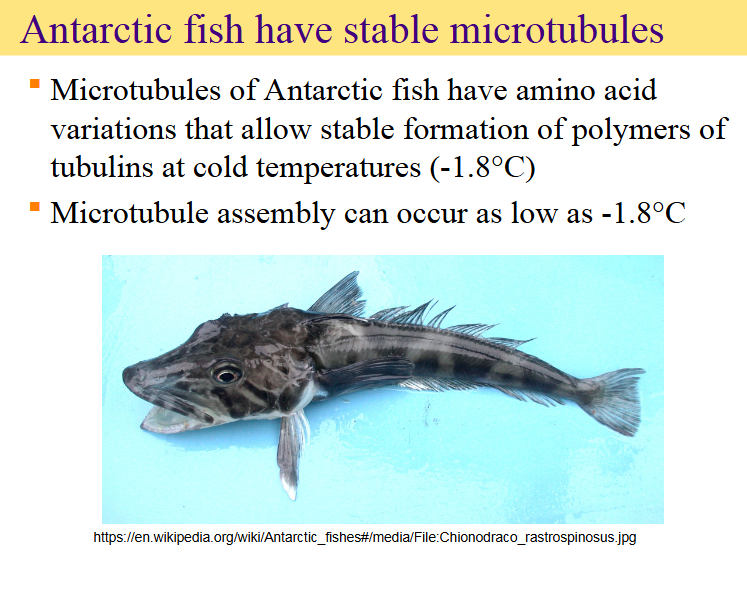

explain this
0: no growth, stable at certain length
concentration of tubulin available (x axis)
negative end tends to shrink, positive end tends to grow
if you exceed Cc (+) you will have growth,
if you exceed Cc(-), you will have growth in negative end.
posititve end is more likely to grow, in the treadmilling range
The main takeaway of this graph is the concept of microtubule treadmilling, which occurs when the concentration of free tubulin falls within a specific window (the shaded range).
The graph illustrates this behavior through three key points:
Different Critical Concentrations: The (+) end has a much lower critical concentration (\(C_c(+)\)) than the (-) end (\(C_c(-)\)). This means the (+) end requires far less tubulin to start growing.
Opposing Behaviors in the Treadmilling Range: When the free tubulin concentration is between \(C_c(+)\) and \(C_c(-)\):
The (+) end is above its critical concentration, resulting in growth.
The (-) end is below its critical concentration, resulting in shrinkage (loss of subunits).
Net Flux of Subunits: Within this shaded range, tubulin is continually added to the (+) end and lost from the (-) end. This creates a "conveyor belt" effect where subunits appear to move down the length of the filament, even if the total length stays relatively constant.
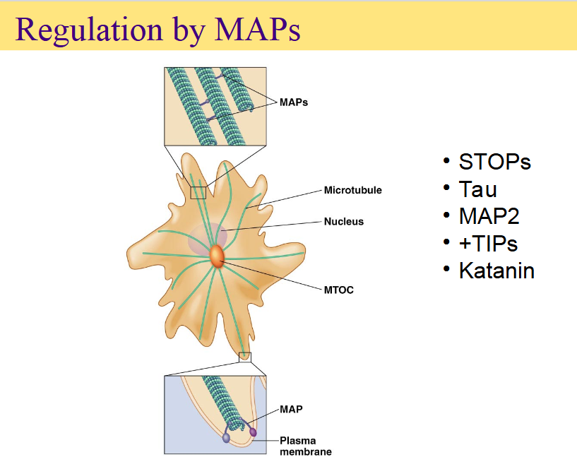
explain regulation of MAPs: microtubule associated proteins
stable tubule only polypeptides
tau protein: have a bundle of microtubules
The main takeaway of this slide is that Microtubule-Associated Proteins (MAPs) are crucial regulators that control the structure, stability, and positioning of microtubules in a cell.
While microtubules can assemble on their own, the cell relies on MAPs to bundle them together, anchor them to membranes, or cut them when needed.
Key Visual Elements
Top Inset: Shows MAPs acting like physical cross-links or bridges that bundle and space parallel microtubules together.
Bottom Inset: Shows a MAP capping and anchoring the dynamic end of a microtubule to the plasma membrane, helping to shape or anchor the cell's skeleton.
Central Cell: Shows microtubules (green lines) radiating out from the MTOC (Microtubule Organizing Center) near the nucleus to fill the cell cytoplasm.
Specific MAPs Listed
STOPs (Stable Tubule-Only Proteins): Proteins that protect microtubules from cold-induced or chemical disassembly, keeping them highly stable.
Tau & MAP2: Structural MAPs that stabilize microtubules and determine their spacing. Tau is heavily found in neuronal axons, while MAP2 is found in neuronal dendrites.
+TIPs (Plus-end Tracking Proteins): Specialized MAPs that bind specifically to the growing (+) ends of microtubules, regulating their growth dynamics and helping them find and attach to specific targets like the plasma membrane.
Katanin: A microtubule-severing protein (named after the Japanese samurai sword, katana). It cuts microtubules into shorter fragments, which can stimulate rapid disassembly or create new templates for microtubule growth.
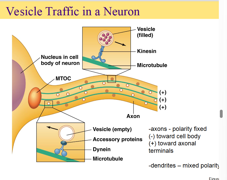
how does the movement along microtubules work:
Motor proteins move along microtubules
Direction is determined by polarity and the type of
motor protein
Kinesin moves in (+) direction
Dynein moves in (–) direction
Movement is fueled by hydrolysis of ATP
Rate of movement is determined by the ATPase
domain of motor protein and regulatory proteins

explain this
fluorescent microscope to poinpoint location of organelles to follow the movement
The main takeaway of this slide is the introduction of FIONA (Fluorescence Imaging with One Nanometer Accuracy), a groundbreaking technique used to measure the exact step size of motor proteins (kinesin and dynein) inside living cells.
This landmark 2005 Science study by Kural et al. solved a major limitation in microscopy to watch molecular motors walk in real-time.
How the Technique Works
Overcoming the Blur: Under a normal light microscope, a single tiny fluorescent molecule looks like a large, blurry blob due to the physics of light (the diffraction limit). This blurry blob is shown in the black-and-white inset box on the graph.
The 3D Gaussian Fit (Graph a): FIONA bypasses this limit mathematically. By capturing a high number of light particles (photons), scientists can fit the blurry blob into a precise 3D Gaussian curve (the colorful peak).
1-Nanometer Precision: While the blob itself is hundreds of nanometers wide, the absolute center (the very tip of the peak) can be pinpointed with 1-nanometer accuracy.
The Biological Setup (Images A & B)
Tracking Cargo: The researchers used this technique to image peroxisomes (small cellular organelles) moving inside a thin cellular protrusion.
Measuring the "Steps": Because these peroxisomes are actively dragged along microtubule tracks by kinesin and dynein motors, tracking the precise center of the peroxisome allowed scientists to see the exact size of the physical "steps" the individual motor proteins take while working inside a living cell.

explain this
The main point of this slide is to show that both kinesin and dynein move along microtubules in discrete increments of approximately 8 nanometers.
This 8-nm step size is highly significant because it perfectly matches the physical distance between adjacent tubulin dimers on a microtubule track, proving that these molecular motors step systematically from one tubulin subunit to the next.
whats the different of speed on walking between kynesin and dynein
speed of wlaking steps of kinesyn
kinesin and dynein have the same speed
they think the aomunt of kinesin increases the speed

how kinesins walk? graph from experiment'?
does it walk like we walk, or inchworm fashion
the label was at the top of the whit stalk,

how do kinesin actually walk?

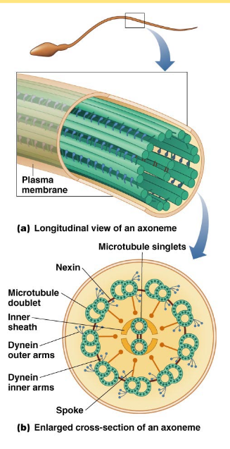
talk about cilia and flagella
what they r composed of and 1 characteristic
Cilia
Numerous, wavelike motion (respiratory epithelium)
Flagella
Single or in pairs, whip-like movement
Composed of microtubules arranged into axoneme
Bundle of parallel microtubules
Nine pairs of microtubules around a central pair
“Nine-plus-two”
Asymmetric activation of dynein causes movement
what r some processes that microtubules r involved in
