U3 medicine - dermatology pt 1
 Call Kai
 Learn
Learn Practice Test
 Spaced Repetition
 Match
Match Flashcards
 Knowt Play
1/118

There's no tags or description
Looks like no tags are added yet.
Name | Mastery | Learn | Test | Matching | Spaced | Call with Kai | Chat |
|---|
No analytics yet
Send a link to your students to track their progress
119 Terms
sunscreen
guidelines
-30 spf or higher
-broad spectrum (protects against UVA and UVB rays)
-reapply every 2 hours
skin
3 layers - epidermis (outer), dermis, hypodermis
function
protection of organs
sensation
regulation of temp

fitzpatrick scale
used to measure the skin types ability to tolerate sun exposure
i - always burns, never tans
ii - usually burns, tans minimally
iii - sometimes mild burn, tans uniformly
iv - burns minimally, always tans well
v - very rarely burns, tans very easily
vi - never burns, always tans

describing a lesion
-distribution
-morphology
-arrangement (annular, serpiginous, dermatomal)
macule
flat color change on skin less than 1cm

patch
flat color change on skin greater than 1 cm

papule
elevated, solid lesion less than 1 cm

plaque
elevated/raised
solid lesion greater than 1cm

nodule
solid, round or oval elevated lesion greater than 1 cm

pustule
circumscribed collection of pus
pus = leukocytes = bacterial infection

vesicle
fluid filled papules (elevated)
no pus
less than 1 cm

bullae
similar to vesicle but greater than 1 cm
superficial in epidermis only, thin walled

wheal
hives
firm, edematous papule or plaque
transient (last less than 24 hours, come and go)

scale
excess dead epidermal cells (aka keratin)

crust
collection of dried serum and cellular debris
a scab
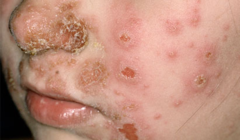
erosion
focal loss of epidermis
heals without scarring bc doesnt penetrate below the dermoepidermal junction

ulcer
afocal loss of epidermis & dermis
heals WITH scarring
usually secondary to peripheral arterial disease which causes impairment of vascular or nutrient supply to the skin

fissure
linear split in the epidermis
sharply defined, nearly vertical walls
>usually also into the dermis as well
chronic fissures may scar

atrophy
thinning of the epidermis or dermis which causes a depression in the skin
skin appears thin, translucent and wrinkled with easily visible blood vessels

scar
an abnormal formation of connective tissue post dermal damage
initially scars are thick & pink but with time become white and atrophic

diagnostic techniques
shave biopsy
punch biopsy
excisional biopsy
shave biopsy
a scalpel or a razor blade is used to scrape the surface of the skin
only shaves epidermis (outer layer)
punch/excisional biopsy
sample from epidermis, dermis, and hypodermis
technique for obtaining diagnostic full-thickness skin specimens
KOH prep
potassium hydroxide prep
add KOH to skin scraping dissolves keratin but not the fungi
examine under microscope
fungal culture
bacterial culture and sensitivity
wood's lamp
>black light
dermatoscope
magnifier with a non-polarized light source
35% more accurate than clinical diagnosis with Melanoma
reduces the number of benign lesions excised
moh's surgery
a technique used to excise tumors of the skin by removing fresh tissue, layer by layer, until a tumor-free plane is reached
microscopically controlled serial excisions of a skin cancer

moh's surgery
gold standard
> highest Cure Rate
used in cosmetically sensitive areas and areas with high rate of metastasis like ears, lips, genitals, nose
used for
-aggressive, invasive subtypes of BCC or SCC
-large lesions (>2cm)
-melanoma (MM)

ED&C
electrodessication and curettage
basic skin procedure
technique of curettage using a sharp curette is followed by electrodessication
used for superficial BCC, SCC in situ
-results in SCARRING

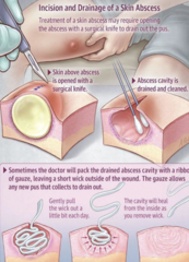
I&D
incision and drainage
with or without a bacterial culture
cryotherapy
superficial skin lesions are frozen with liquid nitrogen for a few seconds
time depends on the desired diameter & depth of freeze
commonly used on actinic keratoses, seborrheic keratoses, warts
medications
beware of combination creams (Anti-fungal/Steroid)
potency can be increased by occlusion x 4 hours (gloves/seran wrap over skin)
caution when applying to areas of thin skin
may cause glaucoma/cataracts when used on eyelids
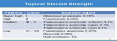
medications
palms/soles (thick skin) - ointment or cream
skin folds (always occluded) - cream or lotion
hairy areas (not vaseline) - lotion, solution, gel, foam
mucosal - non-irritating
enhance efficacy with occlusion

super high potency
class 1
clobetasol propionate 0.05%
high potency
class ii
fluocinonide 0.05%
medium potency
class iii-iv
triamcinolone acetonide oinment, cream, or lotion
low potency
class vi-vii
fluocinolone acetonide 0.01%
desonide 0.05%
hydrocortisone 1%
causes of acne
-bacteria: Propionibacterium acnes
inflammation
-hormones: androgen and their receptors
-excess sebum
-plugging of the follicle
acne vulgaris
in skin of color, can lead to permanent post-inflammatory hyperpigmentation
>treat aggressively to prevent this complication
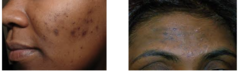
acne vulgaris
appears in areas of sebaceous gland activity
face/chest/back/shoulders
comedonal acne
whiteheads (closed comedrones) and blackheads (open comedones)

inflammatory
inflammatory papules and pustules

nodulocystic
comedones, inflammatory lesions and large nodules with or without scarring

acne vulgaris treatment
creams, gels, washes, and foams
>retinoids, antibiotics, benzoyl peroxide
oral medications
>antibiotics, hormonal, isotretinoin
retinoids
used to treat comedonal and inflammatory acne
-Adapalene
-Tretinoin
-Tazarotene
antibiotics
used to treat inflammatory acne and P. acnes
-Clindamycin
-Erythromycin
-Dapsone
benzoyl peroxide
used to treat P. acne
ongoing research regarding conversion to benzene
antibiotics
oral meds used to treat inflammatory acne and P. acnes
-Tetracyclines
>Doxycycline, Minocycline
hormonal treatment
oral medication that targets androgen receptors to treat acne
contraceptive pills or spironolactone
isotretinoin
acutane - last line treatment
6-9 months
does not work for hormonal acne
vitamin A in high doses
targets excess sebum
teratogenic so has adverse fetal effects
register with Ipledge, monitor BW (Lipid panel, CBC, CMP) n
adverse effects: mood changes, depression, joint pain, dry lips, extreme photosensitivity
no association with IBS
hidradenitis suppurativa
chronic disease that targets apocrine gland skin sites, particularly the axillae and groin
starts at puberty
physical exam shows recurrent tender inflammatory nodules/abscesses, open comedones & sinus tracts with purulent drainage
confirm with bacterial culture
think inverse cystic acne
high incidence of depression/suicide

hidradenitis suppurativa treatment
weight reduction, smoking cessation, chlorhexidine
intralesional triamcinolone
incision & drainage
antibiotics n tetracycline (1st line), topical
prednisone (short term)
excision
biologics
>humira, cosentyx, bimzelx
folliculitis
inflamed hair follicles
-may be superficial or deep
-can affect anywhere there are hairs
tender red perifollicular papule, often with a surface pustule

folliculitis
infection, occlusion (blockage), irritation
if bacterial infection, commonly due to S. Aureus (gram positive)
bacterial treatment
oral cephalosporins
-cephalexin, cefadroxil
and/or
topical mupirocin
if yeast infection, due to malassezia (more itchy)
yeast treatment
topical treatments
-azoles (econazole or ketoconazole)
-selenium sulfide
or
systemic treatments
-fluconazole (Diflucan)
hot tub folliculitis
due to P. aeruginosa, bacterial infection
treatment
oral ciprofloxacin
or
topical gentamicin
rosacea
persistent erythema (redness) of the convex surfaces of the face including nasolabial fold
telangiectasia (blood vessels), flushing, erythematous papules and pustules
cause remains unknown
but
theories = sun damage, mites, abnormal vasomotor response to stimuli
or due to
steroid-induced rosacea: long term or too strong of steroids on face can cause rosacea

types of rosacea
phymatous (skin thickening, nose nodules)
ocular (involves the eye)
papulopustular (papules/pustules within the rosacea)
erythematotelangiectic (flushing)

rosacea treatment
can resemble early lupus
--> differentiate with nasolabial fold involvement, biopsy or labs (ANA)
topical treatment
-metronidazole
-azelaic acid (otc Finacea)
-sulfacetamide
-topical ivermectin (Soolantra)
systemic treatment
- low dose doxycycline
-avoid triggers (spicy foods, alcohol)
-gentle skin care regimen
-lasers for blood vessels
-rhinophyma - surgery, CO2 laser
seborrheic dermatitis
thought to be an abnormal inflammatory reaction to Malassezia (yeast) in sebum rich areas (scalp, central face)
aka dandruff or cradle cap
clinical findings
greasy scale on erythematous base
treatment
-infants = reassurance outgrow by 6 months and use Aquaphor or coconut oil
-adults = mild topical steroids (hydrocortisone cream) for short-term flares
and
topical antifungals (ketoconazole cream) for longterm control
seborrheic dermatitis
in skin of color, affected areas may present lighter in color and have no redness
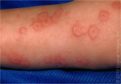
erythema multiforme
aka EM minor
-sharply marginated, erythematous macules which become raised and edematous over 1-2 days
-acral predominance (palms/soles)
-typically a ring of erythema forms = 'target'
-strongly associated with preceding herpetic infection or Mycoplasma pneumonia
SJS
steven johnson syndrome - skin sloughs off
aka erythema multiforme major
-patients have EM like skin lesions that cover 10% BSA or less
-always has mucosal involvement (oral, ocular or genital)
-usually represent adverse reaction to medication
>mostly rxn to Sulfonamides or anticonvulsants
TEN
toxic epidermal necrolysis
same as SJS but with 30% or greater BSA

EM minor
most cases are self limited: 1-2 weeks
recurrence is common
usually no prodromal symptoms
may have orolabial herpetic outbreak 3- 14 days prior to EM rash

EM major
SJS
has 5% mortality rate
recurrence uncommon
prodrome
influenza-like symptoms 1-2 weeks prior to cutaneous lesions

toxic epidermal necrolysis
40% mortality
more than 30% of skin sloughs off'
concerns: secondary infections, sepsis
usually represent adverse reaction to medication
>most commonly Sulfonamides and anticonvulsants
EM minor
self limiting
no mucosal involvement
no systemic involvement
0% detachment of skin
EM major
SJS
5% mortality
mucosal involvement
usually systemic involvement
10-30% detachment of skin
TEN
40% mortality rate
mucosal involvement
systemic involvement
more than 30% detachment of skin
SJS or TEN
requires admission to hospital
TEN managed in burn unit
alopecia
loss of hair
-non-scarring hair loss due to hormonal, hair care/styles, immune system (alopecia areata), telogen effluvium (stress/nutrition/pregnancy/
COVID/secondary cause)
-scarring hair loss due to follicles destroyed by inflammation or discoid lupus
alopecia areata
non-scarring alopecia
auto-immune disorder
-complete hair loss of scalp (totalis)
or
-complete hair loss of body (universalis)
rapid and complete loss of hair in 1 or more round or oval patches (1-5cm) with 'exclamation point hairs' along periphery
treatment
can be difficult
localized hair loss = intralesional or topical triamcinolone
extensive hair loss = topical immunotherapy, oral JAK inhibitors, off-label treatments
androgenic alopecia
genetic balding
hair loss related to aging
sensitivity to DHT (dihydrotestosterone)
female
-hair loss around crown of head
-treatments are Minoxidil or Spironolactone
males
-hair loss in front temporal areas, vertex of scalp
-treatments are Minoxidil or Finasteride

paronychia
inflammation of the nail fold
presents with erythema, swelling, throbbing pain, can have pustular drainage
caused by bacteria, may have secondary candida colonization
treatment - systemic & topical antibiotics

onychomycosis
tinea unguim
subungual hyperkeratosis with scaling, yellow discoloration and onycholysis (end of the nail lifts) ¨
diagnosis: culture (clipping) or biopsy of nail plate
treatment is difficult
-topicals: ciclopirox
-systemic: terbinafine (lamisil)

erythema infectiosum (5th disease)
caused by parvovirus B19
physical exam
-mild fever and headache
-red slapped checks appear for 2-4 days
-lacy patch on limbs/trunk
complications
-spontaneous abortion
-polyarthropathy
treatment
-none

hand foot and mouth
usually caused by Coxsackie virus
common, mild, short lasting
very contagious - spreads by direct contact, nasal/oral secretions
physical exam
-flat pink patches evolve into blisters on heads/feet
-small vesicles in/around mouth
-red macules and papules on buttocks/arms/genitals
treatment
-don't pop blisters
-antiseptic mouthwashes
-pain meds for oral ulcers
-maintain fluid intake
-no vaccine or antiviral meds

measles
rubeola
caused by measles virus
morbillivirus family
high contagious via respiratory droplets
physical exam
-cough, conjunctivitis
-gray-white papules, Koplik spots
-high fever
-rash starts on rash and spreads within 24-36 hours and spread to entire trunk and extremities
-palms and soles have no rash
treatment
-can be prevented with vaccination

cellulitis
bacterial skin infection
acute, spreading inflammation of the dermis & subcutaneous tissue
-if possible send out bacterial culture of any drainage
treatment
start with Abx that covers H.flu, Strep, Staph
--> penicillin (dicloxacillin) or cephalosporin
severe case
--> 1st generation cephalosporin IV
possible surgical intervention

erysipelas
painful, macular rash with well-defined margins
superficial cellulitis (upper dermis) that extends into the superficial cutaneous lymphatics
abrupt onset & rapid progression
rash is typically confined to the face which becomes fiery red, but may progress to the extremities
treatment
-penicillin

impetigo
bacterial skin infection characterized by isolated pustules that become crusted and rupture
highly contagious gram-positive bacterial infection
S. aureus is the most common pathogen
2forms of impetigo = bullous impetigo and nonbullous impetigo
can spread cutaneously and systemically (rare)

impetigo
most common bacterial skin infection
approximately 10% of skin problems observed in pediatric clinics
occurs more frequently in a warm, humid environment
bullous impetigo is most common in neonates & infant
impetigo
bullous impetigo
-rapid onset of blisters that enlarge & rupture
nonbullous impetigo
-erosions covered by moist, honey-colored crusts
-begins as small, easily ruptured vesicles
-most common sites: face & extremities
impetigo treatment
antibiotics to cover gram-positive bacteria
systemic
-cephalexin (Keflex)
-Cefadroxil (Duricef)
topical
-mupirocin (Bactroban) is applied to lesions AND nares in chronic nasal carriers
-get bacterial culture
-encourage proper hand-washing
syphilis
caused by T. pallidum
transmission
T. pallidum enters the body via skin/mucous membranes through abrasions during sexual contact and transmitted transplacentally from mother to fetus during pregnancy
travels via circulatory system, invades the central nervous system
primary syphilis
primary lesion, or chancre develops at site of inoculation
painless, indurated macule/papule
highly infectious
heals spontaneously within 3-6 weeks
serologic tests for syphilis may not be positive during early primary syphilis

secondary syphilis
occur several weeks after primary chancre and may persist for weeks to months, very contagious
physical exam
-rough, red papules or patches on the trunk and frequently affects palms and soles
-the rash does not itch
-general malaise
may affect liver, kidneys, CNS, joints, eyes

latent syphilis
patient suppresses infection but no clinically apparent lesions
only evidence is positive serologic test
tertiary (late) syphilis
rare because of widespread availability of antibiotics
symptoms - gummatous lesions, cardiovascular syphilis

neurosyphilis
may occur at any stage, when T pallidum invades the CNS
symptoms - acute syphilis meningitis, ocular involvement or may be asymptomatic
congenital syphilis
may lead to stillbirth, neonatal death, infant disorders (deafness, bone deformities, neurologic impairment)
syphilis
serologic tests
-treponemal qualitative AND nontreponemal qualitative and quantitative
-perform HIV testing in all patients with syphilis
-public health laws require that all cases be reported to the state department
treatment
-penicillin IM
-alternative - Doxycycline or Tetracycline
candidiasis
opportunistic yeast
-oral infection can be 1st sign of HIV
-often follows antibiotic use, diabetes, obesity, poor self-care
-grows best in warm, moist areas
-body = intertrigo
>beefy, red papules/plaques in skin folds +/- satellite lesions; +/- thick 'cheesy' discharge
-oral = thrush
>whitish plaques on erythematous base of buccal mucosa
intertrigo treatment
keep area dry/clean
discontinue offending agent
topical/oral antifungals

thrush treatment
nystatin suspension
fluconazole (Diflucan)
-if breastfeeding infant must also treat mother's nipples topically
superficial fungal infections
tinea capitis - scalp
tinea corporis - body; ringworm' tinea cruris - groin & gluteal cleft; 'jock itch'
tinea manuum - palms
tinea unguim (onychomycosis) - nails
tinea pedis - soles; 'athlete's foot'
tinea capitis
dry scaling with moth-eaten hair loss
very inflamed mass/abscess (Kerion)
untreated may result in permanent scarring/baldness
may result in swollen lymph glands at the sides of the back of the neck
tinea capitis treatment
take culture
topical antifungals cannot reach infection and are ineffective
ketoconazole shampoo (nizoral) or selenium sulfide shampoo (otc) is moderately effective as adjunctive therapy
oral griseofulvin x 6weeks - 6 months
throw out hairbrush
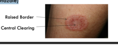
tinea corporis
ringworm
well demarcated annular plaque with central clearing and peripheral scale
contagious
diagnosis
KOH prep (optional)
topical treatment
-azoles (econazole, ketoconazole)
tinea incognito
results when patient uses a potent topical steroid (not antifungal) on tinea corporis
drives fungus deeper into the skin; affects hair follicles
physical exam
regular red, scaly plaque with follicular papules
pustules = more severe and indicates Majocchi's Granuloma ¨
treatment
oral griseofulvin x 4-6 weeks

tinea cruris
jock itch
well-defined scaly plaque with widespread erythema
intensely pruritic
spreads through contaminated towels/sheets or autoinoculation
50% of patients also have on hands or feet
3x more common in men ¨
treatment
-keep groin dry, weight loss
topical antifungals
- azoles x 2-4 weeks
