5. Post-mortem changes in muscle and its conversion into meat
 Call Kai
 Learn
Learn Practice Test
 Spaced Repetition
 Match
Match Flashcards
 Knowt Play
1/35

There's no tags or description
Looks like no tags are added yet.
Name | Mastery | Learn | Test | Matching | Spaced | Call with Kai | Chat |
|---|
No analytics yet
Send a link to your students to track their progress
36 Terms
In a living animal, what is the major function of muscle, and how is the ATP needed to support it produced and used?
Major function = contraction
ATP is required for:
Contraction
Maintaining the muscle's functional integrity
Fuelling the Ca²⁺ pump of the sarcoplasmic reticulum (relaxation)
ATP is produced from:
Free fatty acids
Glucose (from blood)
Glycogen (stored in muscle fibres)
How supply is ensured: enzyme systems in the sarcoplasm and mitochondria deliver ATP to the contractile elements.
Key idea: fuel (fatty acids, glucose, glycogen) → ATP via sarcoplasm and mitochondria → powers contraction, Ca²⁺ pump and muscle integrity
Energy metabolism in muscles
Fed animals have low free fatty acids and therefore glucose is mostly used
Fasting animals use free fatty acids
If FFAs and glucose are not enough than glycogen is used
Glycogen and glucose broken down easily by:
Glycolysis
Oxidative decarboxylation
Oxidative phosphorylation
Aerobic energy metabolism in muscle (post-mortem context: why it stops after death)
Occurs in the mitochondria (the Krebs cycle and electron transport chain), after glycolysis in the sarcoplasm
Requires oxygen: 6 O₂ per 1 glucose
Yields far more ATP than glycolysis alone (about 30-32 vs. 2 ATP per glucose)
No oxygen → only glycolysis → pyruvate becomes lactate → pH falls
Post-mortem acidification
Death → O2 supply (including glucose and FFAs) to muscle ceases when blood circulation stops
Therefore ATP only regenerated through breakdown of glycogen by glycolysis (anaerobic)
Glycogen broken down → LA accumulates as no longer removed by blood = muscle gradually acidifies
If glycogen is not limiting: LA production ceases when enzyme system will no longer function at low pH
pH change post-mortem
Unstressed animal: pH fall from 7.2 to 5.5
importance of acidification on meat quality
Affects: water-holding capacity (WHC) and color
Influence of lower pH on structure of muscle constituents
WHC decreases so that drip/exudate is lost - especially if muscle is cut
Meat that is relatively dark and translucent in living animal will become paler and opaque when dead
How does the water holding capacity change when an animal dies?
When animal dies → pH decreases & Lactic Acid accumulates
This causes pH to fall to isolectric point of myofibrillar proteins causing the electrical charge on these proteins to reach zero
Hence proteins lose the electrostatic repulsion that allows them to hold water
Additionally due to rigor mortis, actin and myosin link irreversibly to form actomyosin cross-bridges.
Because they are packed so tightly together, it causes the myofilament lattice to shrink laterally.
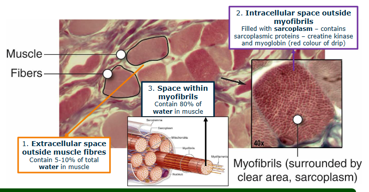
Where is the water bound in muscle?
Extracellular space outside muscle fibres - contain 5-10% of total water in muscle
Intracellular space outside myofibrils - filled with sarcoplasm - contains sarcoplasmic proteins - creatine kinase and myoglobin (red color of drip)
Space within myofibrils - about 80% of water
Acidification effect on meat color
Acidification = protein change
Change in structure = increase light scattering properties of contractile elements of muscle fibre
When animal was still living there was little extracelular space which caused light to be absorbed rather than reflected.
As myofilament lattice shrinks and extracellular space volume increases structure becomes optically more heterogeneous.
As a result meat appears paler/lighter
What happens to water when WHC decreases?
Water is lost from proteins → squeezed out from myofibrils into sarcoplasm increasing “extra-myofibrillar” volume
Eventually flows out of muscle and when meat is cut will flow out.

What is this figure?
Rigor mortis development
Figure: relationship between ATP depletion and the onset of rigor mortis
Rigor mortis development
Rigor mortis is the post-mortem stiffening and loss of extensibility in muscle tissue as it transforms into meat.
In livin muscle, ATP binds to the myosin head to cause its dissociation from actin, keeping the muscle relaxed.
When an animal is slaughtered, oxygen supply ceases and energy metabolism becomes anaerobic.
As ATP levels drop below 5 mmol * kg-1 actin and myosin bind irreversibly to form permanent actomyosin cross-bridges.
Rigor onset time
Relate to factors affecting level of glycogen and creatine phosphate at death and rate of post-mortem muscle metabolsim
Affected by stress and exercise: if there was high stress before death than glycogen and creatine phosphate were used up meaning that ATP cannot be regenerated after death = rigor moris.
However if the animal was relaxed during death: then there would still be a lot of creatine phosphate and glycogen allowing the regeneration for ATP for a little while = later rigor mortis.
Rate of development reduced if carcass cooled quicker: at a temperature between 10C and 15C enzyme activity is minimized and ATP is used up very slowly. Any colder SR stops working = no calcium ion. Any higher ATPase works really fast and rigor mortis occurs very quickly
Discuss the different types of rigor mortis that can be found?
Normal rigor mortis as mentioned :Anaerobic glycolysis converts muscle glycogen into lactic acid, dropping muscle pH from approximately 7.2 down to an ultimate pH of 5.2–5.5. As creatine phosphate (CP) and glycogen are exhausted, ATP resynthesis stops, and permanent actomyosin cross-bridges fix the sarcomere length
Alkaline rigor:
Occurs when animals suffer severe pre-slaughter exhaustion.
This depletes glycogen and CP
Therefore after slaughter no LA can be formed
Gives DFD appearance
Rigor mortis development and meat quality
After rigor muscle can no longer shorten (be stretched) and sarcomere and muscle length fixed
Sarcomere length (reflects myosin and actin overlap) important for tenderness and toughness
After onset of rigor → no danger of cold/heat shortening or compromising meat texture
Temperature at which rigor mortis is entered
Above at least 15C, but mostly 35C = less tender meat due to heat shortening causing the denaturation of proteolytic calpains
At cold temperature = cold shortening
Resolution of rigor and tenderization of meat
You can not completely resolve it
Muscle is not extensible again as pre-rigor
Thick and thin filaments remain locked by mosyin cross-bridges (actomyosin)
Tenderisation not caused by filaments regaining ability to slide
Myofibrils structure begins to break down.
Why do different tenderization rates matter commercially, and how is the problem handled?
The issue: meat tenderizes at different rates, so each type needs a different recommended ageing time.
Why long ageing is a problem: keeping meat at refrigerated temperature is expensive because of:
Storage costs
Refrigeration costs
Weight loss (moisture loss during ageing)
The solution: a compromise is reached commercially, producing meat with acceptable tenderness in a reasonable time rather than waiting for maximum tenderness.
Recommended conditioning time for pork, lamb and beef

Conditioning/aging
the holding of carcasses or meat cuts at various temperatures above freezing to cause improvements in meat quality
What are the steps in the conditioning process
Tenderization occurs in two phaess:
Rapid phase → changes in myofibrillar component
Most important
Attachments of thin (actin) filaments to z-discs show some breakdown
Increase in amount water-soluble nitrogen compounds
Slower phase → structural weakening of intramuscular connective tissue
Only small changes seen in major connective tissue components such as collagen
Two main sorts of enzymes involved in tenderization
Tenderization comes from proteolytic enzymes in muscles
Calpains
Though to be more imporant (at least in red meat species and poultry)
Are released upon increase in Ca2+ concentration
optimal pH: neutral to alkaline (7.5)
Released early post-mortem
Cathepsins
May be more important in post-mortem degradation of fish muscle
Tenderizes meat more than calpains at higher temperatures.
Are released from lysozymes, when lysozyme membrane are distrupted post-mortem
Optimal pH: acidic
Released later post-mortem
Calpains
Activated by Ca2+ ions
Maximum activity in neutral to alkaline conditions (pH 7.5)
Show some activity (±25%) at pHu (pH 5.5, normal meat)
Degrade: myofibrillar component
Actin and myosin
Troponin-T
Titin (connectin)
Desmin
What inhibits calpains
Inhibited by endogenous protein calpastatin - high calpastatin activity reduces extent of proteolysis in muscles
Cathepsins
Occur in lysosomes and sarcoplasm
Released from lysosomes post-mortem
Maximum activity in mildly acidic conditions
Degrade:
Troponin-T
Some collagen cross-links
Mucopolysaccharides of connective tissue ground substance
Only actin and myosin < pH 5 - unlikely under normal conditions
Calpastatin
Inhibits calpains
Calpastatin activity in muscle thought to be more vital than amount of calpains present in controlling development of meat tenderness
Encouraged efforts to identify alleles of calpastatin gene
When animals are stressed during slaughter, elevated adrenaline levels will increase calpastatin activity
Why is meat from Bos taurus cattle more tender than the meat from Bos indicus?
Bos Taurus cattle have more post-mortem proteolysis due to less calpastatin

What evidence shows that calpains are important for meat tenderization?
β-agonists reduce the activity of proteolytic enzymes (including calpains) that drive normal muscle protein turnover (accretion and breakdown)
This reduced activity persists post-mortem
Result: little or no proteolysis during aging, so the meat stays relatively tough
Different forms of calpains
Two main forms:
m-calpain (calpain II) activated at high Ca ion concentration
μ-calpains (calpain I) activated at low Ca ion concnetration: μ- calpain
Which calpain is more crucial regarding post-mortem tenderization?
μ-calpains
Activated earier than m-calpains
correlates with when most breakdown of myofibrilar protein occurs
What is an indicator for the conditioning process?
One most obvious change that occurs in myofibrillar proteins during ageing of meat from all species is breakdown of troponin-T
Small amount of actin and myosin degraded too
Most breakdown occurs in proteins associated with z-disc and cytoskeleton - particularly titin (connectin) and dsemin
Key process of tenderisation:
Gradual breakdown of cytoskeleton framework which holds contractile elements (thick and thin filaments) attached and arranged within structure of muscle fibre
Possible action of calpains post-mortem
After exhaustion of ATP and development of rigor mortis: membrane systems of SR and mitochondria no longer sequester Ca ions
Released into sarcoplasm and bath myofibrils
Increased Ca concentration activates μ-calpains allowing proteolysis
Normally calpains inhibited by being bound to calpastatin
Calpain activity promoted by higher Ca levels → Ca ions remove inhibition
Calpain activity is also promoted by higher pH and temp
Enhance enzyme activity post-mortem by infusing carcass with 0.3 M CaCl
Vitamin D to improve tenderness
Vit D is fat soluble and is essential for calcium and phosphorous metabolism
Therefore elevates calcium concentration in blood and muscle tissue
More Ca also stimulates activity of calpains post-mortem
Disadvantages of giving animals vit D to improve tenderness
May reduce feed intake and growth rate is commercially uneconomic
Very high dietary intake is toxic