GI 1.1 Final
 Call Kai
 Learn
Learn Practice Test
 Spaced Repetition
 Match
Match Flashcards
 Knowt Play
1/53

There's no tags or description
Looks like no tags are added yet.
Name | Mastery | Learn | Test | Matching | Spaced | Call with Kai | Chat |
|---|
No analytics yet
Send a link to your students to track their progress
54 Terms
oral cavity epithelium?
masticatory mucosa → keratinizing strat. squam.
lining mucosa → NK strat. squam.
specialized mucosa → papillae + taste buds
drug absorption → sublingual route bypasses portal circ.
extrinsic and intrinsic tongue muscles? sensory nerves?
genioglossus (protrude), hyoglossus (depress), styloglossus (retrude), palatoglossus (elevate post. tongue; ONLY CNX)
shape change for speech/swallow (CNXII for everything)
ant. 2/3 sensation (lingual) vs taste (chorda tympani), post. 1/3 sensation + taste (CNIX), epiglottis (int. laryngeal)
blood supply of tongue? lymphatics of tongue?
lingual artery (ECA); venous drainage to int. jugular
tip (submental), sides (submandibular) + deep cervical), post. (deep cervical)
lingual papillae? taste buds?
filiform (center ant. 2/3; w/o buds), fungiform (margins), circumvallate (V shape ant. to term. sulcus), foliate (lat.)
neuroepithelial (sensory; synapse w/ CN VII, IX,X), supporting (microvilli, no synapse), basal (stem cells)
teeth tissues? salivary glands?
enamel (ameloblasts), dentin (odontoblasts → neural crest), cementum (covers root)
root nerve supply → inf. alveolar nerve (CN V3)
parotid (serous only), submandibular (mixed, mostly serous), sublingual (mixed, mostly mucus)
pharyngeal muscles? function?
circular (constrictor) → sup., middle, inf. (UES = cricopharyngeus)
longitudinal → stylopharyngeus (CNIX), palatopharyngeus, salpingopharyngeus (CNX)
elevate larynx, shorten pharynx during swallow/speak
GI histology?
mucosa → epi., LP, musc. muc.
submucosa → meissner plexus (sec.), galnds
muscularis → auerbach plexus (motility); inner circ., outer long.
serosa/adventitia → serosa = intraperitoneal; adventitia = retroperitoneal
cells/glands throughout GIT?
esophagus → mucous glands, adventitia
stomach → cardiac (mucus), fundic (parietal, chief, enteroendocrine), pyloric (G cell)
duodenum → villi, crypts, brunner glands
jejunum → villi, crypts, plicae circularae
ileum → goblet cells, peyer’s patches
colon → crypts, goblet cells NO villi
anus → transition to strat. squam.
small intestine segments? structure?
duodenum → sup., desc., hori., asc.; retroperitoneal EXCEPT prox. 1st + distal 4th
major duodenal papilla → opening of hepatopancreatic ampulla (main pancreatic + bile duct)
minor duodenal papilla → acc. pancreatic duct
jejunum → thick wall, fewer arterial arcades, long vasa recta, prom. circ. fold
ileum → thinner walls, more arcades, short vasa recta, peyer’s patches (lymphoid agg.)
large intestine segments? structure? appendix?
cecum → asc. → tran. → desc. → sigmoid → rectum → anal canal
teniae coli (3 muscle bands), haustra (sacculation of colon wall), epiploic appendages (fat filled peritoneal pouch), ileocecal valve (mucosal fold, min. anti-reflux role)
appendix → lymphoid rich, variable position; mcburney’s point = T10 referred pain
rectum location? structure?
begins @ S3, ends @ pelvic diaphragm
rectal ampulla → dilated lower portion
puborectalis sling → forms anorectal angle for continence
transverse folds → perm. mucosal folds
viridans streptococci class? plating? symptoms?
bacteria catalase (-) anaerobes
gram (+), a, B, or y-hemolytic (green on blood agar)
main micro in oropharynx, GIT
dental caries (via sugar fermentation) + endocarditis
LA → low pH (kills microbiota + favors acid res.) → low O2 (anaerobe bind) → polysaccharide = biofilm/plaque → demineralization/lesion

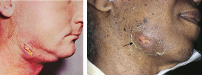
actinomyces israelii class? plating? symptoms?
bacteria catalase (-) anaerobes
gram (+), sulfur granules, filamentous
via poor oral hygiene or trauma/surgery
usually cervicofacial → lumpy jaw

porphyromonas gingivalis class? plating? symptoms?
bacteria catalase (-) anaerobes
gram (-), black on blood agar
uses fimbriae as VF
gingivitis, periodontitis, oral abscesses


fusobacterium necrophorum class? plating? symptoms?
bacteria catalase (-) anaerobe
gram (-)
enters jugular vein (Dx w/ neck CT) → systemic spread (esp. lungs); sore throat
suppurative jugular thrombophlebitis (lemierre)
candida albicans class? plating? symptoms?
fungi
prod. buds/blastoconidia; smooth white, fuzzy, creamy colonies (pseudohyphae)
oral thrush (scrapable), yeast infection; HIV pts

HSV1 class? features? symptoms?
herpesviridae; dsDNA → infect + replicate in mucoepithelial cells
multinucleated giant cells (syncytia) → tzanck smear; cowdrey bodies
latent in trigeminal ganglia
oral (lips, tongue) vascular lesions (recurrent), can progress to encephalitis; gingival ulcer, swollen gums, cervical LAD
EBV class? features? symptoms?
herpesviridae; dsDNA → uses C3d receptor (CR2, CD21)
heterophile antibody-positive infectious mono → stimulates B cell growth; can remain dormant/latent
oral hairy leukoplakia (balloon cells, hyperkeratosis, lat. tongue, non-scrapable)
HPV class? features? symptoms?
papillomaviridae; circular dsDNA, naked
infects host basal epithelial cells → proliferate/grow → stratum spinosum thicken → wart condyloma, papilloma (benign or malignant)
laryngeal cancer, squamous papilloma, nonkeratinizing SCC
mumps class? features? symptoms?
paramyxoviridae; (-) RNA
spreads/mulitplies in local lymphoid tissues + reticuloendothelial cells
meningitis, parotitis, orchitis in unvaccinated child

aphthous ulcer symptoms? histo/micro?
inflammatory; canker sore
associated w/ stress, IBD, behcet, celiac, HIV
painful, shallow, recurrent mucosal ulcer
mononuclear, neutrophilic infiltrate

irritation fibroma symptoms? histo/micro?
inflammatory; trauma-induced @ bite line, gingiva → buccal submucosal nodular mass
fibrous stroma

pyogenic granuloma (lobular capillary hemangioma) symptoms? histo/micro?
inflammatory; vascular proliferation w/ granulation tissue tissue rxn (rapid growth) → red/ulcerated, pregnancy associated
peripheral ossifying fibroma → gingival growth from periodontal lig. following pyogenic granuloma
polyploid lesion resembling mushroom @ skin surface

peripheral giant cell granuloma symptoms? histo/micro?
inflammatory; reactive gingival lesion → fibroangiomatous stroma
DDxs = brown tumor, giant cell tumor
fibrovascular stroma w/ multinucleated giant cells; intact overlying gingival squamous mucosa

squamous papilla features? histo/micro?
neoplastic (most common benign oral cavity neoplasm) via HPV
exophytic lesion w/ branching squamous epithelium + fibrovascular cores
cauliflower, skin/pearly/grey warts
squamous proliferation w/o dysplasia, keratosis, viral cytopathic change

hairy leukoplakia symptoms? histo/micro?
proliferative; white, fluffy, non-scrapable patch on lateral tongue
low malignancy risk
more common in EBV, immunocompromised
balloon cell, hyperkeratosis (expanded corneus layer w/ retained nuclei), acanthosis (expanded stratum spinosum, but orderly)

erythroplakia symptoms? histo/micro?
red, velvety, non-scrapable patch
w/ severe dysplasia → loss of polarity, freq. mitosis, coarse chromatin
carcinoma in situ, minimally invasive CA
high malignancy risk

squamous cell carcinoma (SCC) symptoms? histo/micro?
ulcerated, indurated lesions w/ irregular borders
associated w/ HPV 16/18, tobacco, alcohol, betel quid (asia), sunlight (lip), idiopathic (YA)
better prognosis w/ p16 IHC+
leukoplakia (keratin pearl, hyper keratinization) + erythroplakia = precursors
keratinizing or non-keratinizing (HPV), fibrovascular cores, NO dysplasia
odontogenic cysts? tumors?
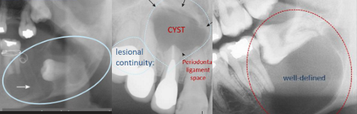
dentigerous cyst → crown of impacted/unerupted tooth (fluid accumulates btwn tooth + dental follicle) = unilocular cyst, NK squamous w/ chronic inflammation
radicular cyst → apex of tooth (long pulpitis = necrosis) = granulation + epithlialization
keratocystic tumor → well-defined unilocular to multilocular = keratinized SSE w/ prom. basal layer + NO RETE ridge
xerostomia features? sialadenitis features?
via sjogren, meds, radiation → inc. risk of candidiasis + caries
salivary gland inflammation (related to mumps/autoimmune) → acinar atrophy w/ fibrosis
mucocele features? ranula features?
trauma induced duct rupture → pseudocyst lined w/ inflammatory granulation or fibrous CT (recurs if incomplete excision)
bluish hue (may contain mucin + MQ)
sublingual (floor of mouth) duct cyst (recurs if incomplete excision)
may plunge into mylohyoid
pleomorphic adenoma features? warthin tumor features? mucoepidermoid carcinoma features?
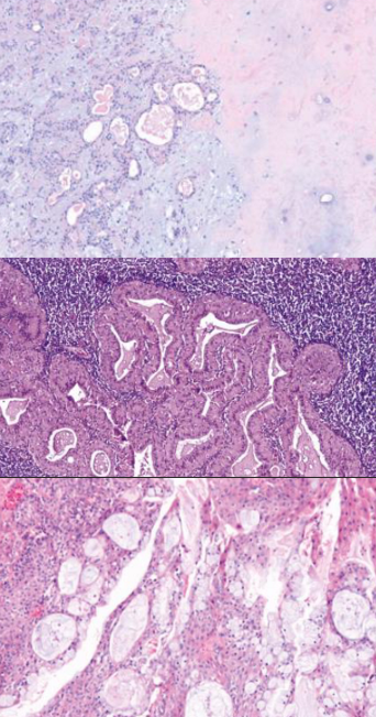
most common (benign), @ parotid gland (recurs if not entirely excised) → epithelial + mesenchymal + cartilaginous
@ parotid gland, papillary cystadenoma lymphomatosum → risk w/ smokers; epithelial + lymphoid stroma
most common (malignant), mucus filled vacuoles → mucinous + squamous cells; grade dependent
esophagus tissue? muscle? innervation?
stratified squamous epithelium → transition to columnar @ GE jxn
barretts esophagus → metaplasia of squamous to columnar w/ goblet cells → low (inc. mit, basal nuclei w/ mild atypia) vs high grade dysplasia (p53 mut, loss of polarity, mit., crowded glands + lose LP) → adenocarcinoma
muscle → upper skeletal, middle mixed, lower smooth
innervation → auerbach (myenteric) + meissner (submucosal) plexus
esophageal atresia?
congenital mechanical esophageal obstructions
segment of esophagus doesn’t develop near tracheal bifurcation w/ fistula (UE or LE connect to bronchus/trachea)
nonbilious emesis, choke, cough @ initial feed (progressive dysphagia)
TE fistula = aspiration, suffocation, pneumonia
acquired esophageal mechanical obstruction?
benign esophageal stenosis → fibrous thicken = progressive dysphagia (solid → liquid)
esophageal webs → mucosal protrusion into lumen @ upper esophagus = poorly chewed, non progressive dysphagia
shatzki ring → thin mucosal ring @ GE jxn via reflux/inflammation = solid food dysphagia
steakhouse syndrome/impaction; Tx via dilation
plummer-vinson syndrome cause? features? risk?
mechanical obstruction
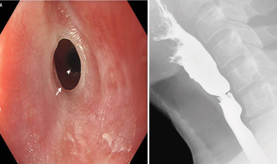
Fe def. anemia (ox. stress + DNA damage = epi. injury) + prox. esophageal webs (squamous mucosal protrusion into lumen)
dysphagia (solids > liquids) w/ hypochromic anemia in middle aged women
risk for SCC of esophagus + pharynx
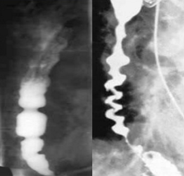
nutcracker esophagus? corkscrew esophagus?
functional obstruction, measure pressure + muscle w/ manometry
high amplitude, hypercontractile sequential @ distal esophagus = dysphagia + chest pain
barium = alt. narrow + distended seg.
high amplitude, uncoordinated + simultaneous/diffuse contraction @ mid + distal esophagus = dysphagia + chest pain
Dx w/ barium swallow; Tx w/ CCBs, botox, myotomy

achalasia cause? features? symptoms?
functional obstruction; neurodegeneration of submucosa myenteric plexus → impaired LES relaxation + impaired peristalsis = food build up/dilation
1= degeneration of NO neurons; 2= chagas/protozoal tyrpanosoma cruzi damage plexus
DM, malignancy
dysphagia (solid + liquid), regurgitation, chest pain, weight loss
bird beak sign/megaesophagus
esophageal diverticula structures? formation? types? symptoms?
epithelial lined mucosal pouch protrude from esophageal lumen
pulsion/false → inc. intraluminal pressure via hypercontractile or LES dysfxn = mucosa + submucosa herniates through muscle (zenker, epiphrenic)
traction/true → ext. pulling via adj. inflammation fibrotic adhesion (gran. lymph.) = fibrosis pulls on all wall layers (midesophageal)
= regurgitation of undigested food, halitosis, aspiration

pharyngoesophageal diverticulum (zenker)? midesophageal diverticulum? epiphrenic diverticulum?
above UES spasm (cricopharyngeal) after swallow → inc. pressure @ distal pharynx → hypopharyngeal herniation @ killians triangle = halitosis, int. dysphagia
true; adhesion btwn inflamed mediastinal LN (via TB, histo) + esophagus → pulls all layers (true)
distal 10 cm/above LES= motor abnormalities (achalasia, hypertensive LES, spasm)

hiatal hernia types?
sliding (common) → GE jxn + gastric cardia up thru diaphragm = GE reflux
paraoesophageal → stomach pouch protrudes through esophageal hiatus adj. to GE jxn (below diaphragm) = more risks
esophageal lacerations causes? types?
via retching, alcohol, inc. pressure (cough, abdominal strain)
mallory-weiss → longitudinal @ GEJ, mucosa + submucosa = hematemesis, melena
boerhaave → transmural w/ perforation, all layers = mediastinitis, pneumothorax, pneumomediastinum (crunching/hamman sign), pleural effusion
surgical intervention

esophagitis causes?
chemical → caustics, alcohol, hot liquid, smoking; necrosis + neutrophils
infectious → HSV (cowdry-A), CMV, candida; ulcer, inclusion, pseudomembranes
GERD (most common) → LES relaxation/incompetence via inc. intra abd. pressure, inc. gastric vol., dec. distance, spice; multinucleate squamous cell, elongated LP papillae, basal cell hyperplasia, eosinophilia
= barrett’s esophagus (patchy red, velvet mucosa w/ columnar + goblet cells above GEJ)
eosinophilic → allergy/immune-med.; >15 eosinophils/HPF, stacked circular rings/furrows/“feline esophagus”

esophageal adenocarcinoma risk factors? features? molecular?
presents late w/ prog. dysphagia, weight loss, pain, hematemesis
GERD, barretts, obesity/high red meat, tobacco/radiation, prolonged proton pump inhibitors
lower 1/3 w/ gastric cardia; nodular mass/patch, poorly diff. glandular cells w/ mucin prod. (ulcerative forms)
tumor suppressor mut. (TP53, CDKN2A =p16/19), proto-oncogene (MET, ERBB2)
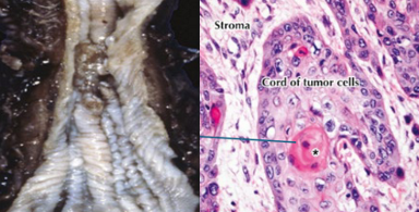
esophageal squamous cell carcinoma (ESCC) risk factors? features? molecular?
most common esophageal cancer; presents late w/ prog. dysphagia, weight loss, pain, hematemesis
alcohol, tobacco, lye ingestion/diet, hot beverage, plummer-vinson, HPV, low SES (irritants)
middle 1/3 w/ small white plaque; invades SM + ME, tumor cells in cords + nests (keratin pearls)
tumor suppressor mut. (TP53, CDH1, NOTCH1/3), proto-oncogene over express (EGFR, cyclin D1, RAS/AKT)
= hoarse voice (recurrent laryngeal nerve) + cough (trachea)
gastric anatomy + cells?
mucosa (gastric pits + glands), SM, M, S
cardia (GEJ) → mucin cells (protective mucus)
fundus/body (above GEJ + middle w/ oxyntic gland) → parietal (HCI), chief (pepsinogen), ECL (histamine)
antrum/pylorus (distal 1/3 + duodenal jxn w/ pyloric gland) → G cell (gastrin), D cell (somatostatin)

acute gastritis features? cause? symptoms?
inflammation + neutrophil infiltration
edema, hemorrhage, erosion
via direct damage, NSAID (inhibit COX → dec. PG), H. pylori/uremia, aging/impaired renewal
asymptomatic w/ epigastric pain, nausea, vomit

chronic gastritis features? types cause, features?
w/ lymphocytes + plasma cells; nausea, upper abdominal pain sometimes vomit
autoimmune atrophic @ body-fundus → parietal cell Ab (hypochlorhydria + hypergastrinemia) + intrinsic factor Ab (dec. Vit B = pernicious anemia)
oxyntic gland flat w/ loss of rugal fold; endo hyperplasia, metaplasia w/ goblet cell (GACA), NEN
H. pylori @ antrum but spreads (common) → inc. HCl sec. (destroys parietal cells = ““ + possible lymphoid agg.)
w/ flagella, urease, adhesins, CagA, VacA
peptic ulcer, MALToma; Dx w/ biopsy (anti-Ab), urease, PCT (DNA)
mixed gastritis

peptic ulcer disease (PUD) cause? features? symptoms? types?
H. pylori, NSAIDs, stress, trauma, ZES (inc. gastrin)
early (fibrin, necrosis, inflammation, granulation) vs chronic (scarring + stenosis)
nausea, vomit, bloat → melena, hematemesis, perforation, nocturnal pain
gastric (punched-out lesion w/ clean base in lesser curvature; pain worse w/ eating + weight loss) OR duodenal (pain relieved w/ eating + weight gain, solitary morph.)
Tx w/ H. pylori+ (triple/quad) + H. pylori- (PPI)
hypertrophic gastropathies types?
menetrier = TGF-a → mucous cell hyperplasia (adenocarcinoma risk), protein loss, enlarged rugal folds
zollinger-ellison = gastrinoma → inc. HCL (hyperchlorhydria), PUD, parietal hyperplasia (adenocarcinoma risk)
gastric adenocarcinoma features? symptoms? metastasis?
poorly diff., large ulcers w/ heaped up rolled margins
epigastric pain, weight loss, early satiety, virchow node, sister mary joseph nodule, krukenberg tumor
liver, peritoneum, lung, bone
types of gastric adenocarcinoma?
ulcerative = H. pylori, salt, PAHs- smoking → flat necrotic base
polypoid = HER2, RAS → cauliflower, papillary
scirrhous = CDH1 mut. → linitis plastica, signet ring
colloid = PAS+ → mucinous
gastric lymphoma (MALT) cause? features?
translocation → t(11;18), t(1;14), t(14;18)
NF-kB act.
CD19, 20, 43(+) & CD5,10(-)
gastric neuroendocrine neoplasm (NEN) features? types?
intramural masses, salt + pepper chromatin, chromogranin(+), synaptophysin(+)
type 1= atrophic gastritis → inc. gastrin + dec. HCl
type 2= gastrinoma MEN1 → dec. gastrin + inc. HCl
type 3= sporadic → normal gastrin + HCl