Ch 9 Enzymatic Catalysis
 Call Kai
 Learn
 Practice Test
 Spaced Repetition
 Match
 Flashcards
 Knowt Play
1/67

There's no tags or description
Looks like no tags are added yet.
68 Terms
General properties of enzymes
biocatalysts
active site
specificity
enzymes increase reaction rate
lower activation energy
x106 to 1012 rate enhancement
enzymes are classified by reaction type (7 classes)
oxidoreductases
transferases
hydrolases
Lyases
isomerases
ligases
translocases
oxidoreductases
oxidation-reduction reactions
e- transport reaction
transferases
transfer of functional groups
hydrolases
hydrolysis reactions
lyases
group elimination to form double bonds
isomerases
isomerization
ligases
bond formation coupled with ATP hydrolysis
translocases
movement of molecules across/within membrane
ex: flipASE
enzymes act on specific substrates
highly specific
lock and key ES model → geometric complementary and electronic complementary
induced fit→ conformational change on substrate binding, facilitates shifts of particular residues and functional groups into proper positions to catalyze reaction
Nearly all enzymes that participate in chiral reactions are
stereospecific
Some enzymes catalyze very stereospecific reactions
enzymes must act on a very stereospecific compound

Some enzymes are more permissive than others
ex: chymotrypsin mediates peptide bond hydrolysis on a wide variety of substrates and can even catalyze related reactions on different functional groups (ester bond hydrolysis)

Transition state
X‡ = point of highest free energy
Reaction rate is proportional to
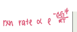
Does ∆G change with catalyst?
No, ∆G does not change with catalyst
catalysts act be lowering free energy barrier by same amount for both forward and reverse reactions
∆∆Gcat‡ =
change in EA
reduction in ∆G‡ by the catalyst
Preferential transition state binding
an enzyme’s active site has greater structural complementarity to the TS than to the substrate or product itself
an optimal set of non-covalent interactions between E and S occurs specifically at transition state (ES‡)
Transition state analogs are enzyme inhibitors
proline racemase is inhibited by planar analogs of proline
planar analogs bind to the enzyme 160x greater affinity than proline
rational basis for drug design

cofactors expand the range of enzymatic reactions
needed for specific catalyst
in order to complete the catalytic cycle, the coenzyme must return to its original state
cofactors can be metal ions or coenzymes
metal ions/metalloenzymes
common ex: Fe2+/3+, Zn2+, Cu+, Mg2+
what happens when Cd2+ or Zn2+ replaces Zn2+
toxic!
apoenzymes
require cofactors
inactive
holoenzyme
active
apoenzyme+cofactor
coenzymes
organic molecules
many derived from vitamins
cosubstrates AND prosthetic groups
cosubstrates
transiently bound
ex: NAD+/NADH, NADP+/NADH, CoQ
prosthetic groups
permanently bound
ex: heme (includes Fe) in Mb + Hb
ex: Thiamine pyrophosphate (TPP) — aldehyde group transfer, active form of thiamine (vit. B1)
Alcohol Dehydrogenase (ADH) Reaction

In hydrophobic environments, acids ____ pKa
increase pKa
it is more stable protonated
In hydrophobic environments, bases ___ pKa
decrease pKa
it is more stable with neutral/no charge in hydrophobic environment
When a like charge is nearby, acids ___ pKa
increase pKa
When a like charge is nearby, bases ____ pKa
decrease pKa
When there are salt bridges or H-bonds, acids ____ pK
decrease pK
When there are salt bridges or H-bonds nearby, bases ___ pK
increase pK
Proteins shift their pKa values to stabilize whatever charge state is energetically favorable in that microenvironment
Hydrophobic protein interior -
Acids increase pKa — the group wants to remain neutral b/c hydrophobic core hates charge
Bases decrease pKa — side chains positive when protonated, to escape this instability, base rapidly kicks off proton to be neutral
Like Charges nearby -
Acids increase pKa — to prevent charge-charge repulsion, want the acid to hang on to its proton
Bases Decrease pKa — positive charge next to protonated base may repel, thus base loses its proton easier
Salt Bridges or H-bonds -
acids decrease pKa — deprotonated form is stabilized in salt bridge/H-bond, thus it is rewarded and acid gives up proton earlier
bases increase pKa — protonated form if stabilized by H-bond/salt-bridge, protein rewards it for keeping its proton by base holding onto proton more
Concerted acid-base catalysis
ability of enzymes to arrange several catalytic groups around their substrates makes concerted acid-base catalysis a common mechanism
ex: Bovine pancreatic RNase A
RNase A Mechanism
RNA molecule is hydrolyzed in 2 steps
2 Histidine residues, both act as acid/base
concerted acid/base catalysis for each step
RNase A step 1
His 12 acts as a general base and His 119 acts as acid
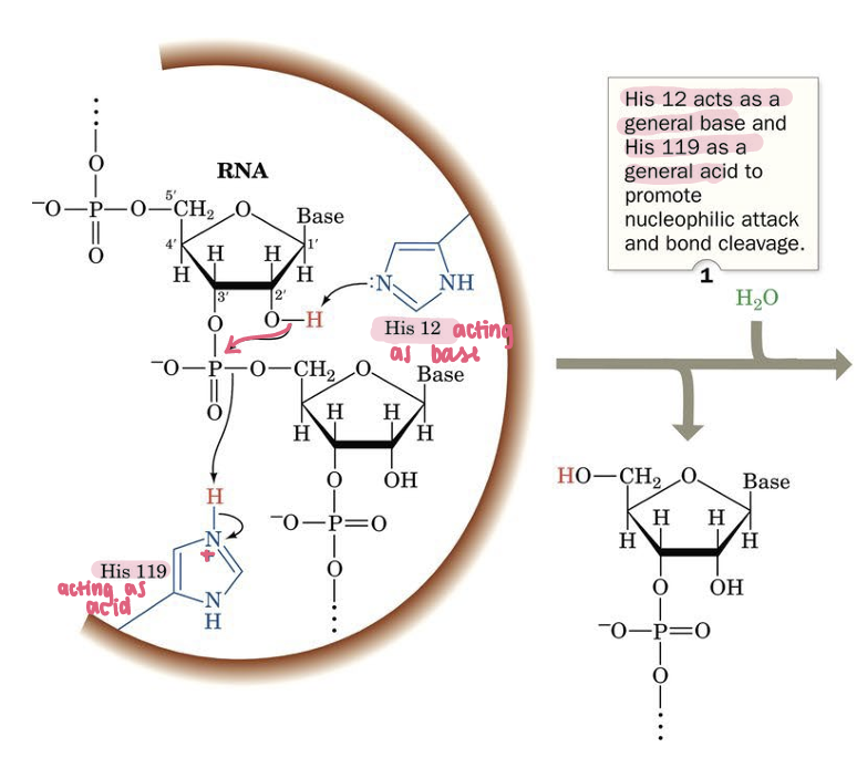
RNase step 2
His 12 acts as a general acid and His 119 as a general base to promote Hydrolysis

Covalent (nucleophilic) catalysis
usually requires a nucleophile
transient formation of covalent bond between E & S
3 stages:
nucleophile reaction between catalyst and substrate to form covalent bond
withdrawal of e- from reaction center by new electrophilic catalyst
elimination of catalyst, a reaction that is essentially reverse of step 1
biologically important nucleophiles
alkoxide (RO-)
Thiolate (RS-)
Neutral 1* amine (RNH2)
Neutral imidazole

Biologically important electrophiles
Protons (H+)
Metal ions
Carbonyl carbon atom
Cationic imine (Schiff base)

Metal ion catalysts contributions
orient structures
redox: transfer electrons
electrostatics: stabilize or shield negative charges
Zn2+ and Carbonic Anhydrase
Zinc orients the OH group for catalysis

How do enzymes facilitate their catalysis reactions
enzymes bring substrates into contact with catalytic groups
enzymes bind their substrates in proper relative orientations for reaction
electrostatic catalysis - distribution of charges around catalytic site lowers free energy of T.S.
enzymes freeze relative translation & rotational motions of substrates
Geometry of SN2 reaction
bimolecular, nucleophilic substitution

Lysozyme
acts on peptidoglycan
breaks B(1→4) bonds between NAM and NAG in cell wall peptidoglycan
can also break B(1→4) in chitin
found in cells/secretions
evolved to ward off gram (+) bacteria
Lysozyme cleavage site
repeating units of (GlcNAc-MurNAc)n

HEW Lysozyme
classic antibacterial enzyme
(NAG)6: hexamer of N-acetylglucosamine acting as an inhibitor, binds to enzyme’s groove without being easily cleaved
enzyme has a long cleft that can accommodate up to 6 residues
Glu35 and Asp52 sit right inside active site cleft — acting as acid-base pair that coordinates cleavage of carb chains
Lysozyme reaction mechanism
acid-based catalysis
Glu acts as an acid and base
covalent catalysis — transition state binding
Lysozyme reaction mechanism step 1
binding with substrate polysaccharide

lysozyme reaction mechanism step 2
general acid base catalysis — protonate glycosidic bond
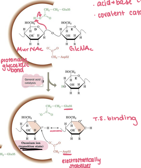
Lysozyme reaction mechanism step 3
covalent catalysis, new E-S covalent bond formation

Lysozyme reaction mechanism step 4
water binding

serine proteases
proteolytic enzyme
peculiarly reactive Ser residue
found in digestive enzymes, blood clotting, inflammatory factors, development
cleave specific for peptide bond
chymotrypsin, trypsin, elastase
chymotrypsin
serine protease cleaving for large bulky hydrophobic groups
Tryptophan, Phenylalanine, Tyrosine
trypsin
serine protease cleaving for basic, large, bulky, positive residues (arginine and lysine)
elastase
serine protease cleaving small hydrophobic residues (alanine, valine, glycine)
catalytic traid
H-bonded Ser, His, Asp residues that participate in catalysis in serine proteases and related enzymes

Specificity pocket of chymotrypsin
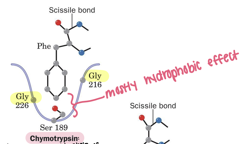
Specificity pocket of trypsin
cationic side chains of trypsin’s preferred residues (Arg, Lys) can therefore form ion pairs with Asp

Specificity pocket of elastase
rapidly hydrolyzes Ala, Gly, and Val-rich protein elastin
binding pocket is largely occluded by side chains of Val and Thr

active site residues in serine proteases are quite different, suggesting-
seems highly improbably they could have evolved from a common ancestor protein
evidence for convergent evolution
Serine proteases Phase 1 — Acylation
Nucleophilic attack & formation of 1st tetrahedral intermediate
His57 acts as a base, deprotonating hydroxyl group of Ser195
Ser195 becomes an alkoxide nucleophile, which attacks the carbonyl C of polypeptide backbone
Breakdown to Acyl-Enzyme Intermediate
His57 acts as a general acid, donating proton back to N atom of peptide bond to be cleaved
peptide bond breaks, splitting protein in half
Release of the First Product (Amine Terminus)
amine product diffuses out of active site
remaining half of substrate covalently attached to Ser195, forming acyl-enzyme intermediate
Serine proteases Phase 2 — Deacylation
Nucleophilic attack by water
His57 acts as a general base, pulling proton off water molecule, making it a hydroxide ion
activated water molecule attacks acyl-enzyme ester bond making 2nd tetrahedral intermediate
His57 acts as a general acid once more, donating a proton to break the bond between Ser195 and remaining substrate
second half of peptide is released
Ser195 is protonated back to its original state, resetting the enzyme so it can catalyze another reaction
TS Stabilization in Serine Proteases
main reason activation barrier is decreased
catalytic efficiency of serine proteases comes from preferential binding of the tetrahedral intermediate (T.S.) over initial ES complex
Before attack, substrate’s carbonyl is sp2 hybridized and flat — sits outside of the oxyanion hole
Once nucleophile (Ser 195) attacks, carbon shifts to sp3 tetrahedral hybridized and a negative charge builds up on oxyanion
geometric change forces oxygen atom to pop into oxyanion hole, forming 2 strong H-bonds with backbone NH of Gly 193 and Ser 195
Why does the barrier drop for chymotrypsin lowering activation energy by binding to TS state rather than ground state?
the 2 extra H-bonds only form when the substrate is forced into high-energy, tetrahedral shape
the enzyme provides a pocket that perfectly matches and rewards the TS’s geometry and negative charge
thus lowers energy required to reach it