OPHTHALMOLOGY PROBLEMS
 Call Kai
 Learn
 Practice Test
 Spaced Repetition
 Match
 Flashcards
 Knowt Play
1/74

There's no tags or description
Looks like no tags are added yet.
75 Terms
what does vision require
light
eyes
brain
which nerve allows us to see
CN II - the optic nerve
‘nerve of sight’
what does the retina do
converts light energy into electrical impulses that travel down the optic nerve to the brain for processing (ultimately to the occipital cortex)

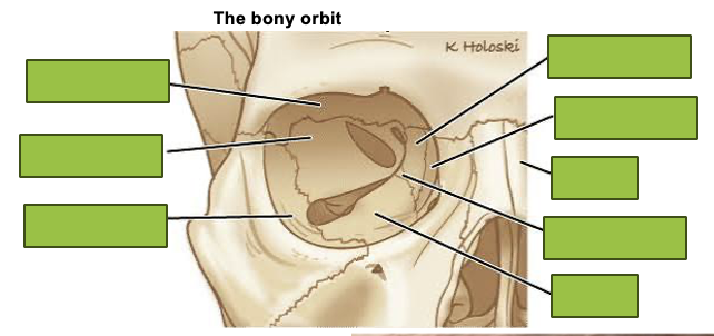


image showing orbital contents
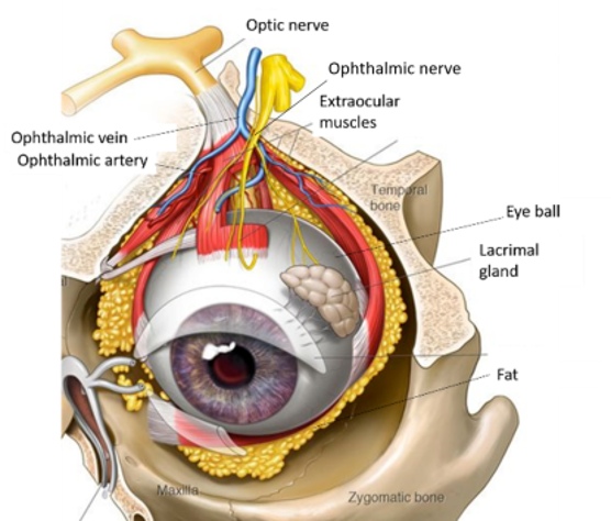
we can see the iris because of the…
cornea - needs to be clear for the iris to be visible (covers iris + pupil)
(CONJUNCTIVA COVERS THE SCLERA)


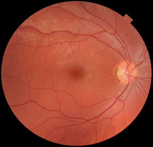
label this image
THIS IS AN IMAGE OF THE RETINA

where are cones concentrated
at the macula

where are rods concentrated
around the periphery of the retina

what structure converges at the optic disc
axons converge at the optic disc
what is the relationship between the retina and the optic nerve
the axons in the retina come together to form the optic nerve
visual disturbance VS visual loss
visual disturbance can be transient
visual loss is not usually transient
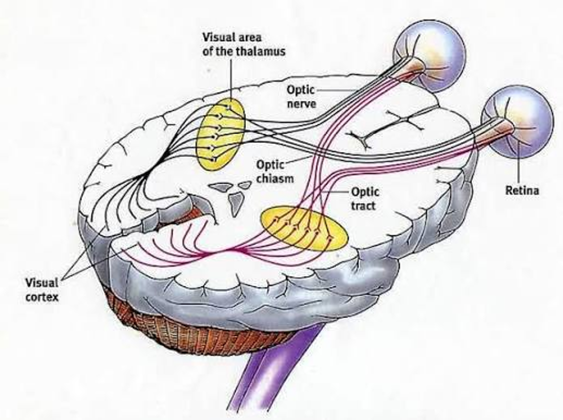
what are causes of visual disturbance
visual disturbance can arise from:
a pathological process affecting the eye
a pathological process affecting brain structures involved in visual processing
how can we narrow down the causes of visual disturbance
how exactly is the vision disturbed?
other features/ signs/ symptoms the patient is experiencing?
give examples of how vision can be disturbed (5)
loss of vision
visual field defect
central vision
peripheral vision
one or both eyes affected
flashing lights
floaters
double vision (diplopia)
what are some other features/ signs/ symptoms of visual disturbance (6)
pain
redness
droopy lid (ptosis)
pupil size inequalities
systemic symptoms e.g. headache
recent surgery
which examples of vision disturbance and other features/ signs/ symptoms of visual disturbance are red flags (6)
visual field defect
central vision
peripheral vision
one or both eyes affected
double vision (diplopia)
—
pain
redness
systemic symptoms e.g. headache
recent surgery
what are ‘eye causes’ of visual disturbance (5)
cataracts
macular degeneration
glaucoma - damage due to raised intraocular pressure
retinal detachment
eye infections
what are ‘brain causes’ of visual disturbance (4)
stroke
n.b. amaurosis fugax
inflammations
migraine
raised intracranial pressure
what are ‘systemic causes’ of visual disturbance (4)
diabetes and diabetic retinopathy
vessel occlusions and atherosclerosis
giant cell arteritis
accelerated hypertension
what are cataracts and what are they caused by
cataracts are an opacification of the lens
typically due to ageing

in which demographic do cataracts occur earlier in
in diabetics and following trauma/ steroid use

what do cataracts result in
a gradual loss of vision

why does diabetic retinopathy cause visual disturbance
chronically high blood glucose levels damages the microvasculature in the eye

what are consequences of diabetic retinopathy
in the eye, retinal vessels can:
become ‘leaky’ » retinal oedema (most significant if macula is affected)
shut down » retinal ischaemia and new blood vessel growth (neovascularisation)
which can then bleed and cause visual loss
worsening diabetic eye disease is associated with…
poor diabetic control
how are diabetics monitored for changes in the eye
the diabetic eye screening service monitors for changes
what are causes of flashes
what are causes of floaters
flashing lights can be produced by mechanical tugging on the retina
floaters can occur from vitreous debris
(both flashes and floaters are very common)
give examples of vitreous debris
embryological remnants
posterior vitreous detachment (PVD)
post inflammation e.g. infection

if someone experiences a ‘sudden shower of floaters’, what is the likely cause of this
likely completed posterior vitreous detachment (PVD)
should check for retinal tear

if someone experiences a ‘floaters and flashing lights’, what is the likely cause of this
likely posterior vitreous detachment (PVD)
should check for retinal tear
if someone experiences a ‘flashing lights followed by a curtain of vision loss’, what is the likely cause of this
suspect retinal detachment
if someone experiences a ‘floaters with darkening of the vision’, what is the likely cause of this
suspect vitreous haemorrhage
what is the cause of visual field defects
a lesion in any part of the visual pathway gives you a particular visual field defect
(therefore you can pinpoint a visual field defect to a particular location in the visual pathway)
diagram showing examples of visual field defects

what is the relationship between the location of the lesion in relation to the optic chiasm and which eye(s) are affected
lesions that happen posterior to the optic chiasm will affect both eyes
lesions that happen anterior to the optic chiasm will affect one eye

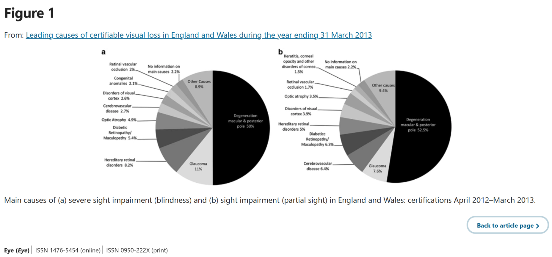
pie chart showing causes of visual loss
state causes of visual loss
macular degeneration
Age-Related Macular Degeneration (AMD)
chronic open angle glaucoma
acute angle closure glaucoma
what are the types of macular degeneration (AMD)
wet AMD
dry AMD

outline wet AMD
new blood vessel growth at the macular area
these blood vessels leak fluid
what is the treatment for wet AMD
intravitreal anti-VEGF injection
VEGF = protein that causes abnormal leaky blood vessels to grow beneath retina
outline dry AMD
deposits called ‘drusen’ are found in the macular region
what is the treatment for dry AMD
there is no treatment for dry AMD
what is chronic open angle glaucoma
a group of conditions that lead to progressive retinal nerve fibre layer thinning and optic disc changes

what is the largest modifiable risk factor for chronic open angle glaucoma
largest modifiable risk factor is intraocular pressure (IOP)
why does chronic open angle glaucoma cause vision loss
there is decreased outflow at the trabecular meshwork
trabecular meshwork is the primary outflow pathway for aqueous humour (eye drainage)
how do medications for chronic open angle glaucoma work
medications can:
increase alternate outflow
decrease aqueous production

what is a normal patient history for someone who has acute angle closure glaucoma
prolonged time spent in dim lighting
positions
laying prone and going from sitting » supine increases IOP
emotional stress
pharmacological
why is darkness a trigger for acute angle closure glaucoma
because darkness causes the pupil to dilate
this can block drainage of aqueous humour
what are signs of acute angle closure glaucoma
decreased visual acuity
IOP 40+
ciliary injection
mid-dilated, non-reactive pupil
synechiae (when the iris sticks to other structures in the eye)

what are the types of management for acute angle closure glaucoma
medical
surgical
what are some conditions that can cause a red eye and which respective structure is affected (8)
pre-septal cellulitis, orbital cellulitis, blepharitis (eyelids)
keratitis, ulcers (cornea)
bacterial/ viral/ allergy/ neoplasia (conjunctiva)
episcleritis (episclera)
scleritis (sclera)
uveitis (uveal tract)
angle closure (angle)
endophthalmitis (vitreous)
outline pre-septal cellulitis
associated with skin commensal bacteria e.g. S.aureus, S.pyogenes
e.g. lid abrasions, insect bites, hordeola (stye), dacryocystitis
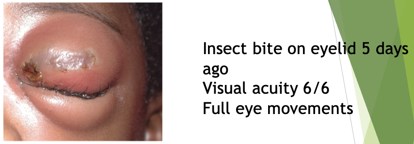
what is the Tx for pre-septal cellulitis
oral antibiotics e.g. co-amoxiclav
outline orbital cellulitis
associated with paranasal sinus infections by S.pneumoniae and Haemophilus
e.g. dacryocystitis, tooth infections
sight and life threatening

what can orbital cellulitis be complicated by
can be complicated by:
abscesses
cavernous sinus thrombosis
what is the Tx for orbital cellulitis
hospital admission
IV ceftazidime/ metronidazole
monitor visual acuity (VA), RAPD, colour vision
what are other causes of a red eye
sub-conjunctival haemorrhage
conjunctivitis
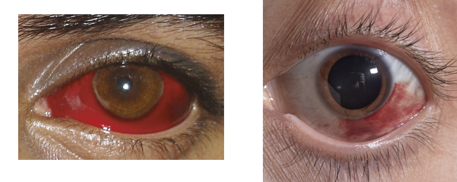
outline sub-conjunctival haemorrhage (causes and recovery period)
typically idiopathic
can result from Valsalva maneuovre (vomiting, coughing, sneezing, straining)
and trauma/ medications/ high BP
resolves in 1-2 weeks without treatment
if sub-conjunctival haemorrhage is found in children, what should be considered
non-accidental injury (NAI)
what are symptoms of conjunctivitis
discharge - watery, mucoid, purulent
chemosis - swelling of conjunctiva
lymphadenopathy
sexual history
(can be viral, bacterial, allergy)
what are symptoms of bacterial conjunctivitis (4)
injection
purulent discharge
lashes stuck together in the mornings
gritty sensation
what is the Tx for bacterial conjuncitivitis
chloramphenicol eye drops/ ointment
what are symptoms of viral conjunctivitis (6)
injection
water discharge
gritty discharge
one eye is affected first then the other
may be associated with prior URTI
lid swelling
what are symptoms of allergic conjuncitivitis (3)
chemosis
itching
mucoid discharge
seasonal, also associated with asthma and eczema
what is the Tx for allergic conjunctivitis
cool compresses
cromoglycate eyedrops
antihistamines
what are symptoms of STI associated conjunctivitis
gonorrhoea
rapidly progressive
mucoid discharge
corneal perforation
chlamydia
watery discharge
how else can conjunctivitis be contracted
neonatal conjunctivitis (from mother)
what is uveitis
an inflammatory process within the front of the eye (the uvea)
incl. iris, ciliary body, choroid
vision can be decreased
what are symptoms of uveitis
pain
photophobia

what is the Tx for uveitis
referral to eye doctor
steroid eyedrops (autoimmune so antibiotics will not be effective)
what other conditions can uveitis be linked to (2)
Crohn’s disease
rheumatoid arthritis
referral pathways for ophthalmic problems
