Vascular Anomalies + HHTs
 Call Kai
 Learn
 Practice Test
 Spaced Repetition
 Match
 Flashcards
 Knowt Play
1/21

There's no tags or description
Looks like no tags are added yet.
22 Terms
Vascular anomaly presence
characterized by endothelial proliferation. Vascular malformations are structural errors with no proliferation, and they don’t regress.
Most of these conditions are caused by somatic (mosaic) variants, with a few germline exceptions. Somatic cases are usually sporadic with low recurrence risk, and testing often needs affected tissue because blood can be negative.
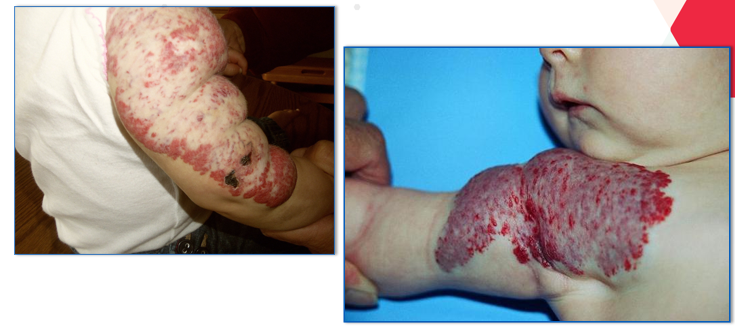
Infantile Hemangiomas
absent at birth but then proliferates
GLUT-1 positive
Large segmental IH should prompt PHACES workup
GLUT1 Role in Proliferation of Hemangiomas
Diagnostic immunohistochemical marker for infantile hemangiomas
PHACES
Posterior fossa anomalies,
Hemangioma, Arterial anomalies,
Cardiac defects,
Eye anomalies,
Sternal defects/Supraumbilical raphe.
It is diagnosed by a large facial/head hemangioma plus major or minor criteria.
Congenital hemangiomas (RICH/NICH)
fully formed at birth and GLUT-1 negative.
They are caused by somatic GNAQ/GNA11 variants.
Port wine stain
usually sporadic.
A PWS in the V1 (ophthalmic) distribution raises concern for Sturge-Weber.


Sturge-Weber
facial capillary malformation
leptomeningeal malformation
refractory seizures
glaucoma/buphthalmos
developmental delay
It is caused by a somatic GNAQ variant

Capillary Malformation-Arteriovenous Malformation (CM-AVM)
multifocal small CMs with fast-flow lesions.
AD caused by RASA1 (CM-AVM1) or EPHB4 (CM-AVM2).
Consider it in patients with multifocal CMs, and screen for fast-flow CNS lesions.
MCAP (macrocephaly-capillary malformation)
macrocephaly
multiple CMs
brain anomalies such as Chiari-like findings
It is caused by somatic PIK3CA and is part of PIK3CA-Related Overgrowth Spectrum.
Venous malformations
Most common vascular malformation
Risk of localized intravascular coagulopathy (elevated D-dimer, phleboliths)
Usually found on the head, neck, extremities, trunk
They are caused by somatic TEK (TIE2) or PIK3CA variants.
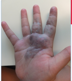
Blue rubber bleb nevus syndrome:
multiple bluish nodules
often with GI involvement
Caused by somatic TEK.
Typically with a double variant in cis.

Glomuvenous malformation
Autosomal dominant, caused by GLMN loss of function.
It is hyperkeratotic, cobblestone-like, and painful
It has reduced penetrance and variable expressivity, explained by a second somatic hit (acquired uniparental disomy of 1p).

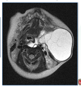
Lymphatic malformations
microcystic, macrocystic, or combined, and often associated with overgrowth syndromes.
PROS (PIK3CA-Related Overgrowth Spectrum)
A spectrum of segmental overgrowth conditions that includes CLOVES, MCAP, KTS, and macrodactyly
It is caused by somatic PIK3CA variants.
Targeted therapy (alpelisib, sirolimus) is an option.
CLOVES
Congenital
Lipomatous
Overgrowth
Vascular malformations
Epidermal nevi
Skeletal anomalies.
AKT1/PIK3CA associated
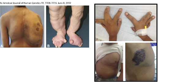
Klippel-Trenaunay
Triad of:
port wine stain
Varicosities (with a persistent lateral marginal vein)
Limb overgrowth
It also falls under PROS.

Proteus syndrome
Progressive
asymmetric overgrowth that is minimal at birth
It is caused by somatic AKT1 c.49G>A (p.Glu17Lys)
It carries a DVT risk and is distinct from PROS.
PTEN Hamartoma Tumor Syndrome (Cowden, Bannayan-Riley-Ruvalcaba):
Autosomal dominant germline PTEN loss of function.
Features include macrocephaly, trichilemmomas, lipomas, penile freckling, GI polyps, and vascular anomalies.
It carries increased risk of breast, thyroid, and endometrial cancers (also colorectal, renal, and melanoma).
AD, PTEN GL variants
Hereditary hemorrhagic telangiectasia (HHT)
Autosomal dominant, about 1 in 5,000, with age-related penetrance.
Diagnosed by the Curaçao criteria:
recurrent epistaxis
mucocutaneous telangiectasias
visceral (inside organs) arteriovenous malformation (AVMs)
first-degree relative w/ HHTs
Definite requires 3 or more criteria.
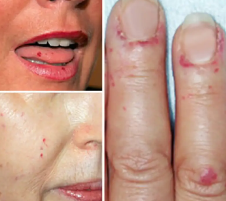
HHT Molecular Testing Consideration
Curacao criteria is age dependent, so kids young enough might not manifest symptoms yet
Targeted familial variant testing in families with an identified variant can be valuable for inclusion and exclusion of screening.
HHT Genes
ENG (HHT1, pulmonary and cerebral AVMs) 44%
ACVRL1 (HHT2, liver AVMs) 52%
SMAD4 (HHT with juvenile polyposis, increased risk for colorectal cancer) 1%
GDF2/BMP9 (HHT-like phenotype) very rare
HHT Treatments and Complications
Pulmonary AVMs can lead to stroke, hypoxemia.
Pregnancy AVMs may lead to rupture, screening prior to conception needed
Hepatic AVMs cause high output heart failure, secondary pulmonary hypertension
Cerebral AVMs: seizures or hemorrhage