Anatomy 6511 Histology Pictures Exam III 낱말 카드
 Call Kai
 Learn
 Practice Test
 Spaced Repetition
 Match
 Flashcards
 Knowt Play
1/251

There's no tags or description
Looks like no tags are added yet.
252 Terms
Artery
Would this be an artery or vein?

Vein
Would this be an artery or vein?
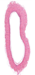
Vaso vasorum
What structure are the venule and arteriole part of in the adventitia of this large vessel?
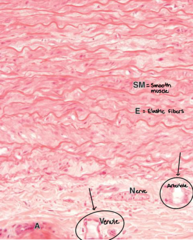
Nervi vascularis
What is the nerve in the adventitia of this large vessel called?

Artery (more open)
What is labeled 1?

Vein (more collapsed, lumen larger)
What is labeled 2?

Elastic
What type of artery is shown?

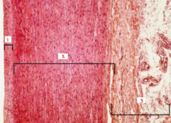
Tunica externa
What layer of the elastic artery is labeled 1?
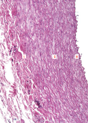
Tunica media
What layer of the elastic artery is labeled 2?

Tunica intima
What layer of the elastic artery is labeled 3?
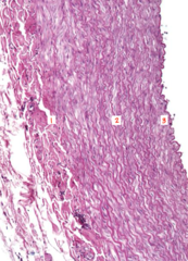
Elastic
What type of artery is shown?

Tunica intima
What layer of the elastic artery is labeled 1?

Tunica media
What layer of the elastic artery is labeled 2?

Tunica externa
What layer of the elastic artery is labeled 3?
Vaso vasorum
What structure are the circled venules and arterioles part of?

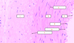
Muscular
What artery type is shown?

Endothelial
What cell type is labeled 1 on the muscular artery?

Internal elastic lamina
What is labeled 2 on the muscular artery?
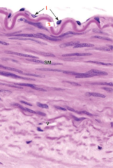
Adventitia
What layer is labeled 1 on the muscular artery?

Smooth muscle
What cell is labeled 2 on the muscular artery?

Media
What layer is labeled 3 on the muscular artery?

Intima
What layer is labeled 4 on the muscular artery?

Endothelial
What cell is labeled 5 on the muscular artery?
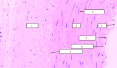
Internal elastic lamina
What is labeled 6 on the muscular artery?
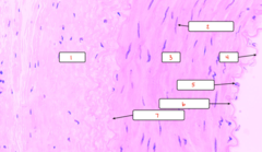
External elastic lamina
What is labeled 7 on the muscular artery?
Arteriole
What artery type is shown?

Arteriole
What artery type is shown?

Intima
What layer of the arteriole is labeled 1?

Media
What layer of the arteriole is labeled 2?

Adventitia
What layer of the arteriole is labeled 3?

Arteriole
What type of artery is shown?
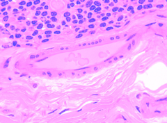
Arteriole
What type of artery is shown?
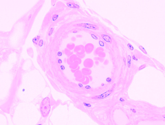
Vein
What is labeled 1?

Artery
What is labeled 2?

Capillary
What is labeled 3?

Pericytes
What cell type is being pointed to in the capillary?

Pericytes (periphery of capillary)
What cell type is pictured?

Continuous
What type of capillary is pictured?

Fenestrated
What type of capillary is pictured?

Sinusoidal
What type of capillary is pictured?

Artery
What is labeled A?

Venule
What is labeled B?

Venule
What is labeled 1?

Arteriole
What is labeled 2?

Pericyte
What cell type is labeled 3?

Venule
What is labeled 1?

Arteriole
What is labeled 2?

Medium
What type of vein is shown?

Medium
What type of vein is shown?

Medium
What type of vein is shown?

Adventitia
What layer of the medium vein is labeled 1?
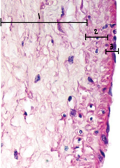
Media
What layer of the medium vein is labeled 2?

Intima
What layer of the medium vein is labeled 3?

Large
What type of vein is shown?
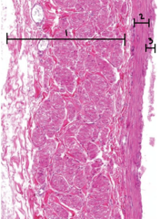
Adventitia
What layer of the large vein is labeled 1?

Media
What layer of the large vein is labeled 2?

Intima
What layer of the large vein is labeled 3?

Atherosclerosis
What condition is shown in this image?

Adult
Is this cross section of the thymus from an adult or child?

Child
Is this cross section of the thymus from an adult or child?

Cortex
What are the arrows pointing to in the thymus (stained darker)?

Medulla
What are the arrows pointing to in the thymus (stained lighter)?

Capsule
What are the arrows pointing to in the thymus?
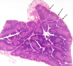
Septa
What are the arrows pointing to in the thymus?

Thymic epithelial cells
What cell type are the arrows pointing to in the cortex of the thymus?

Lymphoblasts
What cell type are the arrows pointing to in the cortex of the thymus?

Thymic (Hassall's) Corpuscles
What structures are the arrows pointing to?

Thymic (Hassall's) Corpuscles
What part of the thymus is shown in the image?

Thymic (Hassall's) Corpuscles
What part of the thymus is shown in the image?

Capsule
What part of the spleen is labeled 1?

Trabeculae
What part of the spleen is labeled 2?

Splenic nodules
What part of the spleen is labeled 3?

Trabecular vein
What part of the spleen is labeled 4?

Red pulp
What part of the spleen is labeled 5?
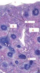
Capsule
What is labeled 1 in the stroma of the spleen?

Trabeculae
What is labeled 2 in the stroma of the spleen?

Trabecular blood vessels
What is labeled 3 in the stroma of the spleen?

White pulp
What is labeled 1 in the spleen?

Red pulp
What is labeled 2 in the spleen?

Trabeculae
What is labeled 3 in the spleen?

Capsule
What is labeled 4 in the spleen?

Red pulp
Is red or white pulp of the spleen labelled?

White pulp
Is red or white pulp of the spleen labelled?

Trabecular veins
What in the spleen is labeled 1?

Germinal center
What in the spleen is labeled 2?

Red pulp
What part of the spleen is labeled 1?

White pulp
What part of the spleen is labeled 2?
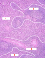
Germinal center
What is arrow 1 pointing to in the spleen?

Central arteriole
What is arrow 2 pointing to in the spleen?
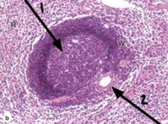
B cells (germinal center)
What cell type is produced at this site?

T cell maturation (thymus)
What is the primary function of this organ?

White pulp
What are the darker stained regions called?
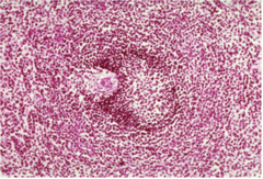
Reticular network
What is prominently shown in this staining of the spleen?

Macrophages
What cell type is shown surrounded by RBC (arrow is pointing to RBC)?

Diffuse lymphatic tissue
In this image of the GI tract, the lymphocytes clustered in the middle is an example of what?

Germinal center
What is the arrow pointing to in the spleen?
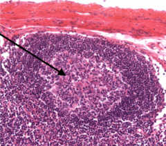
MALT in tonsils
What is shown in this image?

Tonsil
What tissue is this?

Peyers patches in intestine
What is shown in this image?

MALT in appendix
What is shown in this image?
