Eicosanoid Biosynthesis
 Call Kai
 Learn
Learn Practice Test
 Spaced Repetition
 Match
Match Flashcards
 Knowt Play
1/20

There's no tags or description
Looks like no tags are added yet.
Name | Mastery | Learn | Test | Matching | Spaced | Call with Kai | Chat |
|---|
No analytics yet
Send a link to your students to track their progress
21 Terms


Eicosanoids are signaling molecules derived from 20 carbon, poly-unsaturated fatty acids, primarily arachidonic acid.
These include prostaglandins, thromboxanes, Leukotrienes,
One example prostacyclin (PGI2), which is a very vasodilator and inhibits platelet aggregation.
These molecules add locally and are not stored but synthesized on demand from arachidonate acid released from membrane phospholipid.


eicosanoid names
eicosanoids are named based on origin and structure.
Prostaglandins (PG) was originally found in prostate tissue
Thromboxanes (TX) found first in platelets
Leukotrienes originally found in leukocytes.
-the number following their names in indicates the number of double bonds in each molecule, thus understanding this naming convention system helps in identifying their biological functions and their pathways.
eicosanoid sites of synthesis
eicosanoids are synthesized from polyunsaturated fatty acids as we mentioned (arachidonic acid) (arachidonic acid itself comes from linoleic acid).
eicosanoids are produced rapidly in demand in various tissues, including: endothelial cells, leukocytes, platelets and kidneys.
unlike neurotransmitters or hormones, these are not stored in granules, which allows for quick, localized utilization and response.


Eicosanoid Functions
• Important roles in
• inflammation & fever
• blood clotting
• blood pressure regulation
• sleep/wake cycle
• tissue growth
• reproductive processes
• Prostaglandins serve as a prototype
• To have function, bind to receptors
• G-protein coupled receptor (GPCRs)
• Peroxisome proliferator-activated receptor (PPARs)
eicosanoids mediate both immediate and long term responses from these receptors.
Release of arachidonic acid from precursors
• Phosphatidyl inositol is cleaved by phospholipase A₂ to yield arachidonic acid
This first step is crucial because it provides the primary substrate for the downstream eicosanoid synthesis.
The enzyme activation is tightly regulated and is often treated by external stimuli like cytokines from the immune system.
(Text inside the diagram):
Site of cleavage by Phospholipase A₂
Site of cleavage by Phospholipase C
Phosphatidyl inositol


in this slide, we will continue discussing the mechanism of arachidonic acid release.
R2 refers to the fatty acid side of the chain on membrane phospholipids.
The action of phospholipase A2 on the SN2 position liberates arachidonic acid.
Release of arachidonic acid from PIP₂
• PIP₂ is cleaved by phospholipase C to yield DAG (di-acyl-glycerol) as well as inositol triphosphate (IP3).
While IP3 triggers calcium release from the endoplasmic reticulum, DAGs role is to remain in the membrane and is a precursor to arachidonic acid via additional enzymatic steps that we will be discussing.
(Text inside the diagram):
cleavage by Phospholipase C
PIP₂ phosphatidylinositol-4,5-bisphosphate


DAG (di-acyl-glycerol) is further processed by DAG lipase and monoacyl glycerol lipase, which are in charge of liberating arachidonic acid.
This two step process is another route for generating arachidonic acid for biosynthesis of eicosanoids
Activation of phospholipase A₂
• Ligand binding to GPCR leading to PLC activation
• PLC cleaves PIP₂ → DAG + IP₃, opens ER Ca²⁺ channels
• Ca²⁺ activates PLA₂ which is translocated to the plasma membrane and where it cleaves arachidonic acid for eicosanoid biosynthesis


Corticosteroid action
• Corticosteroids mediate anti-inflammatory effects by blocking action of phospholipase A₂ (can’t produce arachidonate) (and in turn will also stop the production of eicosanoids).
This explains their potent anti-inflammatory effects in clinical settings and the clinical examples of asthma and autoimmune diseases.
(Text inside the diagram):
Phospholipid
Corticosteroid inhibits this step
Arachidonic Acid
to continue discussing the reactions that are taking place,
once arachidonic acid is free, it can follow TWO major metabolic pathways: 1. cyclic pathway (enzyme cyclooxygenase or COX) 2. linear pathway (enzyme lipooxygenase or LOX).
each of these pathways lead to distinct classes of eicosanoids that are produced with different physiological roles.
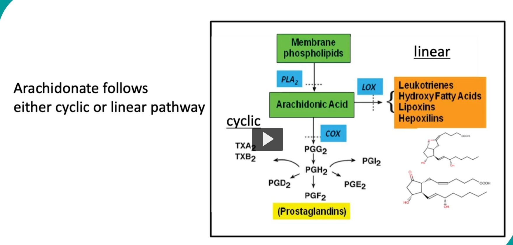

Cyclic/cyclooxygenase (COX) pathway
first, we will be discussing the cyclic pathway.
• The first and committed enzyme (conversion of Arachidonic Acid to prostaglandin H2, PGH₂) is mediated by prostaglandin H₂ synthase
• PGH₂ synthase is a heme-containing oxygenase that mediates two reactions:
it is important to remember that PGH2 synthase is the committed step in prostaglandin and thromboxane biosynthesis.
Two active sites in PGH₂ synthase
• cyclooxygenase (cyclooxygenase is the first active site) converts AA to PGG₂
• Peroxidase (peroxidase is the second active site) converts PGG₂ to PGH₂
each step is essential for proper biosynthesis.


In this slide we can look at the first part of PGH2 synthase action.
a tyrosine residue in the COX site will form a radical that initiates the oxygenation of arachidonic acid, converting it into the hyperperoxy intermediate, PGG2.
then the peroxidase domain will use heme group and glutathione to reduce PGG2 into PGH2.
the PGH2 will then serve as the branching point to produce the multiple biologically active prostanoids.
PGH₂ Synthase mechanism - 1
PGH2 synthase is the universal precursor for prostaglandins and thromboxanes.
so we will be looking at two main mechanisms of the PGH2 synthase in detail.
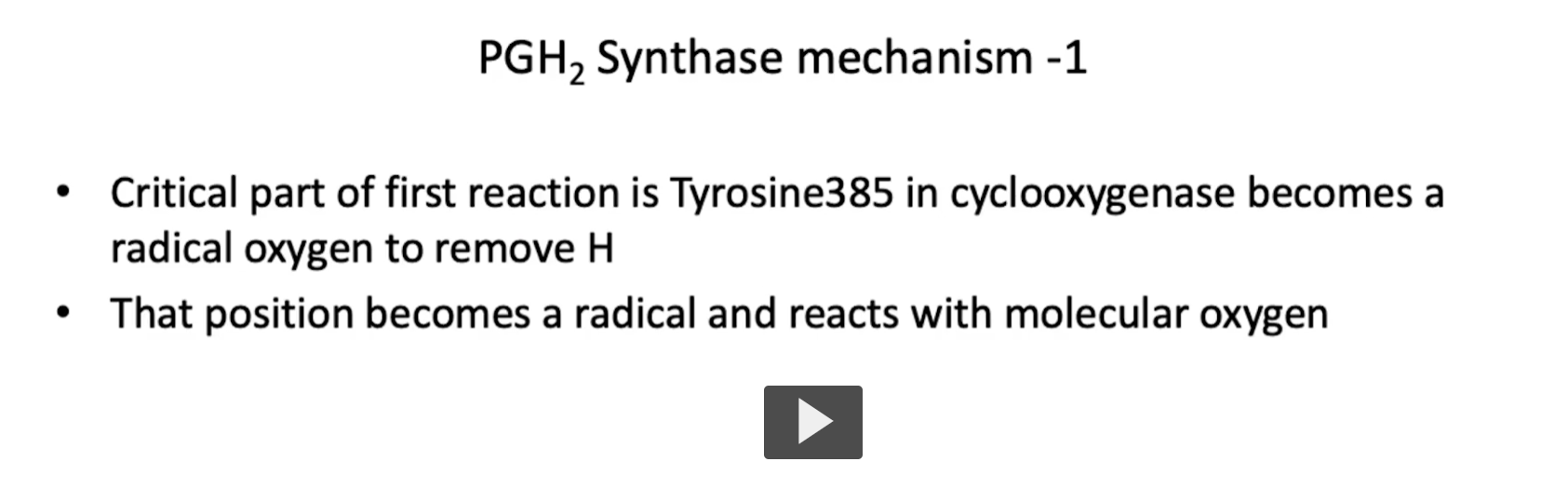
• First, the Critical part of first reaction is Tyrosine385 in cyclooxygenase becomes a radical oxygen to remove H
• That position becomes a radical and reacts with molecular oxygen

PGH₂ Synthase mechanism - 2
Second:
• Peroxidase contains a heme prosthetic group, the iron is oxidized
• 2 molecules of glutathione reduce the heme yielding H₂O and PGH₂
• PGH₂ is the precursor to all series 2 PGs (prostaglandins) and TX (thromboxane’s).
Th PGH2 Synthase mechanism concludes with water and PGH2 as the final products.
It is important to remember that PGH2 acts as the precursor for other series 2 prostaglandins and thromboxanes as well.


more clinically, we can look at the role of NSAIDs, and their role in inhibiting PGH2 synthase specifically.
• Non-steroidal anti-inflammatory drugs (NSAIDS) (like ASPIRIN) work by preventing formation of PGG₂ (and thus PGH₂)
• Aspirin irreversibly acetylates a Serine to inactivate COX
• Ibuprofen (and related NSAIDS) blocks entry of arachidonate into active site (and thus preventing prostenoid synthesis).
Ibuprofen also acts reversibly.
(Text on left diagram):
Aspirin (acetylsalicylic acid)
Endoperoxide synthase (active)
↓
Salicylic acid
Endoperoxide synthase (inactive)
Synthesis of PGH₂ derivatives
once PGH2 is formed from these reactions, each tissue specific enzyme will generate final products.
• Specific enzymes dictate the derivative produced, some tissue specificity
• In platelets, thromboxane synthase → TXA₂ (thromboxane A2) and TXB₂ (thromboxane B2)
• In endothelial cells (blood vessels), prostacyclin synthase yields PGI₂
This explains the opposite actions of these molecules, while some work on platelet aggregation, others work on platelet inhibition.


now we will look at the linear pathway. the linear pathway starts with the action of lipoxygenase enzymes on arachidonic acid. These enzymes will convert it to hydroperoxy-eicosatetraenoic acid (HPETE)
HPETEs are precursors for leukotrienes and other linear eicosanoids.
The Linear Pathway
• Catalysis starts with conversion of AA to one of several hydroperoxy-eicosatetraenoic (HPETE) acid forms
• Reaction catalyzed by lipoxygenase
– Mammals have different lipoxygenases named for double bond they oxygenate (5, 12, or 15-)
In this image, we can look at a downstream effect of the linear pathway acting on arachidonic acid.
The downstream products of the HPETE include lipoxins and hepoxylins, these compounds are involved inflammation regulation.


Additional steps produce derivatives
-additional steps in this cascade will produce other derivatives.
• Lipoxins and hepoxilins are unique pathways
• All leukotrienes come from 5-HPETE
– 5-Lipoxygenase (5-LOX) requires special membrane protein, FLAP (5-lipoxygenase-activating protein)
• Clinically, Antiasthma drugs work by binding FLAP, directly inhibiting 5-LOX, or by blocking leukotriene receptors directly.
pathway of peptidoleukotrienes
in this image, we can further observe the pathway for peptoleukotrienes.


FLAP and 5-lipoxygenase (5-LOX)
• Production of 5-HPETE leads to inflammatory leukotrienes (from leukocytes)
• FLAP is a protein bound to the nuclear membrane and activates 5-lipoxygenase
• Mechanism of activation is unclear but requires binding of AA and contact to 5-LOX
enzymes are all present at nuclear membrane
all enzymes required for the synthesis of leukotrienes are located on or near the nuclear membrane.
This facilitates the coordinated production of inflammatory mediators at the sites of immune activation.


Peptidoleukotriene biosynthesis
• Peptidoleukotrienes are short lived
• They are potent mediators of inflammation, hypersensitivity, vasodilation, and respiratory disorders
• Derived from leukotriene A4
• Requires glutathione (cysteine donor) and glutathione-S-transferase, which adds glutamic acid with γ-glutamyl transferase
It is important to remember that peptidoleukotrienes such as LTC4, LTD4, and LTE4 are all derived from LTA4, these compounds are short lived, but extremely important in promoting inflammation, hypersensitivity and bronchoconstriction. Their synthesis requires glutathione, gamma glutamyl transferase and glutathione-S-transferase.
Now, let’s walk through the initial synthesis of Peptidoleukotriene
starting with the molecule 5-HPETE
5-HPETE is converted into LTA4 (leukotriene A4) by the enzyme 5-lipoxygenase (5-LOX)
From here, LTA4 has two main fates. 1. If acted upon the enzyme LTA4 hydrolase, it becomes LTB4, which has potent chemotactic properties. 2. however, the enzyme glutathione-S-transferase conjugates glutathione to LTA4, forming LTC4.


Once LTC4 is released, it is sequentially processed into LTD4 and LTE4. This will occur via enzymatic cleavage of peptide bonds in glutathione molecule.
First the enzyme gamma-glutamyl transferase will remove the glutamic acid portion from LTC4, producing LTD4.
and next, the enzyme dipeptidase will cleave off the glycine residue from LTD4, yielding LTE4 below
It is important to remember that both LTD4 and LTE4 are biologically active molecules, and together, with LTC4 above, they are called the cystenyl leukotrienes

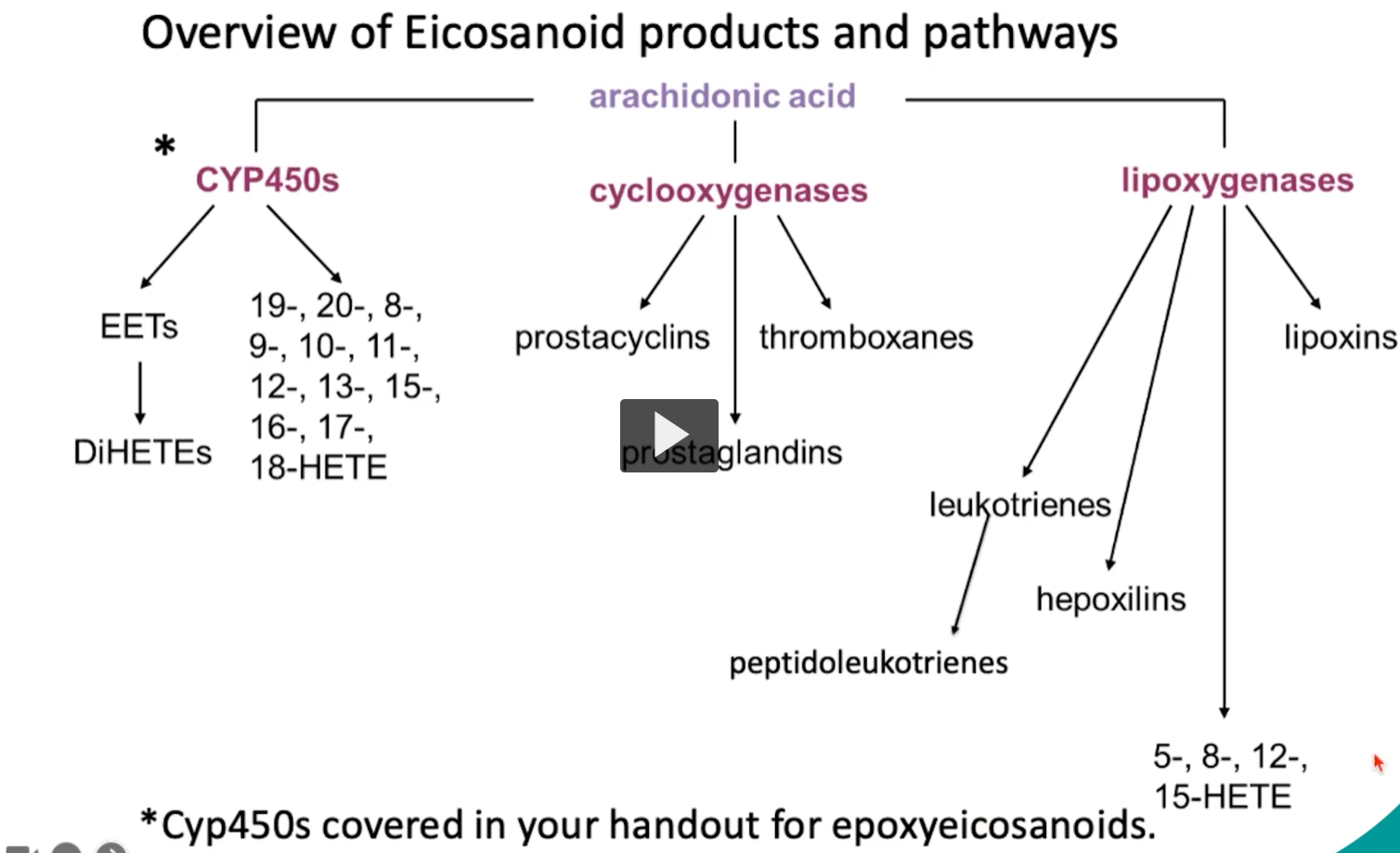
Here we have the overview slide of the major enzymes involved in eicosanoid synthesis synthesis.
Students should recall the enzyme phospholipase A2, which releases arachidonic acid.
the COX enzymes, producing prostaglandins
the 5-lipoxygenase enzymes
and the FLAP making leukotrienes
and glutathione-S-transferase, to help form the peptidoleukotrienes
Which enzyme catalyzes the committed step in prostaglandin biosynthesis?
Please choose from the list below. More than one may be correct.
A. phopholipase A2
B. prostaglandin H2 synthase
C. 5-lipoxygenase
D. 5-lipoxygenase activating protein
E. glutathione-S-transferase

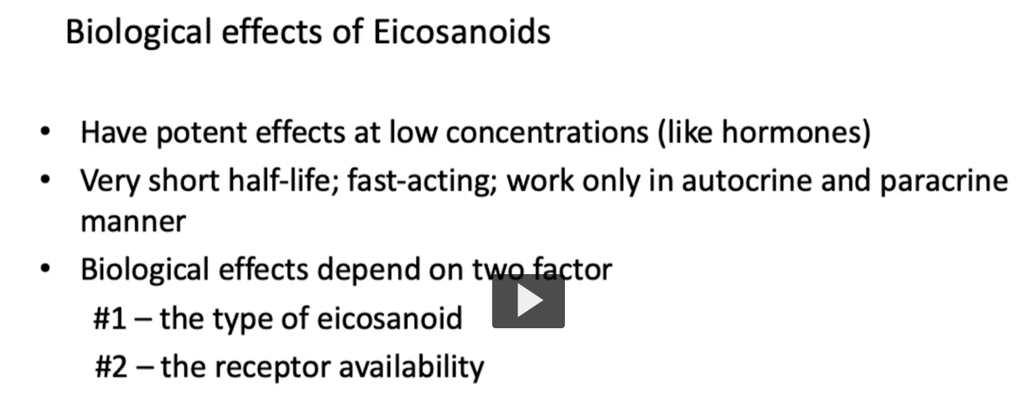
biological effects of eicosanoids
eicosanoids act like local hormones, but are either paracrine or autocrine in nature, meaning that they act at low concentrations and are quickly degraded.
Biological effects of Eicosanoids
• Have potent effects at low concentrations (like hormones)
• Very short half-life; fast-acting; work only in autocrine and paracrine manner
• Biological effects depend on two factor
#1 – the type of eicosanoid (which eicosanoid is produced)
#2 – the receptor availability (which receptors are present in the target tissue)
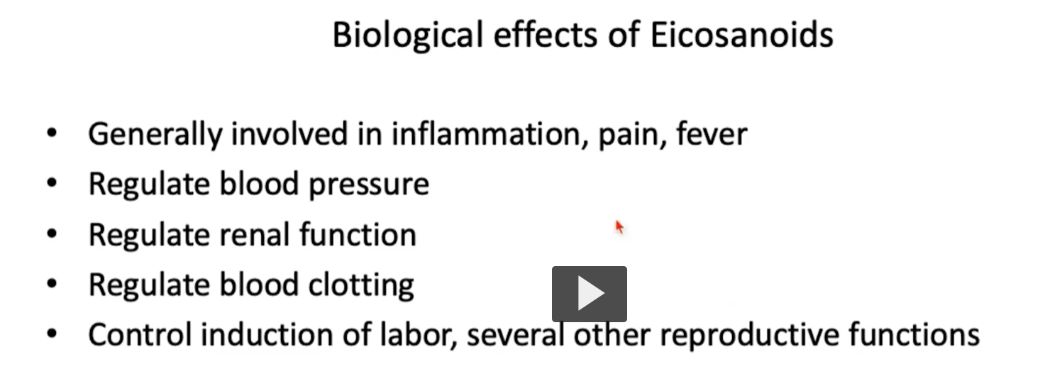
Biological effects of Eicosanoids (overall)
• Generally involved in inflammation, pain, fever
• Regulate blood pressure
• Regulate renal function
• Regulate blood clotting
• Control induction of labor, several other reproductive functions

Prostaglandins (PG)
• Characterized structurally by cyclopentane ring
• Mediate inflammation:
– vasodilation → redness & heat (PGE₁, PGE₂, PGD₂, PGI₂)
– increase vascular permeability → swelling (PGE₂, PGD₂, PGI₂)
Prostaglandins (PG)
• Regulate pain and fever (PGE₂ specifically)
• Regulate blood pressure: vasodilator prostaglandins PGA and PGI₂ lower systemic arterial pressure
• Regulate platelet aggregation: PGI₂ = potent inhibitor
• (kidney) PGE₂ inhibits reabsorption of Na⁺ and water in the collecting duct.
• PGI₂ works through vasodilation, regulation of glomerular filtration rate
• PGE₂, PGF₂ work in reproductive system to stimulate uterine muscle contractions during labor


thromboxane molecules
Thromboxanes (TX)
• Characterized structurally by 6-member, oxygen-containing ring (yellow)
• Synthesis by platelets
• Stimulate vasoconstriction and platelet aggregation
• TXA₂ is also produced in the kidney → causes vasoconstriction and mediates the response to angiotensin 2
Leukotrienes (LT)
• Characterized structurally by three conjugated double bonds, plus one unconjugated double bond (yellow)
• Very potent bronchoconstrictors (LTC₄, LTD₄, LTE₄)
• Increase vascular permeability
• Attraction and activation of leukocytes (LTB₄)

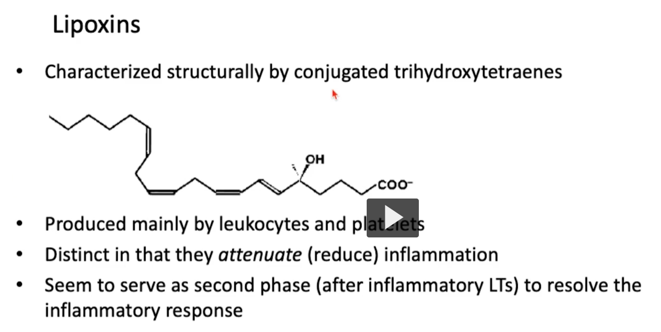
Lipoxins
• Characterized structurally by conjugated trihydroxytetraenes (picture here)
• Produced mainly by both leukocytes and platelets
• Distinct from all the previous molecules we discussed because these help attenuate (reduce) inflammation
• Seem to serve as second phase (after inflammatory LTs) to resolve the inflammatory response
Which compound is a source of arachidonic acid?
Please choose from the list below. More than one may be correct.
A. oxaloacetate
B. inositol phosphate
C. cholesterol
D. phosphatidyl choline
Phosphatidyl choline is a membrane phospholipid and source of arachidonic acid, when cleaved by phospholipase A2,
