The Skeletal System (Bones/Bone Tissue): Bone Structure and Function
 Call Kai
 Learn
 Practice Test
 Spaced Repetition
 Match
 Flashcards
 Knowt Play
1/64

There's no tags or description
Looks like no tags are added yet.
65 Terms
What are the components of the skeletal system?
-Bones of the skeleton
-Cartilage
-Ligaments
-Other connective tissues
What are the functions of the skeletal system? (4)
-Support and protection: serve as framework for entire body and protect delicate tissues from injury and trauma
-Movement: serve as attachment sites for skeletal muscles, other soft tissues, and some organs; acts as a system of levers
-Hemopoiesis: process of blood cell production occurring in red bone marrow
-Storage of mineral and energy reserves: calcium and phosphate are stored within and released from bone
Compact bone (cortical bone)
-Relatively dense connective bone tissue
-Appears white, smooth, and solid
-80% of bone mass

Spongy bone (cancellous or trabecular bone)
-Located internal to compact bone
-Appears porous
-20% of bone mass

1. What is the difference between compact bone and spongy bone?
Compact bone consists of a solid, dense mass of bone tissue, whereas spongy bone contains a more porous arrangement of the tissue.

Ligaments
Dense regular connective tissue that anchors bone to bone

Tendons
Dense regular connective tissue that connects muscle to bone

Long bones
-Greater in length than width
-Elongated, cylindrical shaft (diaphysis)
-Most common bone shape
-Found in upper and lower limbs (arm, forearm, thigh, leg, toes)
-Vary in size

Short bones
-Length nearly equal to width
-Carpal bones, sesamoid bones (bones along tendons of muscles), and patella (largest sesamoid bone)

Flat bones
-Flat, thin surfaces, may be slightly curved
-Provide surfaces for muscle attachment
-Protect underlying soft tissues
-Form roof of skull, scapulae, sternum, and ribs

Irregular bones
-Elaborate shapes
-Vertebrae, ossa coxae (hip bones), and several skull bones (ethmoid, sphenoid)

Diaphysis
-Elongated, usually cylindrical shaft
-Provides leverage and major weight support
-Compact bone with thin spicules of spongy bone extending inward

Medullary cavity
-Hollow, cylindrical space within diaphysis
-Contains red bone marrow in children
-Contains yellow bone marrow in adults

Epiphysis
-Knobby region at the ends of long bone
-*Proximal epiphysis*: end of the bone closest to trunk
-*Distal epiphysis*: end furthest from trunk
-Composed of outer thin layer of compact bone and inner region of spongy bone
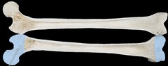
Articular cartilage
-Thin layer of hyaline cartilage covering joint surfaces of epiphysis
-Helps reduce friction and absorb shock in moveable joints

Metaphysis
Region of mature bone between diaphysis and epiphysis

Epiphyseal plate
-Within metaphysis
-Thin layer of hyaline cartilage
-Provides for continued lengthwise bone growth
-Remnant in adults called *epiphyseal line*

Periosteum
-Tough sheath covering outer surface of bone
-Outer fibrous layer of dense irregular connective tissue
-Inner cellular layer: osteoprogenitor cells, osteoblasts, and osteoclasts
-Attached to bone by numerous collagen fibers called *perforating fibers*

What are the functions of the outer fibrous layer of dense irregular connective tissue of the periosteum? (3)
-Protects bone from surrounding structures
-Anchors blood vessels and nerves to bone surface
-Attachment site for ligaments and tendons
Endosteum
-Covers all internal surfaces of bone within medullary cavity
-Incomplete layer of cells
-Contains osteoprogenitor cells, osteoblasts, and osteoclasts

Short, flat, and irregular bones
-External surface composed of compact bone
-Interior composed of spongy bone
-Lack a medullary cavity

Diploë
Spongy bone in the flat bone of the skull

How do short, flat, and irregular bones differ in their gross anatomic structure from long bones?
-External surface composed of compact bone
-Interior composed of spongy bone
-Lack a medullary cavity
Describe blood supply and innervation of the bone.
-Bone is highly vascularized, especially in regions with spongy bone
-Vessels enter from periosteum from small holes called *nutrient foramen*
-Nerves accompany blood vessels through nutrient foramen and innervate bone as well as periosteum, endosteum, and marrow cavity --> sensory nerves that signal injury
Nutrient foramen
-Small opening or hole in bone
-One nutrient artery enters and one nutrient vein exits

Red bone marrow (myeloid tissue)
-Hemopoietic (blood cell forming)
-Contains reticular connective tissue, immature blood cells, and fat
-In children, located in spongy bone and medullar cavity of long bones
-In adults, located in portions of axial skeleton (skull, vertebrae, ribs, sternum, ossa coxae), and proximal epiphyses of humerus and femur

Yellow bone marrow
-Product of red bone marrow degeneration
-Fatty substance
-May convert back to red bone marrow during severe anemia in order to facilitate production of additional erythrocytes

3) What two minerals are stored in bone, and what are their functions in the body?
Calcium is an important component in muscle contraction, nerve conduction, and blood clotting. Phosphate is required for the production of ATP and nucleotides.

4)What are several examples of flat bones in the body?
Most of the bones of the roof of the skull, the scapulae, the sternum, and the ribs are flat bones.

5) How do the diaphysis and epiphysis of a bone differ in structure?
The diaphysis is the shaft of a long bone. It consists of a relatively thick cylindrical arrangement of compact bone, lined with spicules of spongy bone, usually surrounding a central medullary cavity.
Theepiphysiss are the knobby ends of long bones. They consist primarily of spongy bone, surrounded by a thin crust of compact bone

6) What is the function of a nutrient foramen in bone?
The nutrient foramen is an opening within a bone for the nutrient artery and vein as well as innervation.

7) Where is red bone marrow found in the adult skeleton?
The adult skeleton contains red bone marrow within spongy bone of flat bones in the skull, vertebrae, ribs, sternum, and ossa coxae.

Bone connective tissue (osseous connective tissue)
-Primary component of bone
-Composed of cells and extracellular matrix
4 types of bone cells
osteoprogenitor cells, osteoblasts, osteocytes, osteoclasts
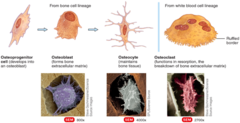
Osteoprogenitor cells
-Stem cells derived from mesenchyme
-Divide through process of cellular division --> another stem cell is produced along with a "committed cell" that matures to become an osteoblast
-Located in periosteum and endosteum

Osteoblasts
-Formed from osteoprogenitor stem cells
-Positioned side by side on bone surfaces
-Function to synthesize and secrete *osteoid*
-Become entrapped within matrix they produce and secrete where they differentiate into osteocytes

Osteoid
-Organic component of bone matrix
-Produced by osteoblasts
-Initial semisolid organic form of bone matrix
-Composed of collagen protein, semisolid ground substance of proteoglycans, and glycoproteins
-Contribute to bone flexibility
-Give bone tensile strength by resisting stretching
-Calcifies as a result of salt crystal deposition
Osteocytes
-Mature bone cells derived from osteoblasts
-Maintain bone matrix
-Found in small spaces between concentric lamellae (*lacunae*)
-Detect mechanical stress on bones --> Trigger new bone formation

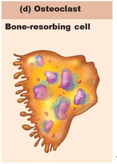
Osteoclasts
- Large, multinuclear, phagocytic cells
-Derived from fused bone marrow cells
-Exhibit ruffled border where they contact the bone, increasing surface area exposure to bone
-Often located within or adjacent to *resorption lacuna*
-Involved in breaking down bone in process called bone resorption
-Destroy matrix and remove calcium salts
Bone formation
-Begins with secretion of osteoid from osteoblasts
-Proceeds with *calcification*
-Requires vitamin D: enhances calcium absorption from GI tract
-Requires vitamin C: needed for collagen formation
-Requires calcium and phosphate for calcification

Bone resorption
-Bone matrix destroyed by substances released from osteoclasts
-Proteolytic enzymes released from lysosomes within osteoclasts to chemically digest organic matrix components (collagen fibers and proteoglycans)
-Hydrochloric acid (HCl) dissolves calcium and phosphate ions, which enter the blood
-May occur when blood calcium levels are low

Osteons (Haversian systems)
-Basic functional and structural unit of mature compact bone
-Oriented parallel to bone diaphysis
-Components include: central canal, concentric lamellae, osteocytes, lacunae, canaliculi

Central (Haversion) canal of osteon
-Cylindrical channel
-Lies at center of osteon and runs parallel to it
-Blood vessels and nerves that supply bone travel here

Concentric lamellae of osteon
-Rings of bone connective tissue
-Surround central canal and form bulk of osteon
- gives bone part of its strength and resilience

Lacunae of osteon
Small spaces that each house an osteocyte

Canaliculi of osteon
-Tiny, interconnecting channels within bone connective tissue
-Extend from each lacuna, travel through lamellae, and connect to other lacunae and the central canal
-House osteocyte cytoplasmic projections permitting intercellular contact and communication
-Allow travel of nutrients, minerals, gases, and wastes between blood vessels and osteocytes

Perforating canals (Volkmann canals)
-Component of compact bone
-Contain blood vessels and nerves
-Run perpendicular to central canals
-Help connect multiple central canals within different osteons
Trabeculae
-Component of spongy bone
-Open lattice of narrow rods and plates of bones
-Bone marrow filling spaces between
-Form a meshwork of crisscrossing bars and plates of small bone pieces that provide great resistance to stresses
Describe the structure of hyaline cartilage.
-Population of cells scattered through matrix of protein fibers
-Embedded in gel-like ground substance that includes proteoglycans, but not calcium
-Resilient and flexible
-High percentage of water
-Highly compressible and good shock absorber
-Avascular and contains no nerves
-Includes chondroblasts, chondrocytes, and perichondrium
Chondroblasts of hyaline cartilage
-Derived from mesenchymal cells
-Produce cartilage matrix
Chondrocytes of hyaline cartilage
-Chondroblasts that have become encased within the matrix
-Occupy small spaces (*lacunae*)
-Maintain the matrix
Perichondrium of hyaline cartilage
-Dense, irregular connective tissue
-Covers cartilage and helps maintain its shape
8) What are the functions of the osteoprogenitor cell, osteoblast, osteocyte, and osteoclast?
Osteoprogenitor cells - adult stem cells from which the remaining cells of bone tissue are produced.
Osteoblasts produce osteoid, which gives rise to the matrix of bone tissue.
Osteocytes are differentiated osteoblasts that have become trapped ,continue to maintain their matrix.
Osteoclasts participate in bone resorption, breaking down bone tissue and releasing calcium and phosphate.

Endochondral ossification
-Begins with hyaline cartilage model
-Produces most bones of the skeleton, including those of upper and lower limbs, pelvis, vertebrae, and ends of clavicle

Steps of endochondral ossification (6)
1. Fetal hyaline cartilage model develops.
2. Cartilage calcifies, and a periosteal bone collar forms.
3. Primary ossification center forms in the diaphysis.
4. Secondary ossification centers form in the epiphyses.
5. Bone replaces almost all cartilage, except articular cartilage and epiphyseal cartilage.
6. Lengthwise growth continues until epiphyseal plates ossify and form epiphyseal lines.

Bone remodeling
-Continual addition of new bone tissue (*bone deposition*) and removal of old bone tissue (*bone resorption*)
-Bone continues to renew and reshape itself
-20% of skeleton is replaced yearly --> does not occur at same rate everywhere on skeleton
-Dependent on osteoblasts, osteoclasts, and osteocytes --> activities of these cells influenced by hormones and mechanical stress

Mechanical stress
-Occurs in form of weight-bearing movement and exercise
-Required for normal bone remodeling
-Detected by osteocytes and communicated to osteoblasts
-Bone strength increases over period of time in response to mechanical stress
Hormones (for bone growth)
-Molecules released from one cell into the blood and travel throughout the body to affect other cells
-Hormones affect bone composition and growth patterns by altering rates of chondrocyte, osteoblast, and osteoclast activity
-Growth hormone, thyroid hormone, calcitonin, parathyroid hormone, sex hormones, glucocorticoids, serotonin
15) How does a bone grow in width?
15. Bones are widened through appositional growth at the periosteum by osteoblasts laying down rings of matrix called circumferential lamellae.
16) What is bone remodeling, where does it occur, and when does it occur?
Bone remodeling is the removal of old bone tissue and its replacement by new bone tissue. It occurs primarily in response to mechanical stress upon the bone or to stimuli from hormones such as growth hormone, thyroid hormone, or sex hormones.
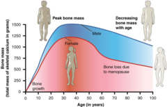
Osteoporosis
-Reduction in bone mass sufficient to compromise normal function
-Women lose more skeletal mass every decade

21) Explain why women are more likely than men to develop osteoporosis.
21. Estrogen is involved in protection of women from osteoporosis. As estrogen levels drop in postmenopausal women, they are more likely to experience osteoporosis
Fractures
Breaks in bone
Describe the process of bone fracture repair. (4)
1. A *fracture hematoma* forms.
2. A *fibrocartilaginous (soft) callus* forms.
3. A *hard (bony) callus* forms.
4. The bone is remodeled.

22) What are the four basic steps in fracture repair?
Initially, broken blood vessels in a broken bone form a hematoma. The hematoma is replaced by a fibrocartilaginous callus, which is then replaced by a compact bone, and then finally remodeled.