Humerus/Shoulder Critique
 Call Kai
 Learn
 Practice Test
 Spaced Repetition
 Match
 Flashcards
 Knowt Play
1/6

There's no tags or description
Looks like no tags are added yet.
7 Terms

1. This is an AP humerus projection demonstrated by the greater tubercule profiled laterally, although not very sufficient.
2. The patient's elbow was not externally rotated as shown by the epicondyles are not parallel to the IR and the radial head is superimposing with the ulna. The greater tubercule is not well profiled laterally. Externally rotate the patient's humerus until the epicondyles are parallel against the IR.
3. The CR looks correct. The collimation should include the soft tissue borders of the humerus and the shoulder, including the elbow joint and 1 inch minimum of the proximal forearm.
4. The technique looks correct, as the trabeculae is fairly visualized.
5. This image is not legal. Patient identifiers, facility number, and the technologist initials are not shown. However, anatomical marker is visualized
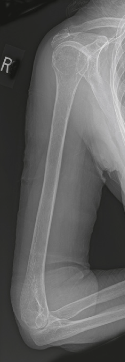
1. This is an lateral humerus projection, specifically a lateromedial humerus projection, demonstrated by the flexion of the elbow with the distal forearm situated towards the abdomen.
2. The patient's epicondyles are not perpendicular to each other against the IR. A figure "8" can usually be seen on the distal humerus to verify a true lateral projection. The capitulum is posterior to the medial trochlea, causing distortion to the proximal forearm. The lesser tubercle is not well visualized as well. Internally rotate the arm until the humeral epicondyles are positioned perpendicular to the IR.
3. The CR looks correct. The collimation should include the soft tissue borders of the humerus and the shoulder, including the elbow joint and 1 inch minimum of the proximal forearm. The tube angle can be adjusted to the position of the humerus.
4. The technique looks correct, as the trabeculae is fairly visualized.
5. The patient's hair shows up artifact and is superimposing on the proximal area of the humerus. Instruct or assist the patient to tying their hair or moving it away from the target anatomy.
6. This image is not legal. Patient identifiers, facility number, and the technologist initials are not shown. However, anatomical marker is visualized

1. This is an left AP oblique projection (Grashey) demonstrated by the profiled glenoid cavity and the open glenohumeral joint space.
2. The patient is rotated slightly too obliqued indicated by the foreshortening of the clavicle. The coracoid process and acromion angle must be superimposed. The greater tubercle of the humerus must be visualized as well (tray holding is also an external rotation). Rotate the patient towards their affected shoulder until the midcoronal place is 35-45 degrees angle with the IR.
3. The CR is centered to low and must be placed at the glenohumeral joint. The IR alignment was taken in portrait instead of landscape. Collimation must close in longitudinally to image only target anatomy and avoid additional patient dose.
4. Exposure technique is good.
5. This image is not legal. Patient identifiers, facility number, anatomical markers, and the technologist initials are not shown.

1. This is an right AP external rotation projection of the shoulder demonstrated by the profiled greater tubercle and the medial head of the humerus.
2. The patient has a fracture on the acromion process and the area of the anatomical neck of the humerus. Given their condition, an external rotation can be extremely painful and unsafe for a patient to be positioned in. Assess the patient carefully before putting them in an uncomfortable position.
3. The CR is too low and and should be 1 inch inferior of the coracoid process to avoid clipping of the T1 rib. Collimation should open up slightly more transversely to include the sternoclavicular joint.
4. Technique can be improved with decreasing kVp. There is slight overexposure in the most crucial areas of target anatomy.
5. This image is not legal. Patient identifiers, facility number, and the technologist initials are not shown. However, anatomical marker is visualized but could be placed better in the available open space.

1. This is a left PA oblique projection (Scapular Y) of the shoulder demonstrated by the visual representation of the letter "Y' formed by the coracoid, acromion process, and the scapular body.
2. The patient's shoulder is dislocated indicated by the detachment of the humeral head to the glenoid fossa. The patient is also not obliqued enough. The vertebral border superimposes the thorax more than the lateral border. However, the patient may likely not have been able to withstand prolonged and uncomfortable positioning due to their condition, causing insufficient obliquity.
3. CR looks correct (medial border of the scapula, specifically at the level of the glenohumeral joint). The collimation can be opened up slightly more transversely to avoid clipping of surrounding soft tissues.
4. Exposure technique is good.
5. This image is not legal. Patient identifiers, facility number, anatomical markers, and the technologist initials are not shown.

Spot a positioning error
The lateral and vertebral borders of the scapula are demonstrated without superimposition, and the glenoid cavity is not demonstrated. The lateral scapular borders are demonstrated next to the ribs. The torso was rotated more than needed to superimpose the borders of the scapular body.
Explain how to correct it
Decrease the degree of torso obliquity