Block 2 Human Physiology P225 IU
 Call Kai
 Learn
Learn Practice Test
 Spaced Repetition
 Match
Match Flashcards
 Knowt Play
1/226

There's no tags or description
Looks like no tags are added yet.
Name | Mastery | Learn | Test | Matching | Spaced | Call with Kai | Chat |
|---|
No analytics yet
Send a link to your students to track their progress
227 Terms
CNS
brain and spinal cord

PNS
Sensory, efferent neurons, somatic motor, autonomic: parasympathetic, sympathetic
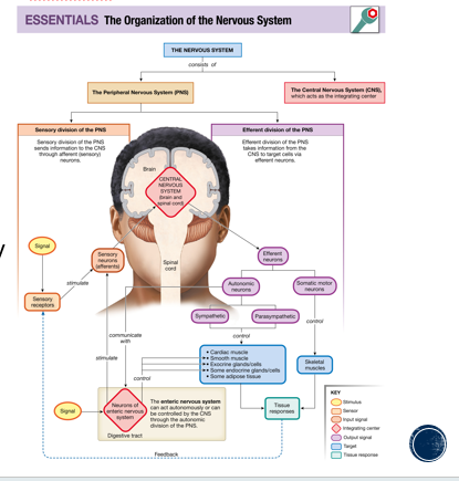
Sensory neurons
Afferent neurons
Motor neurons
Efferent neurons
Somatic motor division
voluntary
autonomic division
involuntary
Dendrites
Projections coming out of the cell body, detect input signal

Cell body
integration center of neuron

nucleus
middle of the cell body

axon hillock
connects axon to cell body

Initial segment
beginning of axon between hillock and first myelin sheath
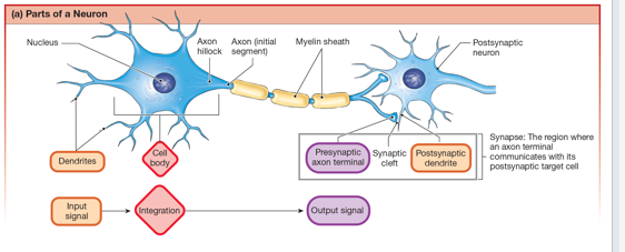
myelin sheath
lines the axon for faster communication

Collaterals
Extra axon branching off of main axon
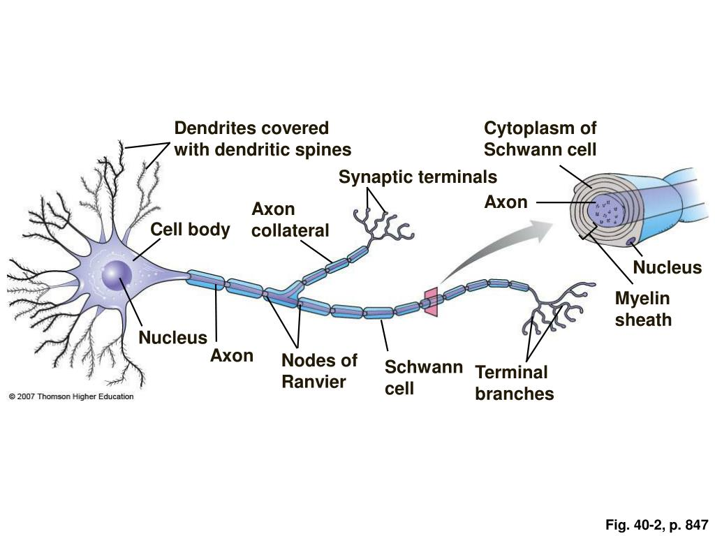
axon terminal
End of the axon that is the beginning of the synapse

Synapse
presynaptic axon terminal (first axon), synaptic cleft (space between), postsynaptic dendrite

Axo-dendritic
axon connects to dendrite

Axo-somatic
axon connects to soma
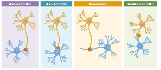
axo-axonic
axon connects to axon
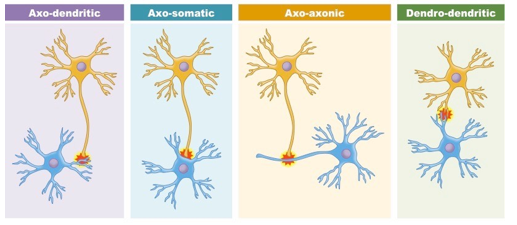
Dendodendritic
dendrite connects to dendrite

Neurotransmitter production
in the soma

Neurotransmitter transport
axonal transport

neurotransmitter storage
axon terminal

Slow axonal transport
cytoplasmic streaming
fast axonal transport
protein “walking”, anterograde and retrograde, vesicle recycling

Neurotransmitter secretion
axon terminal, exocytosis, vesicle recycling
Key factors determining resting membrane potential
K+ concentration gradient, K+, Na+ and Cl- resting membrane permeability
leak channels
always open, K+ goes out, Na+ goes in, counteracted by Na+/K+ ATPase pump

gated channels
chemical, mechanical, voltage, chemical and voltage are most common in neurons

Graded potential characteristics
input signal - change in membrane potential, location: dendrites and soma, type of ion channel: chemically, mechanically, occasionally voltage, ions involved: Na+ K+ and Ca2+. EPSP’s and IPSP’s

EPSP
Excitatory post-synaptic potential, causes depolarization
IPSP
Inhibitory post-synaptic potential, causes hyperpolarization

Graded potential characteristics: signal strength
variable, no minimum strength required, summation
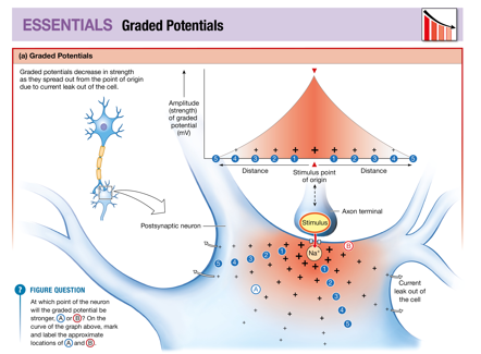
Graded potential characteristics: Current flow
local, not propagated, decremental, spreads away from stimulus in all directions. towards trigger zone (axon hillock and initial segment), subthreshold - No AP. Suprathreshold - get AP

Action potential characteristics
type of signal, regenerative signal within the axon, location: axon hillock through axon terminal, type of ion channels: voltage gated, Ions involved: Na+ and K+ (some Ca2+)
action potential characteristics: signal type
EPSP’s only, no IPSP’s in action potentials
Action potential characteristics: signal strength
all or none, refractory period between action potentials: cannot sum
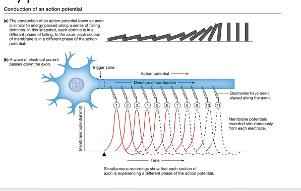
Action potential characteristics: current flow
one way flow, propagated, trigger zone to axon terminals

Action potential sequence: 1st half
Resting membrane potential, depolarizing graded potential: EPSP, EPSP to threshold, rapid depolarization: Na+ voltage gated channels, overshoot: Na+ voltage gated channels
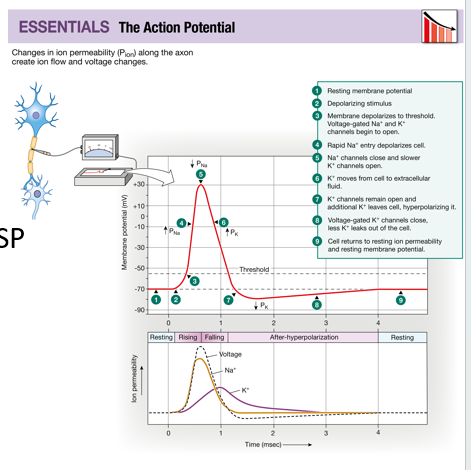
Action potential sequence: 2nd half
repolarization: K+ voltage gated channels, hyperpolarization undershoot: K+ voltage gated channels, returning to resting membrane potential: K+ voltage gated channels, resting membrane potential

Voltage gated Na+ channels
closed, open, inactive. RMP - closed. Threshold - open. Threshold to equilibrium Na+ - open. At equilibrium Na + - inactive. Repolarization - closed

voltage gated Na+ and K+ channel feedback
Na+ voltage gated channel reinforced to completion (equilibrium of Na+) - positive feedback regulation. K+ voltage gated channel returns to RMP - negative feedback regulation.

Refractory periods
limit # of action potentials, forces one way propagation, type depends on Na+ voltage gated channel position, absolute and relative

Absolute refractory period
Cannot generate another action potential, Na+ voltage gated channels are open, they close during repolarization

Relative refractory periods
Na+ voltage gated channels are closed, only a larger than normal stimulus can stimulate a new action potential. Returning to RMP

Action potential propagation: trigger zone
initial segment, EPSP graded potential, voltage gated channels

Action potential propagation: Adjacent area
stimulated: depolarize, non-decremental

Action potential propagation: previous area
absolute refractory area, one directional flow

Rates of conduction
Diameter: larger diameter, less resistance, faster. Myelination: lipid insulation, much faster rates. Saltatory conduction, metabolic efficiency

Demyelination
CNS: heavy metal poisoning, multiple sclerosis. PNS: Guillain-Barre’ syndrome, copper deficiency
hyperkalemia
excess intake of K+, depolarizes membrane potential - makes it easier to get to threshold for an action potential
Hypokalemia
can happen with water toxicity, concentration gradient of K+ to move out of the cell, hyperpolarizes membrane potential, harder to stimulate action potential
Electrical Synapse
gap junction, direct transfer of action potential, bidirectional flow from point stimulation, CNS, cardiac muscle, smooth muscle

Chemical synapse
neurotransmitters, indirect transfer of action potential, one direction of flow, most of PNS, skeletal muscle

Neurotransmitters
direct effect on signal transmission: EPSP or IPSP. Paracrine agents: communication between pre and post synaptic cell. Autocrine agents: may provide self regulation or feedback to pre-synaptic cell
Neuromodulators
alters synaptic activity at pre or post synaptic cell
Ionotropic receptors
fast, ligand gated ion channels: ion specific. EPSP or IPSP

Metabotropic receptors
slow, G protein coupled receptor with 2nd messengers. EPSP or IPSP, intracellular response

Acetylcholine (nicotinic)
Cholinergic, ion channel receptor, skeletal muscle, autonomic neurons, CNS, agonist: nicotine, antagonists: curare
Acetylcholine (muscarinic)
Cholinergic, G protein coupled receptor, smooth and cardiac muscle, endocrine and exocrine glands, CNS, agonist: muscarine, antagonist: atropine
Norepinephrine
beta 1 and beta 3. G protein couple receptor, smooth and cardiac muscle, glands, CNS, adipose tissue, antagonists: alpha receptors: ergotamine, phentolamine, beta receptors: propranolol
Epinephrine
alpha 1, beta 2. G protein couple receptor, smooth and cardiac muscle, glands, CNS, adipose tissue, antagonists: alpha receptors: ergotamine, phentolamine, beta receptors: propranolol
Synapse Component Actions: Presynaptic Cell
Axon terminal, active zone, neurotransmitter vesicles. Voltage-gated Ca2+ channels, docking proteins, exocytosis of neurotransmitter

Synapse Component Actions: Synaptic cleft
interstitial space: ECF, Diffusion
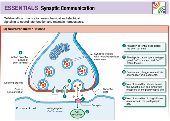
Synapse Component Actions: Postsynaptic Cell
Postsynaptic density, neurotransmitter receptors, enzymes

Termination of neurotransmitter action: stop release
stop simulation, no action potentials

Termination of neurotransmitter action: remove
presynaptic cell re-uptake, enzyme degradation, AChE: acetylcholinesterase, diffusion away from cleft

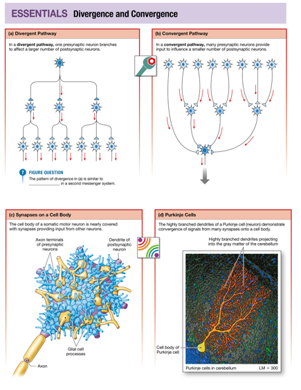
Integration of Neural Information Transfer: Divergence
Spread message, one to many
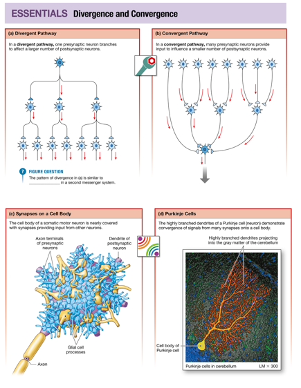
Integration of Neural Information Transfer: Convergence
multiple message input, many to one

Integration of Neural information transfer: back talk
across synapse, neuromodulators, modifying what’s happening at axon terminal
Integration of Neural Information Transfer: Plasticity
mostly CNS, enhance or decrease synaptic activity, can regulate activity at a synpase

Integration of Neural Information Transfer: Stimulus strength
number of APs and frequency of APs. One EPSP can generate multiple APs by staying above threshold for long enough time, needs to be in relative refractory period to start another AP. Stronger EPSP can result in more frequent APs, a higher threshold means more likely to initiate AP and frequency of APs. More neurotransmitter release from stronger stimulus

Information of Neural Information Transfer: temporal summation
Additive EPSP + EPSP, separated in time from the same synapse

Information of Neural Information Transfer: Spatial Summation
Additive EPSP + EPSP, separated by space but all at the same time

Integration of Neural Information Transfer: Inhibitory summation
EPSP + IPSP

Integration of Neural Information Transfer: Global Inhibition
No AP generated, all targets of the postsynaptic neuron are inhibited equally
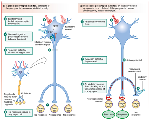
Integration of Neural Information Transfer: Selective Inhibition
Some APs, an inhibitory neuron synapses on one collateral of the presynaptic and selectively inhibits one target

Integration of Neural Information Transfer: Presynaptic Factors
Neurotransmitter production, re-uptake, and breakdown. Amount of Ca2+ entering cell. Amount and rate of Ca2+ removed from cell or put into storage

Integration of Neural Information Transfer: Postsynaptic factors
enzyme activity: removal of neurotransmitter. Receptor up or down regulation. Receptor desensitization. Receptor agonists and antagonists. Alter signal action. Signal transduction pathway and signal amplification

Sensory Info: Conscious
Perceived, special senses and somatic senses, some proprioception
Sensory Info: Unconcious
not perceived, visceral, some proprioception, muscle length and tension
Sensory Info: System components
Stimulus, receptors (transduction), transmission (1st degree sensory neuron: PNS, 2nd degree sensory neuron: CNS decussation, 3rd degree sensory neuron: brain). Integration: cerebral cortex: conscious. Other areas of the brain: unconscious
Sensory Information: Receptors: Structure
Simple: free nerve ending. Complex: specialized receptor area. Specialized: non-neural receptor cells (gustation, vision, hearing, equilibrium). No AP needed in specialized receptors

Sensory Information: Receptors: Adequate stimulus
Chemoreceptors, Mechanoreceptors, Photoreceptors, Thermoreceptors, Nociceptors - pain
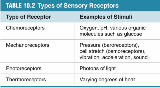
Sensory Information: Receptors: Function
Transduction. Stimulus energy converted to a change in the membrane potential. Receptor potential = graded potential (neuron vs specialized receptor cell). Ionotropic receptor (ion channel). Metabotropic receptor (second messenger opens ion channel)
Receptive Fields: 1st degree receptive field
large vs small, field overlap, 1st degree neuron convergence. Also a 2nd degree receptive field, and the 2 point discrimination test

Sensory Info: integration: unconscious
Spinal cord, brainstem, and hypothalamus
Sensory Info: integration: conscious
Cerebral cortex, perceptual threshold, filtering and habituation, CNS modification of threshold for perception, inhibitory modulation
Sensory Info: Integration: Olfaction
Olfactory cortex, limbic, hypothalamus

Sensory Info: integration: Vision
Midbrain → thalamus → visual cortex
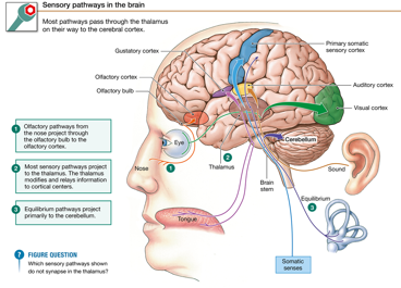
sensory info: integration: Sound and taste
medulla → thalamus → auditory and gustatory cortexes

Sensory info: integration: Equilibrium
medulla → cerebellum and thalamus → cerebral cortex
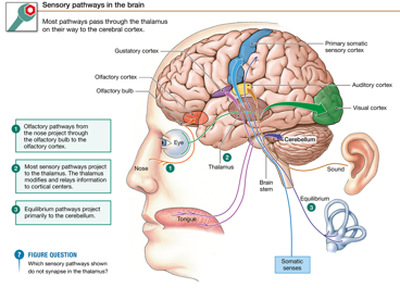
Sensory Information: integration: Somatic senses
Spinal cord → thalamus → 1st degree somatosensory cortex

Sensory Info: sensory coding: Modality
Type of receptor and adequate stimulus, 5 submodalities of taste

Sensory info: sensory coding: labeled line coding
Location of integration determines perception

Sensory info: sensory coding: location of receptor field and labeled line coding
somatosensory and the homunculus, phantom limb pain, crosstalk among neurons, pitch and auditory cortex

Sensory Info: sensory coding: timing
interaural time difference

Sensory Info: sensory coding: population coding and lateral inhibition
number of receptors stimulated, 1st degree receptive fields, axoaxonic inhibition. enhances contrast and makes stimulus easier to perceive

Sensory info: sensory coding: population coding
number of receptors stimulated, receptor threshold low → need weak stimulus. Receptor threshold high → need strong stimulus

Sensory info: sensory coding: frequency coding
frequency of APs, tells how often one sensory neuron fires to one receptor
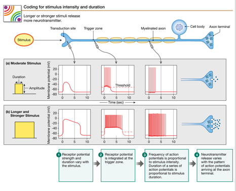
Sensory Info: sensory coding: Duration of stimulus
duration of sending APs, suprathreshold receptor potential, must persist into the relative refractory period
