unit 2 - anthro 3621
 Call Kai
 Learn
 Practice Test
 Spaced Repetition
 Match
 Flashcards
 Knowt Play
1/95

There's no tags or description
Looks like no tags are added yet.
96 Terms
general depression rates in women
approximately 12% of all women experience depression in a given year
25% for low-income women
5-25% for pregnant, postpartum, and parenting women
40-60% for low-income mothers
prenatal depression
affects 10-20% of pregnant mothers
presents as: crying, fatigue, appetite disturbance, loss of pleasure, guilt anxiety, irritability, and poor fetal attachment
how does untreated depression affect pregnancy?
can lead to…
low birth weight and size of infants
higher rates of miscarriage, preterm labor, placental abruption, and preeclampsia
irritable babies
lack of adequate prenatal care
higher use of alcohol and drugs
postpartum depression
suicide
antidepressants and pregnancy
pregnancy puts women with a history of depression at a higher risk for recurrence
pregnant individuals on antidepressants should be aware of the risk of relapse and the risk/benefit ratio of continued use

FDA labeling of antidepressants
pregnancy and lactation drugs include
risk summaries
clinical considerations
data sections
“Use the medication in pregnancy when clearly needed” → physician discretion is relied on over medication data
no distinctions are made between human and animal data, and drugs often see delays in updates of new clinical safety reports
antidepressant use during pregnancy
SSRI’s are some of the most commonly used antidepressants (ex: Prozac, Celexa, Lexapro, Zoloft)
research indicates SSRIs can be used during early pregnancy
absolute risk of SSRI exposure in pregnancy is small
late-trimester exposure to SSRIs is associated with transient irritability, agitation, jitteriness, and tahcypnea
SSRIs increase risk of preterm birth by 5 days
stopping SSRIs during pregnancy is associated with increased rates of spontaneous abortions
benzodiazepine use during pregnancy
anxiety is a risk factor for preeclampsia, preterm labor, low birth weight, PPD, and long-term behavioral, cognitive, developmental, and medical problems in the exposed fetus
benzo use
evidence for slight increases in oral clefts with first-trimester exposure
infants may show signs of toxicity if mothers take high doses in late pregnancy
stress from racism and discrimination during pregnancy
contributes to hypertension and pre-eclampsia, which are especially high in African American women
rates of pre-eclampsia decreased in AA women during Covid
black women were the only group that saw a decrease
racial myths in healthcare
pervasive racial bias and unequal treatment against Black women in the healthcare system result in inadequate treatment for pain and the dismissal of cultural norms
myth = Black patients have less-sensitive nerve endings and thicker skin than Whites
case study: 2011 nursing textbook
all “facts” about Africa in a 2011 nursing textbook were based on an 1980s ethnography written on research from the 1970s
only followed one study about one group of people (!Kung/Ju)
~13,000-44,000 !Kung/Ju were used to represent the 1.2b people in Africa
some of the ideas in the book were rejected by the !Kung/Ju people, but still cited as factual
institutionalized racism
structural barriers that act to limit care and increase risks in the health care system
racialized pregnancy stigma in everyday contexts (Mehra et al., 2020)
regardless of SES, participants encountered the assumption that Black pregnant women had low incomes and were dependent on government resources
Black women were considered welfare queens
regardless of age, education, income, insurance coverage, marital status, or number of children
participants were assumed to be single mothers with multiple children
effects of medical racism
minority women of all SES report experiencing medical racism
effects are associated with preterm birth and contribute to racial disparities for infants
healthcare providers disproportionately recommend tubal ligation to Black women
encountering racial stereotypes or prejudice can overshadow good treatment
nonjudgemental interactions lead to more comfort in sharing important health information, which leads to improved prevention, detection, and management
racism and prenatal care graphs and data

prolaction
responsible for breast development and lactation
released by the pituitary
placental lactogen
facilitates placental growth; higher binding affinity for prolactin receptors than maternal prolactin
released from the placenta into maternal circulation
how does prolactin and placental lactogen affect the maternal brain?
leads to cardiovascular and respiratory changes in the mother
leads to altered glucose metabolism of the mother
insulin resistance in the brain
leads to neurogenesis
increased aggression to protect the young
reduced anxiety and stress response
reduced fever response
mammary gland changes during pregnancy
nonpregnant state = small fat pad, smaller number and size of ducts
pregnant/lactating state = larger nipple and areola size, larger scent glands to orient infants, larger number and size of ducts, increased vascularization, larger alveoli, increased overall size
before birth, placental lactogen stops milk production
after birth, prolactin causes lactogenesis
uterine changes during pregnancy
significant increase in blood flow; approaches 500-750cc/minute at term
involves the uterine venous plexus, uterine veins, uterine artery, vaginal venous plexus, and vaginal arteries
hemorrhaging can lead to total blood loss within 10 minutes
changes to the blood during pregnancy
slight increase in WBCs
minimal change in platelet count
increased pH, decreased pCO2, decreased pHCO3
increased cardiovascular activity (HR, SV, and CO)

changes in pulmonary activity during pregnancy
increased tidal volume (increased lung size and rib cage volume)
decreased expiratory reserve volume and residual volume
increase in O2 demand —> O2 is prioritized in the placenta
breathlessness (elevation of diaphragm)
changes to the CNS during pregnancy
increased pituitary size and activity
estrogen and progesterone enter the brain, changing the balance between inhibition and stimulation
decreased oxytocin release
changes to the kidneys during pregnancy
increased renal blood flow; increased size of the kidneys
increased GFR (creatinine clearance)
decreased serum BUN, creatinine, and uric acid
increased urination frequency
increased risk of UTIs
evolution of pregnancy
mammals can become pregnant and have internal gestation because the mammalian genome has incorporated the syncytin gene
syncytin
a protein produced by placental cells that facilitates trophoblast invasion and transfomration; necessary for placental formation
appears to have originated in viruses
placental classifcation
depends on the number of cellular layers between fetal and maternal blood supplies
hemochorial
found in rodents and primates
3 layers
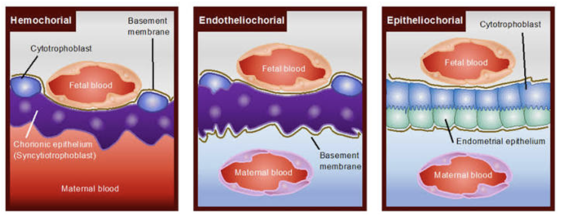
endotheliochorial
found in dogs and cats
5 layers
epitheliochorial
found in pigs and horses
6 layers
diffuse placenta
uniform distribution of chorionic villi covering the chorion’s surface; found in pigs

cotyledonary placenta
have numerous, discrete, button-like structures called cotyledons that join with the maternal caruncle, forming a placentome; found in ruminants (grazing herbivores)

zonary placenta
have a band-like zone of chorionic villi with both edges acting as exchange zones; found in cats and dogs
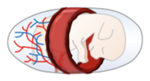
discoid placenta
have a regionalized disc-like structure and singular umbilical cord

placentophagy
the practice of eating placentas
common in most non-human animals, even those that don’t eat meat
theories
hides evidence of birth and blood
recover nutrients used for the placenta
makes it difficult to study placentas across species
chorionic plate
the fetal side of the placenta
contains fetal blood vessels and is derived from embryonic tissue
covered by smooth and glistening amnion overlaying the chorion
umbilical cord is attached at/near the center


basal plate
the maternal side of the placenta
contains maternal blood vessels and is composed of endometrium (uterine lining)
has a rough and spongy appearance; divided into 15-20 cotyledons
perforated by the spiral arteries, allowing entry of maternal blood into the intervillous space


cellular layers of the placenta

average human placenta
diameter = 22cm
depth = 2-2.5cm
average weight = 613g (varies by country)
placenta weight: baby weight = 1:5.6g
umbilical cord = 50cm x 2cm, central cord insertion
how does umbilical cord length affect the fetus?
too short
early severing
constrained fetal movement
too long
excessive fetal movement
poor fetal positioning during birth
primate placentas
slightly smaller and lighter than human placentas
typically have longer umbilical cords and non-central insertion
overview of placental function
nutrient exchange (fatty acids, glucose, amino acids, vitamins, minerals)
oxygen exchange
waste exchange
water exchange
hormone release

human placental lactogen (HPL)
hormone produced by the placenta
antagonizes insulin → spares glucose for use by the fetus
enhances lipolysis → provides an alternate fuel source for the mother
human chorionic gonadotropin (hCG)
hormone produced by the placenta
stimulates corpus luteum to secrete estradiol and progesterone
respiratory function of the placenta
although fetal respiratory movement occurs, no active exchange of gases takes place
intake of O2 and output of CO2 take place by simple diffusion across the fetal membrane
O2 delivery to the fetus is at the rate of 8 mL/kg, which is achieved by cord blood flow of 160-320 mL/min
nutritional function of the placenta
fetus obtains its nutrients from the maternal blood
glucose → transferred to the fetus by facilitated diffusion
lipids → transferred across the fetal membrane or synthesized in the fetus
amino acids → transferred by active transport
water and electrolytes → Na, K, Cl cross by simple diffusion; Ca, P, and Fe cross by active transport
water-soluble vitamins → transferred by active transport, but the fat-soluble vitamins are transferred slowly
immunological function of the placenta
fetus is considered a foreign body to the birthing person
aids in successful function
despite foreign paternal inherited antigens in the fetus and placenta, there is no graft rejection due to immunological protection provided by the placenta
placental transfer of immunity (IgG)
umbilical cord
approx 20cm long at full term
contains umbilical veins, arteries, and wharton’s jelly
develops from and contains remnants of the yolk sac and allantois

umbilical vein
brings nutrients from placenta to baby (only one vein)u
umbilical arteries
bring waste back to the placenta (two arteries)
wharton’s jelly
a mucoid connective tissue that fills and serves as vascular support for the umbilical cord
prevents kinking of the vessels during movement of the fetus in the womb
how does the placenta affect maternal arteries
remodels the smooth muscle and endothelium of spiral arteries with syncytiotrophoblast and trophoblast, impacting blood flow and pressure

anterior placenta location
anterior → can interfere with imaging and feeling fetal movement, but does not impact birth

marginal placental location
marginal → within 2cm of the cervical opening; vaginal birth is tried first, C-section if necessary
partial/total placental location
partial/total → placenta blocks the cervix, making C-sections medically necessary
bilobal placental shape
two lobes

battledore placental shape
marginal cord insertion
associated with small size for gestational age, low birth weight, and emergency C-sections
velamentous cord insertion
umbilical cord inserts into the fetal membranes instead of the placental disc
vessels travel between the amnion and chorion before reaching the placenta
elevated risk of damage to the cord (fetal hemorrhage)
furcate cord insertion
umbilical cord inserts normally into the chorionic plate of placental disc, but the umbilical vessels lose their protective covering of Wharton’s jelly before insertion
elevated risk of damage to the cord (fetal hemorrhage)

interpositional insertion
the umbilical vessels remain protected by Wharton’s jelly as they reach the chorionic plate
very rare, no added risks to fetal development

monochorionic-monoamniotic placenta
occurs with monozygotic twins (identical; share a placenta)
one placenta, one amniotic sac, one chorionic sac, two fetuses
twins do not get a 50/50 split of the placenta
each twin has an umbilical cord

monochorionic-diamniotic placenta
typically occurs with monozygotic twins (identical; share a placenta)
one chorionic sac and two amniotic sacs in one placenta

dichorionic-diamniotic placenta
occurs in dizygotic twins (fraternal; two eggs and two sperm)
two placentas, two amniotic sacs, two chorionic sacs
all independent of each other
placentas are typically smaller in size

gastrointestinal changes during pregnancy
placental vein makes direct contact with the liver

how is the fetus positioned during birth?
at the onset of labor, the fetal skull is oriented sideways
the skull rotates to avoid bone, flexing as it emerges
the fetus is born looking at the mother’s sacrum (spine side)

contractions
squeezing of the uterus to push the fetus out of the cervix
measured from the start of one contraction to the start of the next contraction
triggered by prostaglandins and oxytocin
become more intense and regular as labor progresses
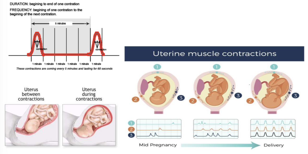
braxton-hicks contractions
false/non-labor contractions
characterized as uncoordinated, isolated, and less frequent
non-labor inducing
cervical effacement
thinning and shortening of the cervix during labor

cervical dilation
increasing size of the cervix during labor
low amniotic fluid
normal volume = 600-900mL at delivery; mostly fetal urine
low volume can put the fetus at risk for cord compression due to lack of blood flow to the fetus
water breaking
spontaneous rupture of membranes
typically occurs at/during the onset of active labor contractions
artifical rupture can be used to speed up or induce labor
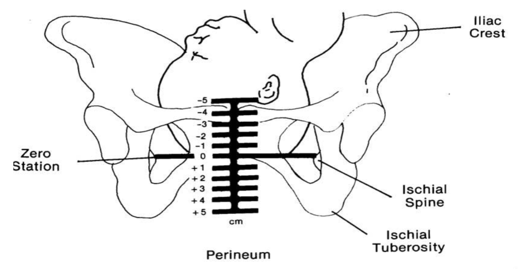
pelvic stations
-5 = not ready for birth
0 = fetus head is at the bottom of the pubic symphysis
+5 = imminent delivery, head has reached the cervix
early stage of labor
gradual dilation and effacement of the cervix
averages 6-12 hours
active stage of labor
increased intensity and frequency of contractions, typically beginning at ~4cm dilation
averages 4-8 hours (for a first pregnancy), but can range from minutes to days
precipitous labors
very short labors (<3 hours)
potential concerns
increased risk of vaginal tears
birth can occur anywhere → potentially bad location, may not be someone with sufficient medical knowledge around
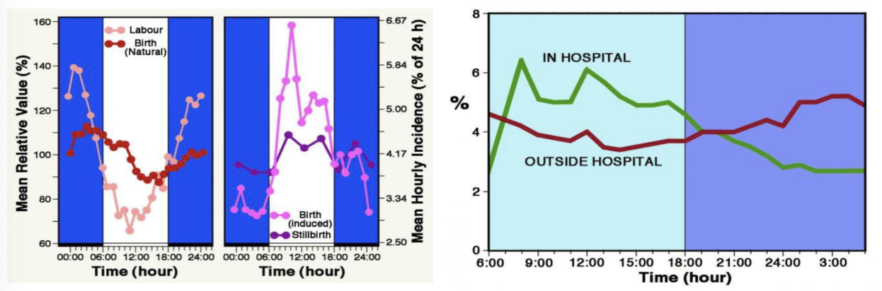
timing of birth
historical trend → most births occur closer to midnight
ancestral form of predator evasion
modern trend → inductions change trends
labor support
support person can be a partner, family member, friend, or trained doula
support for the laboring person has been shown to have positive effects on health and well-being
doula
a trained professional who provides continuous physical, emotional, and informational support to a mother before, during, and shortly after childbirth
not a nurse or physician (although some may have medical degrees)
complete training by a national or international accrediting professional board; adhere to professional standards
typically not covered by insurance in the US
labor induction
stimulation of uterine contractions during pregnancy before labor begins on its own to achieve a vaginal birth
can be done after 39 weeks
commonly done for medical reasons
methods:
prep the cervix using synthetic prostaglandins
insertion of a catheter & balloon into the cervix
rupture of the amniotic sac (amniotic fluid is a natural labor inducer)
use of intravenous medication
misoprostol and labor induction
misoprostol was originally used an ulcer medication, but was found to be very effective at inducing labor
increases the risk of uterine ruptures if the mother has had past uterine trauma (e.g., C-sections, car accidents, etc.)
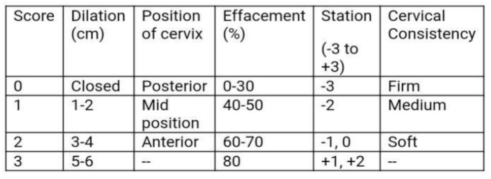
bishop scoring system
based on a digital cervical exam of a patient to determine if induction will be successful for vaginal delivery; ranges from 0-13 points
utilizes cervical dilation, position, effacement, consistency of the cervix, and fetal station
higher score = higher likelihood of a successful induction
augmentation
enhancement of contractions in a pregnant woman who is already in labor
cervical ripening
the use of pharmacologic or other means to soften, efface, and/or dialte the cervix
pushing and delivery
begins at full dilation (10cm)
fetus head moves through the vaginal canal and stretches (or tears) the perineum; most infants deliver face down
ends with the delivery of the baby
fetal skull
made of more bones than adult skulls, allowing parts of the fetal skull to slide and compress as it passes through the vagina
compression may lead to bumps and dents at birth, but they quickly recover
contains 6 fontanelle (soft spots)

fontanelle
an area with no bone in the fetal skull; “soft spot”
location of fetal brain scans using ultrasound probes
epidurals
use a combination of medications for local pain relief; causes numbing of (typically) the entire lower body
epidural needle is inserted into the epidural space (between two vertebrae); typically L3 and L4
performed between 4-6cm dilation
may alter fetal heart rate, leading to a lower APGAR score
interferes with fetal breathing and latching
requires more fetal monitoring
63.4% of US mothers have epidurals
fetal monitoring
fetal monitoring belts are used to track changes in the fetus and mother during pregnancy
bluetooth options are becoming more popular
can be intermittent or continuous
intermittent = recommended by manufacturer
continuous = used for serious complications or when interventions may be necessary, reduced neonatal seizure rate
upright birth position
the historical norm
allows for greater pelvic dimensions and greater ease of delivery
no sacrum interference
gravity assistance
typically involves squatting with hanging supports or horizontal support bars
lithotomy birth position
horizontal/semi-upright position
narrows the pelvis inlet by up to 25%
limited sacrum movement
cord clamping
current recommendations = wait 1-5 minutes (or until pulsing ends) to cut the umbilical cord
allows blood in the placenta to diffuse to the infant
associated with long-term iron levels in the infant
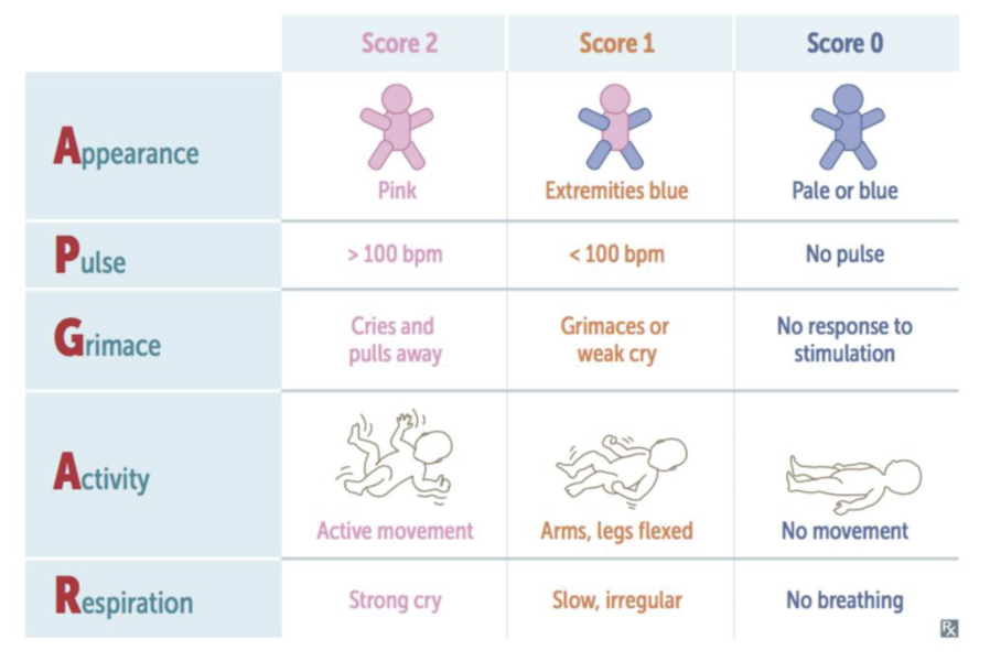
APGAR score
standard physical assessment given to a newborn at 2 minute and 5 minutes after birth to evaluate their overall physical condition and how well they are tolerating the transition to life outside the womb
scored from 0-10
perineum
space between the anal and vaginal opening
perineal tears
rips in the perineum during delivery
90% of first-time deliveries involve tears
usually repaired with stitches as part of routine care, but may require surgery
scaled from 1 (lightest) to 4 (most severe)

episiotomy
incisions made in the perineum to enlarge the vaginal opening
performed to prevent, control, or direct tears
assisted vaginal delivery (AVD)
facilitation by forceps or vacuum extraction inserted through the cervix to guide and pull the fetal head during delivery
associated with increased pelbic floor trauma
may include…
unmedicated labor
VBAC
pain management
episiotomy

vaginal birth after cesarean (VBACs)
individuals who choose to have a vaginal birth after previously having a C-section
historically believed that people who had C-sections before always need to have C-sections for later births