INTEGRATIVE PHYSIOLOGY EXAM 2 : MUSCLE AND MOVEMENT
 Call Kai
 Learn
Learn Practice Test
 Spaced Repetition
 Match
Match Flashcards
 Knowt Play
1/70

There's no tags or description
Looks like no tags are added yet.
Name | Mastery | Learn | Test | Matching | Spaced | Call with Kai | Chat |
|---|
No analytics yet
Send a link to your students to track their progress
71 Terms
Skeletal Muscle
Voluntary, striated, attached to the bone → Attached to the bone, contains sacromeres, T-tubules. Main Calcium source is sacroreticulum. fastcontraction with Troponin +Tropomyosin
Cardiac Muscle
Striated, involuntary, heart muscle with sacromeres and T-tubules which Main Calcium source is sacroreticulum/extracellular fluid. Moderate contraction with Troponin +Tropomyosin, moderate fatigue, limited regeneration
Smooth Muscle
Not striated, involuntary, hollow organs with no sacromeres with Calcium source being sacroreticulum and extracellular fluid. Typically has slow contraction with Calcmodulin + MLCK being regulatory proteins so less fatigue, and considerable regeneration
myosin/actin → filaments → Sacromere → Myofibril → Muscle Fiber → Muscle Fascicle → Whole skeletal muscle
How is the skeletal muscle organized?
Muscle
Made up of many muscle cells/gibers
Muscle Cell
Packed full of long fiber-like contractile organelles known as myofibrils
Myofibrils
Made up of contractile proteins known as myosin and actin and act as long fiber-like contractile organelles
Sacromere
A muscle unit in overlapping stripes with myosin and actin that forms light and dark striations
T-tubules
carry action potentials into the interior of the muscle fiber
Myosin
Thick filaments within a myofibril
Actin
Thin filaments within a myofibril
Z-line
Boundary of sarcomere where actin attaches to adjoining sacromeres
A-band
Stretches end to end of myosin which contains both actin and myosin filaments
I-band
Space between myosin and contains only actin
H-band
Non=overlapped regions of myosin only with no actin
M-line
Center of sacromere holding adjacent myosin together with supporting proteins
The myosin ATPase site
What can activate the myosin head?
Titin
provides elasticity and stabilizes myosin.
Nebulin
helps align actin.
Thin Filaments
When multiple actin molecules form together, they will form a double helix known as this.
Tropomyosin
Binds to actin to block the myosin binding sites on actin
Troponin
Binds to tropomyosin and can move it out of the way
1) Action potential
2) Calcium Release
3) Troponin activated where Calcium binds
4) Release of Tropomyosin for Troponin to pull off
5) Cross bridge between myosin and actin forms
6) Power Stroke when myosin heads pull actin inwards
7) reset
What are the quick muscle contraction steps?
Through a motor neuron that contacts the muscle at the neuromuscular junction acetylcholine is released which binds to a ligand ion receptor to depolarize the muscle membrane
How is an action potential released from the muscle cell membrane?
Sarcoplasmic Reticulum stores and releases Calcium into the cytosol of the muscle cell once an action potential occurs (depolarization will spread down T-tubes)
How is calcium released in the muscle cell?
Due to the release of Calcium, Troponin releases from Tropomyosin, making actin active
How does Tropomyosin get off of the muscle cell membrane
Cross-Bridge
When myosin binds to actin
Power stroke
A methoid requiring ATP to move actin inwards towards the M-line
Sarcoplasmic Reticulum
Where is calcium sent to after power stroke?
DHP receptor
Opens RyR Ca2+ release channels in sarcoplasmic reticulum and Ca2+ enters cytoplasm.
twitch
a single contraction-relaxation cycle
Creatine kinase
helps rapidly regenerate ATP using phosphocreatine.
phosphocreatine
This provides a rapid source of ATP during intense muscle activity.
Fatigue
reversible condition in which a muscle is no longer able to generate or sustain the expected output.
Slow oxidative
Have a high resistance to fatigue due to having low creatine kinase
Fast oxidative-glycolytic
Have a low resistance to fatigue due to having intermediate creatine kinase
Fast glycolytic
Lowest resistance to fatigue (i.e., easily fatigued) high creatine kinase
Isotonic contractions
create force and move load → because elastic elements are already stretched, the entire muscle must shorten.
Concentric action
a shortening action
Eccentric action
a lengthening action
Isometric contractions
create force without moving a load → sarcomeres shorten while elastic elements stretch resulting in little change in overall length
elastic elements
stretch resulting in little change in overall length
IP3-receptor channel
is the primary calcium channel in smooth muscle
calmodulin (CaM)
In smooth muscle, Calcium binds to this where it activated myosin light chain kinase (MLCK).
myosin light chain kinase (MLCK)
phosphorylates light chains in myosin heads and increases myosin ATPase activity
Myosin phosphatase
removes phosphate from myosin, which decreases myosin ATPase activity
myogenic contraction
Smooth muscle cells contain stretch-activated calcium channels → Open when pressure or other force distorts cell membrane
Proprioceptors
Located in skeletal muscle, joint capsules, and ligaments → carry input sensory neurons to CNS
monosynaptic reflex
has a single synapse between the afferent and efferent neurons.
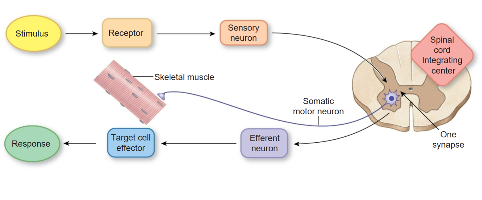
Polysynaptic reflexes
have two or more synapses.

Autonomic Reflexes
Some visceral reflexes are spinal reflexes

Stretch reflex
Propriopretors that is in the position of muscle spindle → will cause a contraction of the skeletal muscle in response to the stretching of a muscle, which occurs monosynaptically

Tendon reflex
Propriopretors that is in the position of Golgi tendon organ → operates as a feedback mechanism to control muscle tension by cuasing muscle relaxation before muscle force becomes so great that tendons must be torn

Joint receptors
Are found in capsules and ligaments around joints
Muscle Spindles
monitor muscle length and prevent overstretching

Alpha-Gamma Coactivation
Stretch on centers of intrafusal fibers unchanged. Firing rate of afferent neuron remains constant. (without it, there would be: Less stretch on center of intrafusal fibers)
Alpha
actual muscle contraction in skeletal muscle fibers
Gamma
keeps the spindle "tuned" during contraction
Crossed Extensor Reflex
a polysynaptic reflex that helps maintain balance when you withdraw from a painful stimulus.
Somatic Reflex
Seen in skeletal muscles, and could be voluntary but not too often (withdrawal reflex)
Autonomic Reflex
Smooth muscles and glands that is under the control of autonomic nervous system but not voluntary
Somatic is seen as voluntary and only using two neurons with post and preganglionic neurons
Compared to autonomic and somatic motor rflexes, what can make both of them unique?
CNS
What type of integrating center can autonomic reflexes use?
1) Alpha motor neuron fires and gamma motor neuron fires
2) muscle begins to contract
3) Stretch on centers of intrafusal fibers unchanged where firing rate of afferent neuron remains constant
Steps of Alpha Gamma Coactivation
There is less stretch on the center
What would happen if gamma neurons weren’t in the alpha-gamma coactivation?
golgi tendon organ
Sensory reflexes of tendon reflexes which lies near its junction with a muscle (response to muscle tension is to relax)
1) Stretching of muscle stimulates snesory receptors in the muscle known as muscle spindles
2) muscle spindle can generate a receptor potential, which could become an action potential when threshold is reached
3) sensory neurons can make an excitatory synapse to activate a motor neuron
4) when excitation is strong enough, one or more action potentails arise int eh motor neuron and propagate along its axon, which extends from the spinal cord to the ventral root
5) Acetylcholine released by action potentials triggers action potnetials int he stretched muscles ot cause contraction
stretch reflex steps
1) As tension is applied to a tendon, tendon organ is stimulated
2) action potentials arise and propagate into spinal cord along in a senosry neuron
3) within the spinal cord, sensory neuron activates inhibitory itnerneuront aht synapes with motor neuron
4) inhibitory neurotransmitter
Tendon reflex steps
Flexor (withdrawal) reflex
Causes flexion of a limb in order to withdraw from a painful stimulus
1) Stimulus is stepping on a tack to send a siganlt ot a sensory neuron
2) Sensory neuron generates action potentials to propagate into spinal cord
3) Wtihin the spinal cord, sensory neurons activates interneurons to extend to several spinal cord segments
4) interneurons activate motor neurons in several spinal cord segments which causes motor neurons to generate action potnetials to propagate towards axon terminals
5) AcH in motor neurons can cuase flexor muscles to contract, causing withdrawal
Withdrawal reflex steps
1) stepping on tack stimulates sensory receptor of a pain sensitive neuron int he right foot
2) Sensory neuron will generate action potentials which can propagate into spinal cord
3) sensory neuron activates several interneurons that synapse with motor neurons ont he self side of the spinal cord
4) interneurons excite motor neurons in several spinal cord segements that innervate extensor muscles to generate more action ptoentials
5) AcH caues extensor muscles of the unstimulated left limb to contract and extends at left foot
Cross Extensor Reflex Steps