Special Senses (Ear)
 Call Kai
 Learn
 Practice Test
 Spaced Repetition
 Match
 Flashcards
 Knowt Play
1/33

There's no tags or description
Looks like no tags are added yet.
34 Terms
External Ear
Consists of the auricle (pinna), the external acoustic meatus (ear canal), and the tympanic membrane (eardrum). Collects funnels and filters, and protects sound before it reaches the middle ear
Auricle (Pinna)
The visible, outer portion of the ear composed of elastic fibrocartilage covered by skin. Captures sound waves, amplifies specific frequencies, and helps the brain locate where sound is coming from

Lobule
Ear lobe, soft fleshy lower portion of the external ear (Auricle)

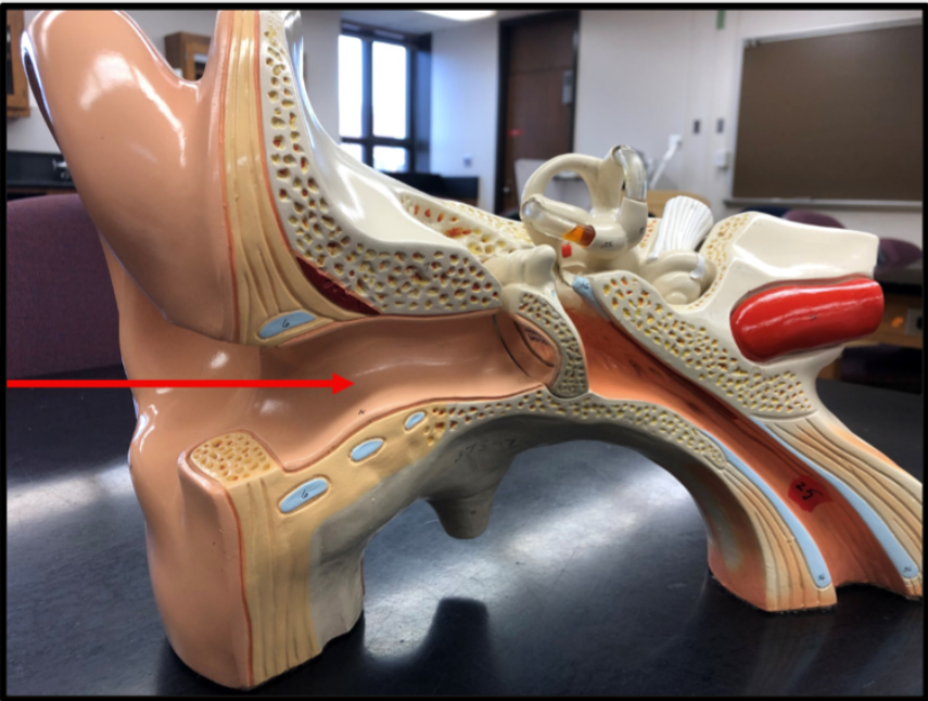
Auditory canal
Alighty curved tube that funnels sound waves from the outer ear (pinna) to the tympanic membrane (eardrum)
Ceruminous Glands
Apocrine sweat glands that produce cerumen (earwax)
Tympanic Membrane
thin, oval, semi-transparent connective tissue membrane that vibrates when struck by sound waves and transfers those mechanical vibrations to the middle ear's tiny bones (ossicles)

Oval Window
Primary mechanical entry point that transfers sound vibrations from the middle ear bones into the fluid-filled inner ear

Round Window
membrane-covered opening between the middle ear and the cochlea that acts as a pressure release valve for inner ear fluids.

Auditory (Eustachian) Tube
The narrow canal that connects the middle ear to the upper throat and nasopharynx. helps with air pressure and fluid and mucus drainage

Malleus
(hammer) first and largest middle ear bone. catches sound vibrations from the eardrum and passes them to the incus
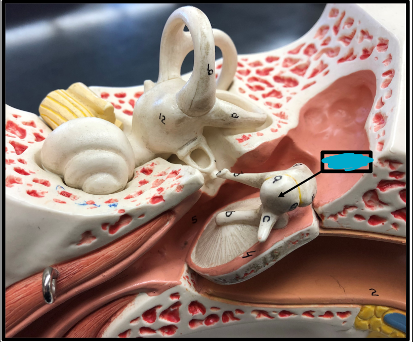
Incus
(anvil) middle ear bone that acts as a bridge between the malleus and the stapes

Stapes
Transmits sound from the eardrum to the inner ear (oval window)

Osseous Bony Labrynth Labyrinth
Protects and supports the fluid-filled spaces of the membranous labryinth
Perilymph
extracellular fluid inside that cushions inner ear structures, transmits sound waves, and helps maintain electrochemical balance (between the ossicles, labyrinth, and the membranous labryinth)

Membranous Labryinth
system of fluid-filled soft tissue sacs and ducts inside the osseous labyrinth that control hearing and balance
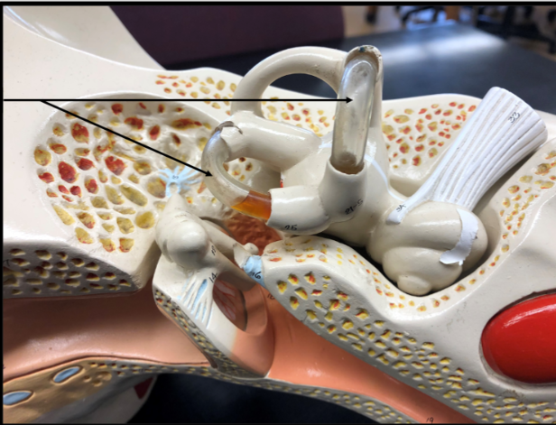
Endolymph
potassium-rich fluid inside the inner ear’s membranous labyrinth that coverts sound waves and head movements into neural signals for hearing and balance

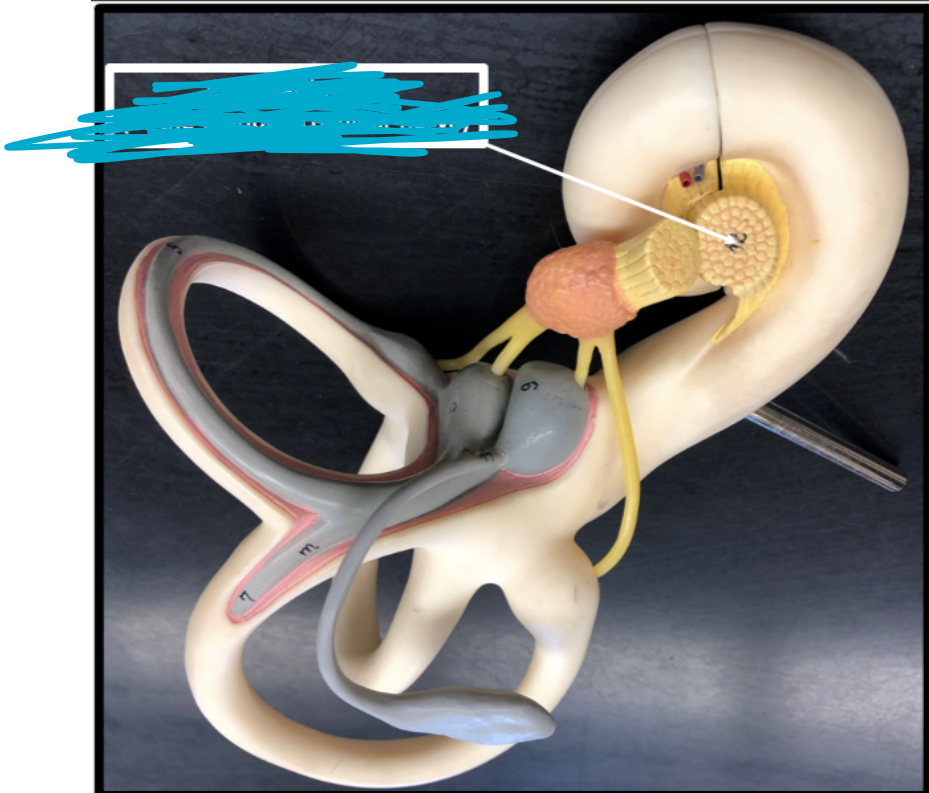
Cochlea
snail-shaped, fluid-filled structure in the inner ear that converts mechanical sound waves into electrical nerve signals for the brain

Vestibular Membrane
The thin two-layered tissue barrier inside the cochlea of the inner ear. Separates the vestibular duct from the cochlear duct

Basilar membrane
Acts as a mechanical frequency analyzer vibrating in response to sound waves. It separates high-frequency sounds near its stiff base from low-frequency sounds near its floppy apex

Scala vestibuli (Vestibular Duct)
Upper perilymph-fluid-filled chamber of the cochlea that transmits sound-induced pressure waves from the oval window towards the apex of the cochlea

Cochelar Duct (Scala Media)
converts mechanical sound vibrations into electrical nerve signals by allowing sensory hair cells to bend against the tectorial membrane, sending auditory data to the brain via the vestibulocochlear nerve
Spiral Organ (Organ of Corti)
primary receptor organ for hearing

Hair cells
Convert physical sound vibrations and head movements into electrical signals

Tectorial membrane
Overlies the sensory hair cells and stimulates them alongside the basilar membrane

Scala tympani (Tympanic Duct)
lower fluid-filled chamber of the cochlea that transmits sound waves through perilymph

Vestibule
Bony chamber of the inner ear that controls balance, spatial orientation, and head posture
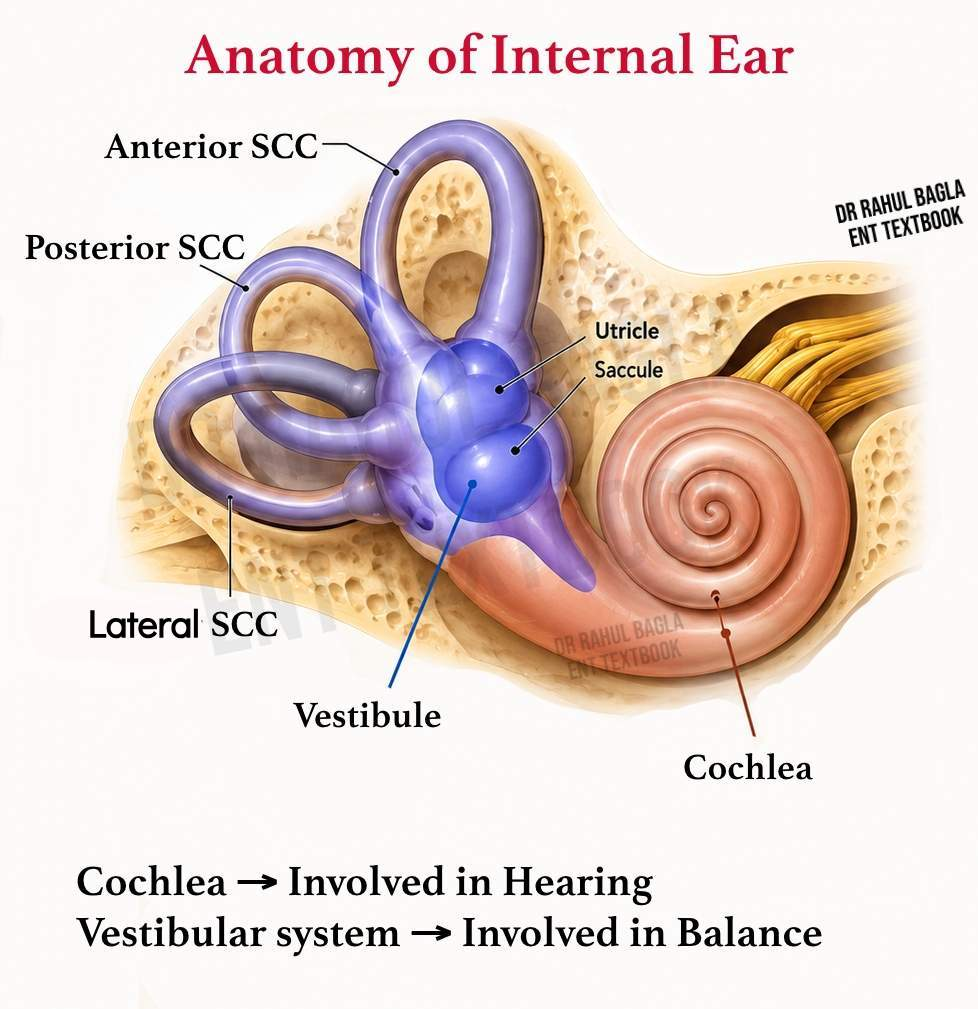
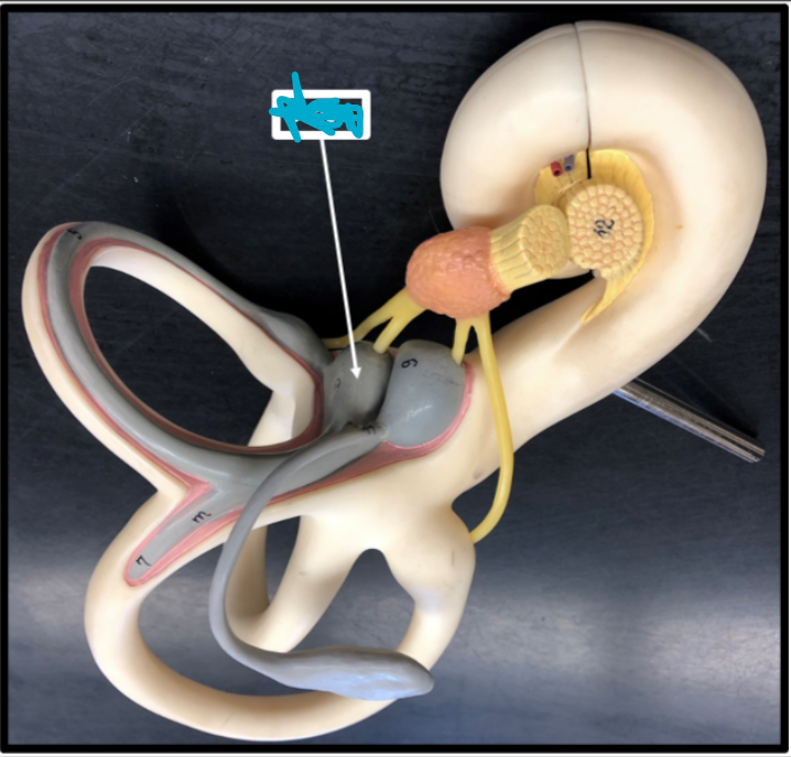
Utricle
A fluid-filled sensory organ in the inner ear that detects horizontal linear acceleration and head tilts relative to gravity
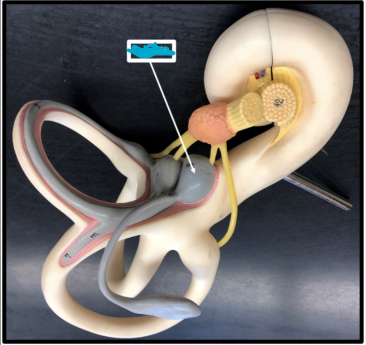
Macula
Specialized sensory patch inside the utricle and saccule of the inner ear that detects gravity, head position, and linear acceleration

Saccule
Sensory organ in the inner ear that detects vertical linear acceleration and head tilting in the vertical plane
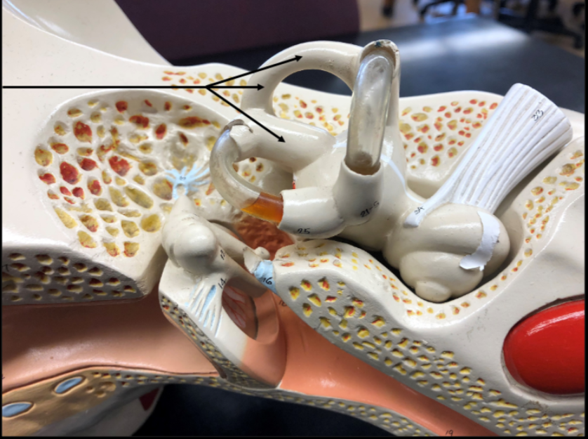
Semicircular Canals
Detect rotational head movements and angular acceleration to help maintain balance, posture, and gaze stability

Ampullae
Detects rotational and angular head movements to help maintain balance and visual stability

Cristae
Senses angular acceleration, rotational movement, and dynamic equillibrium

Vestibular Nerve
Carries balance, gravity, and movement signals from the inner ear to the brainstem

Cochlear Nerve
Sensory nerve responsible for transmitting sound information from the inner ear to tthe brain