L6 - Schizophrenia I
 Call Kai
 Learn
 Practice Test
 Spaced Repetition
 Match
 Flashcards
 Knowt Play
1/48

There's no tags or description
Looks like no tags are added yet.
49 Terms
what is psychosis
Thought disorder characterised by:
- disturbances of reality and perception.
- impaired cognitive functioning
- inappropriate affect
what diseases are associated with psychosis
- schizophrenia
- dementia
- depression
- drug/alcohol abuse
- cannabis use
what are some facts about schizophrenia prevelence
- Effects 1% of the population.
- Higher incidence in males.
- Probably not one disease.
- Adolescent onset but prodromal symptoms.
what are the positive symptoms of Sz
- Hallucinations - mainly auditory.
- Delusions of grandeur, persecution etc.
- Thought disturbances.
- Bizarre behaviour.
what are the negative symptoms of sz
- Social withdrawal.
- Flat affect.
- Anhedonia.
- Reduced motivation.
- Alogia.
what are the cognitive abnormalities seen in sz
Possibly the core feature of the disease:
Include disruptions in :-
information processing/sensory perception
And deficits in:-
Attention
Short-term memory
Behavioural flexibility
What is the prodromal phase of schizophrenia, and how can symptoms progress?
80–90% of patients with schizophrenia experience a prodromal phase before full psychosis develops.
During this phase, early symptoms appear, causing disruption in reality testing.
Possible outcomes:
Progression to full psychosis (orange).
Maintenance of subthreshold symptoms (blue).
Remission of prodromal symptoms (green).

describe the genetic aspect of sz
Clear genetic link:
Genetic variation can cause a change in the structure of a
protein or change the amount of a protein.
But in schizophrenia there is
- not one gene
- not a simple inheritance

what genes may be involved in sz
Certain genes may increase your risk of getting schizophrenia
128 common single nucleotide polymorphisms i.e a C to A switch and 15 CNVs
Highest genetic risk = deletion of 30 genes on chromosome 22- 22q11 deletion syndrome (Found in 30% sz patients now )

what are the 6 types of genes involved in sz
Genes linked to development – myelination, axonal guidance, neuronal migration.
ARC signalling – link neuronal activity to synaptic remodelling.
Group of genes linked to synaptic plasticity, eg neuregulin - alters activity of AMPA and NMDA receptors - function is disrupted in schizophrenia.
VGCC (voltage gated Ca2+ channels) – excitability and synaptic plasticity.
Groups of genes linked to dopamine metabolites and signalling eg. COMT (catechol-O-methyltransferase)- alters DA release.
Genes linked to neuroinflammation.
what are the environmental causes of sz
- family stress
- poor social interactions
- poor maternal nutrition
- infection/viruses at early age
- trauma at early age.
But can have all of those things and not get sz
how do we know genetics doesnt make up the sole cause of sz
monozygotic only 50% so cant be whole story
genetic makeup combines with non genetic factors
1% of population will have sz
what are the 2 main brain regions involved in Sz
the prefrontal cortex (PFC)-
Last part of brain to develop- until 25the hippocampus- reduced size in sz

what are the 2 main structural abnormalities seen in sz
enlargement of cerebral ventricles by 20%
decreased grey matter volume (neurones)
describe the relevance of ventricle enlargement in patients with sz
Enlargement of the cerebral ventricles, observed even in patients experiencing their first psychotic episode (before medication).
Ventricles are involved in CSF production and ion regulation in the brain.
Suggests that structural brain changes can be present early in schizophrenia and are not necessarily caused by medication.

what does decreased grey matter volume relate to
changes continue during disease course
correlates with disease duration
Loss of neurones- impact cognitive and sensory processing

what does grey matter look like
Dendrites that go up to just under scalp
Loss of grey matter means structural changes in neurones
Losing cells, processes and synapses

What is grey matter, and what changes are observed in schizophrenia?
Grey matter: Contains neuronal cell bodies (soma), processes and synapses.
PSD95 and synaptophysin: Synaptic proteins reduced in post-mortem brains of patients with schizophrenia.
Suggests loss of synapses, which is thought to be developmental rather than neurodegenerative.

what structural abnormalities are seen in the PFC in patients with Sz
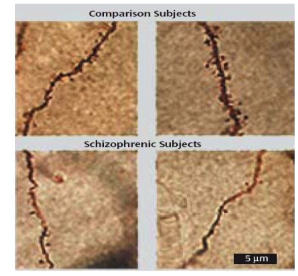
Loss of dendritic processes
Less branches, shorter, don’t spread as far, less info input in sz
decreased dendritic field size (40%) of layer 5 pyramidal cells in human post mortem Sz patients brains

how does spine density change in sz
several studies have shown decreased spine density in pyramidal cells
What structural abnormalities in hippocampal pyramidal cells are associated with schizophrenia?
Normal: Pyramidal cells are organised and aligned, with dendrites projecting in an orderly arrangement.
Schizophrenia: Pyramidal cells become disorganised, with abnormal alignment and dendritic organisation.
This disrupts communication and information processing between neurons in the hippocampus.

give an overview of DA receptors
5 different subtypes of DA receptor
D1 = D1 and D5;
G protein coupled – stimulate adenylyl cyclase - Postsynaptic
D2 = D2, D3, D4; G protein coupled -inhibit adenylyl cyclase -Pre- and postsynaptic

what are the DA pathways in the brain
DA neurons present in two main regions:-
A10 = Ventral tegmental area
A9 = Substantia nigra

what is the dopamine hypothesis, what is its evidence
original idea- SZ due to an abnormal increase in central DA levels (but too simple)
drugs that increase DA (amphetamine) - psychosis
Antipsychotics - all act as D2 receptor antagonists.
what is the mesolimbic dopamine pathway, what was the proposed change in sz
Neurons in VTA = ventral tegmental area (A10) projects to septum, amygdala, hippocampus, ventral striatum and frontal cortex.
Proposed this system is hyperactive in schizophrenia- That’s why antipsychotics help manage positive symptoms
?? accounts for positive symptoms.
what is the mesocortical dopamine pathway, what was the proposed change in sz
Neurons in VTA project to neocortex - prefrontal cortex (PFC).
Proposed this system is hypoactive in schizophrenia- So antipsychotics wont help negative symptoms, may make it worse
?? accounts for negative symptoms.
what is the nigrostriatal dopamine pathway, what was the proposed change in sz
- neurons in substantia nigra - project to dorsal striatum
Dorsal striatum
- part of extrapyramidal motor system
- involved in initiation and control of movement
what is the tuberoinfundibular dopamine pathway, what was the proposed change in sz
Hypothalamus - anterior pituitary
inhibits prolactin - tonic inhibition of lactation
DA released into perivascular spaces,
taken up by capillaries and transported to the pituitary gland,
acts on cells to inhibit the release of prolactin,
regulates milk production in lactating mammals.
Important later for side effects of antipsychotic treatment.
what are the 2 types of receptors in the glutamatergic system
1. Ionotropic receptors (iGluR) = Fast transmission, ions flow in/out of neuron, millisecond responses.
2. Metabotropic receptors (mGluR) = Slow synaptic transmission, activation of second messenger cascades, seconds for responses to arise

What are the main types of ionotropic glutamate receptors and what ions do they allow through?
NMDA receptors: Allow Ca²⁺ and Na⁺ influx; subunits include GluN1, GluN2A–D and GluN3A–B.
AMPA receptors: Allow mainly Na⁺ and some Ca²⁺ influx; subunits GluA1–4.
Kainate receptors: Allow Na⁺ and Ca²⁺ influx; subunits GluK1–5.
These are postsynaptic ionotropic receptors; KA-R and NMDA-R can also act as presynaptic receptors.

what lead to the hypothesis that there is a glutamate hypofunction in Sz
Drugs that block NMDA receptors can induce psychosis eg ketamine (special K) and phencyclidine PCP (angel dust).
PCP and ketamine cause:
NMDA receptor antagonists
Also causes the cognitive dysfunctions seen in sz
- acute psychosis & cognitive deficits in normal subjects.
- exacerbates symptoms in schizophrenic patients.
Which genes and proteins regulate NMDA receptor activity, and how are they linked to schizophrenia?
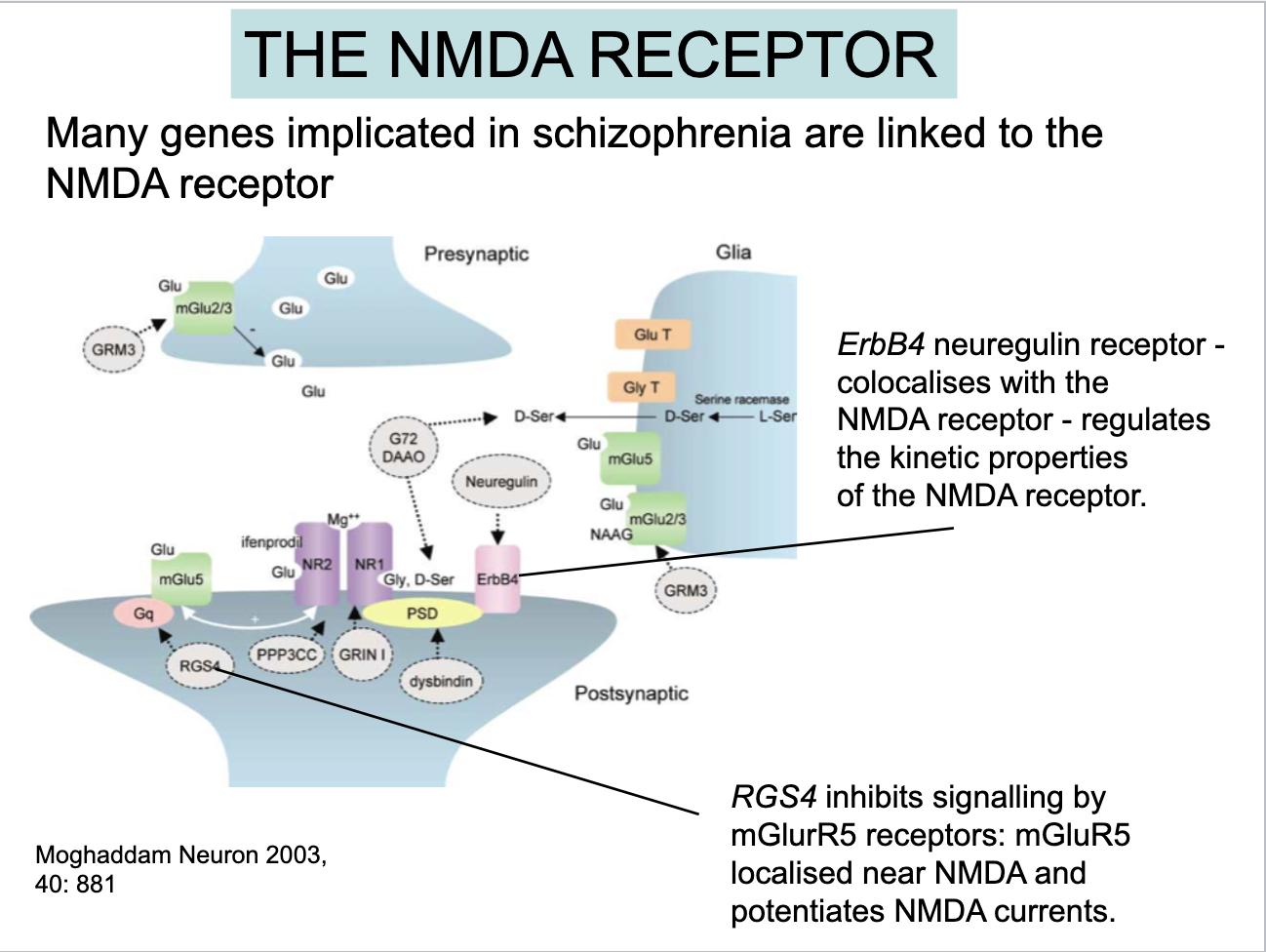
Many genes implicated in schizophrenia are linked to NMDA receptor regulation.
ErbB4: Neuregulin receptor that colocalises with NMDA receptors and regulates their kinetic properties.
RGS4: Inhibits signalling through mGluR5 receptors, which are located near NMDA receptors and can enhance NMDA currents.
what is the structure of a GABAA receptor
5 subunits round a central pore
About 2000 possible combinations but only about 20 expressed
Most commonly expressed Ɑ1, β2, and 𝜸2
Diversity - channel kinetics, GABA affinity, desensitization, phosphorylation, cell type specific expression.

What changes in GABA metabolism are associated with schizophrenia (SZ)?
Reduced GABA levels in post-mortem brains.
Increased GAT (GABA transporter), which removes GABA from the synaptic cleft for recycling.
This may be a compensatory response to low GABA levels.
Note: Increased GAT would remove GABA from the cleft more quickly, rather than allowing it to remain there for longer.
What changes occur in the GABAergic system in schizophrenia?
Also decreased mRNA concentrations for GAD67
- glutamic acid decarboxylase (enzyme that synthesizes GABA)
Also decreased GABA membrane transporter 1 (GAT1)
- responsible for reputake of GABA
describe the diversity between interneurons
GABAergic cells and their axons
Many axons, all release gaba
Inhibitory cell releasing GABA onto soma
Synapse in thousands of pyramidal cells
V important in controlling activity

How does the GABAergic system inhibit pyramidal cells?
PV (parvalbumin) basket cells release GABA onto pyramidal cells.
GABA binds to postsynaptic GABA receptors, opening Cl⁻ channels and allowing chloride ions to enter the cell.
This inhibits pyramidal cell activity and reduces neuronal firing.
GABA receptors can be synaptic or extrasynaptic.

What changes occur in parvalbumin (PV) interneurons in schizophrenia?
Decreased parvalbumin (PV) expression in fast-spiking interneurons in the hippocampus and prefrontal cortex.
The neurons are not necessarily lost; they show reduced PV expression.
Reduced PV mRNA is observed in schizophrenia.

What are perineuronal nets (PNNs), and what is their role around PV cells?
PNNs are a condensed form of the extracellular matrix.
They form a dense mesh around mainly PV interneurons.
They protect neurons and help maintain their connections in place.

What do WFA and PV label, and what is the relationship between PNNs and PV cells?
WFA (wisteria floribunda agglutinin): Labels perineuronal nets (PNNs).
PV: Labels parvalbumin interneurons.
PNNs surround a proportion of PV cells, with regional variations in the brain.

What changes in perineuronal nets (PNNs) are associated with schizophrenia?
Reduced PNN density around interneurons in certain brain regions.
Associated with potential dysfunction, including:
Cortical excitation/inhibition imbalance and thalamocortical dysfunction → cognitive impairments.
Amygdalar hyperexcitability and sensory processing abnormalities → altered perception.

what is the link between GABA and NMDA
NMDA hypofunction particularly on the interneurons
biological feedback control loop
Very active healthy interneurons and reasonably controlled pyramidal neurone
Pyramidal cell fires but doesn’t excite interneuron as much
Release a lot less gaba onto pyramidal cell- so becomes more excitable , more AP fired
Impairs feedback loop

what happens to GABA and glutamate in sz
Excitation inhibition balance
Too much excitation in sz
what are the links between dopamine, gaba and glutamate
Current antipsychotic treatments all act on D2R and target DA system.
Can manage the positive symptoms
Increased drive to striatum increase DA
Changes in COMT activity in PFC decrease DA?
Focus mainly on hippocampus and PFC and cognitive functions
what is neuroinflammation
Microglia and astrocytes become activated – change shape.
Microglia release pro-inflammatory cytokines including IL-6, IL-10 and TNF-alpha.
acute can be good
chronic is bad

what are glial cells
Glial cells- make up 50% of cells in brain
Complex cells , NT release and signalling
When there's an injury/infection/inflammation these are activated and release inflammatory cytokines
Normally clear up damaged neurones and connections
Sz- chronic neurone inflammation
Causes too many synapses being taken up and does more damage
what is the evidence for neuroinflammation playing a role in human sz
Microglia-
Sz- more blob like, shorter branches, hallmark of an inflamed state
but neuroinflammation is not unique to sz- many conditions

What is the role of neuroinflammation in schizophrenia?
Many schizophrenia (SCZ) patients have increased serum inflammatory markers, which correlate with grey matter loss in the medial prefrontal cortex (mPFC).
Activated microglia can engulf and prune synapses, reducing synapse numbers.
How can environmental factors and neuroinflammation contribute to schizophrenia?
Environmental factors (e.g. air pollution, toxins, maternal immune activation and early-life stress) may increase neuroinflammation and affect brain development.
Altered microglia activity can cause abnormal synaptic pruning and disrupt brain function.
The gut–brain axis and immune system may also contribute.
Schizophrenia involves an interplay between environmental factors and genetic risk factors.
