Lumbar Spine anatomy
 Call Kai
 Learn
 Practice Test
 Spaced Repetition
 Match
 Flashcards
 Knowt Play
1/50

There's no tags or description
Looks like no tags are added yet.
51 Terms
How many of each vertebrae?
7 cervical
12 thoracic
5 lumbar
5 sacral (fused)
4 conccygeal (fused
total = 33

General Features of Lumbar Verebrae
largest vertebral bodies
broad, square spinous processes
long transverse processes
Nerve roots and discs?
5 nerve roots (L1-L5)
5 vertevral discs (between L1-S1)

What is the significance of the L5-S1 (Lumbosacral) area
most common site for problems due to amount of stress that gets placed on it
bears more weight than any other vertebral joint
has most amount of movement of the lumbar joints
has the greatest angle than any other joint
increases the lordosis
transitions from the mobile L5 to the stable S1

Common L5-S1 injuries
Spondylolysis
Spondylolisthesis
Spondylosis
Spondylolysis
a defect (fracture) in the pars interarticularis of the arch of the vertebra
scottie dog with collar
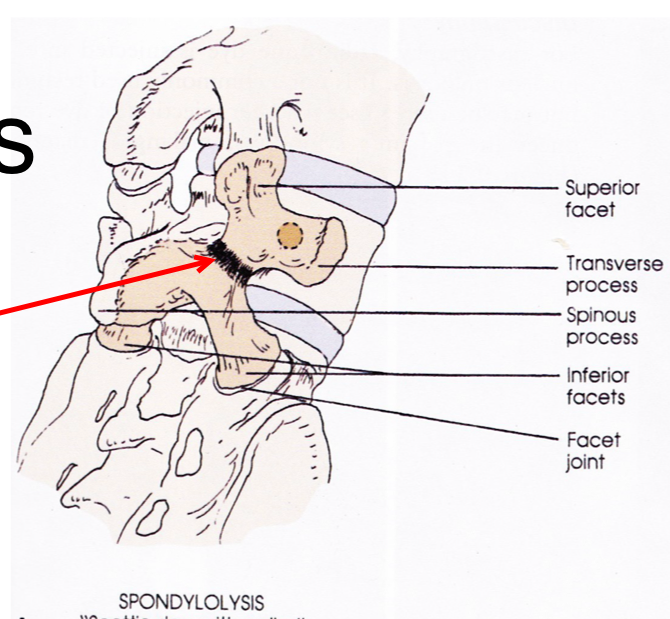
Spondylolisthesis
a forward displacement of one vertebra over another (bilateral fractures of pars)

Spondylosis
degeneration of the intervertebral disc
Lumbar facet joint orientation
along the sagittal plane
C- and J-shaped joints force the orientation 30-45 degrees into coronal plane

Lumbar facet processes orientation
superior processes face posterior and medially and are concave
helps with flexion/extension while limiting side flexion and rotation
Inferior processes face anterior and laterally and are convex

Correct Posture vs faulty posture
Correct: position in which minimum stress is applied to each joint and follows the line of gravity
Faulty: any position that increases stress to the joints
Classificaion of increased Lordosis (Lumbar curve deformity)
Exaggerated/Pathological Lordosis
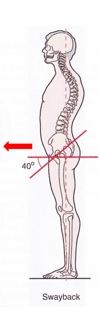
Swayback Deformity
Exaggerated/Pathological Lordosis
patient has a forward head posture, rounded shoulders and medially rotated legs
causes an increased lordosis to maintain the center of gravity

Swayback Deformity
the entire pelvis shifts anteriorly causing the hips to move into extension and the thoracic spine to flex on the lumbar spine
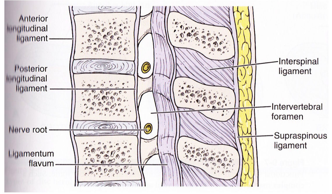
Lumbar Ligaments
anterior longitudinal ligament (ALL)
posterior longitudinal ligament (PLL)
ligamentum flavum
supraspinous ligament
interspinous ligament
intertransverese ligament
they are present throughout the entire vertebral column
Iliolumbar and lumbosacral ligaments
bands that extend from the L5 (L4) transverse processes to the iliac crests, or from the L5 transverse processes to the sacrum
both prevent forward displacement of L5 on the sacrum
both provide stabiliy to the lumbosacral junction

Lumbar Intervertebral Discs
thickest in the lumbar region
give space between verebrae for passage of nerve roots out from the spinal cord through the intervertebral foramina
allow more freedome of movement between the bones
discs are thicker anteriorly
only have pain fibers in the outer layer

Common Discs Injuries
Annular tears
Disc herniation
Annular Tears
Rim lesions
horizontal tears in outer lamellae
Concentric Tears
splitting apart of concentric lamellar layers due to torsion overload forces

How do Annular tears occur?
Can be trauma induced or result of osteophytes (bony spurs) and can lead to progressive degeneration of disc
Disc Herniation Injuries
Disc Bulge
Disc Protrusion
Disc Extrusion
Disc Sequestration
Disc Bulge
The disc bulges posteriorly without rupture of the Annulus Fibrosus

Disc Protrusion
the disc bulges and the nucleus is being contained only by the outermost fibers of the Annulus Fibrosus

Disc Extrusion
the Annulus Fibrosus is perforated and discal material (nucleus) moves into the Epidural space

Disc Sequestration
discal fragments from the Annulus Fibrosus and Nucleus Pulposus are found outside of the outer edge of the disc

What impacts the severity neurological impact?
it is dependent on the amount of pressure on the Nerve Roots or Spinal Cord
If the disc injury/ disc bulge does not compress on neurological tissue then they are not the cause of the patient’s pain/symptoms
Why does the spinal cord end at L1-L2
Because the vertebral column grows much faster
the consus medullaris is the distal end of the cord

Causa Equina Syndrome
pressure on the cauda equina causing server loss of neurological function
BIG RED FLAG need immediate medical help to prevent permanent damage
symptoms
difficulty with micturition (urination)
loss of anal sphincter tone or fecal incontinence
saddle anesthesia (anus, perineum, genitals)
multiple nerve root or progressive motor weakness in the legs or gait disturbance
Peripheral Nerve Distribution

Sciatic Leg Pain
More leg pain than back pain
leg pain below the knee
sharp and shotting
can have radiculopathy
positive SLR and Slump
Somatic Leg Pain
more back pain over leg pain
leg pain above knee
dull and diffuse pain
no radiculopathy
negative SLR and Slump
SOMATIC Pain: meaning pain coming from the body tissues (joints and soft tissues, not neurogenic) also known as Non-Specific Low Back Pain

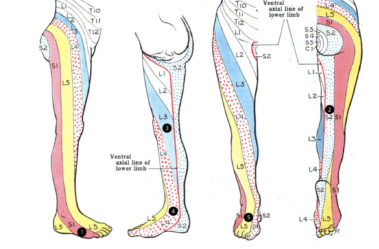
What are Lumbar Dermatomes
an area of skin supplied by a single spinal nerve root
**dont need to memorize
-L2 starts on the ASIS and comes medially at the mid-thigh level
-L3 starts over the TFL, comes medially across the thigh, over the vastus medialis, to the medial shin (half way down shin)
-L4 starts over vastus lateralis, comes over the patella, along the tibia, over the medial malleolus, and into the great toe
-L5 starts at the greater trochanter, goes directly inferiorly over the lateral knee, comes across the tibialis anterior, into the middle 3 toes, under the foot and into the later heel
-S1 comes along the lateral hamstrings, the lateral calf, comes over the lateral malleolus, and into the pinky toe
-S2 comes along the medial hamstrings, the medial calf, and into the medial heel
What are Lumbar Myotomes
A skeletal muscle innervated by a single spinal nerve
To assess myotomes, we will ask (or guide) the patient to get into the position and then say, “HOLD THE POSITION AND DON’T LET ME MOVE YOU OUT OF IT”
Then, we need to resist the action of the myotome for 5 seconds
What are the specific lumbar myotomes?
L1-L2: Hip flexion (and/or adductors)
L3: Knee extension
L4: Ankle dorsiflexion (and/or invertors)
L5: Great toe extension (and/or glut medius)
S1: Ankle plantar flexion, ankle eversion, and/or hip extension
S2: Knee flexion (and/or foot intrinsics)
Movement of the Lumbar Spine
Flexion: 40-60 D
Extension: 20-35 D
Side Flexion: 12-20 D each direction
Rotation: 3-18 D each direction

What two things control the Lumbar Spine
Global Muscle System
Local (Segemental) Muscle System
Global Muscle System

Local (Segmental) Muscle System

What are Prime Movers responsible for
lumbar spine range of motion and alignment
non-specific contribution to spinal control
What are prime stabilizers responsible for
fine-tune control of intervertebral motions
control the stiffness of spinal segments
connections to joint capsules and ligaments for proprioceptive input are activated in anticipation of movement to provide stiffness during motion
Abdominal Wall Muscles
Anterior component
rectus abdominus
Lateral component
external oblique
internal oblique
trasnversus abdominus
Posterior component
Quadratus lumborum
Psoas major
Iliacus
Rectus Abdominus
Compress abdominal contents, flex vertebral column, tense abdominal wall
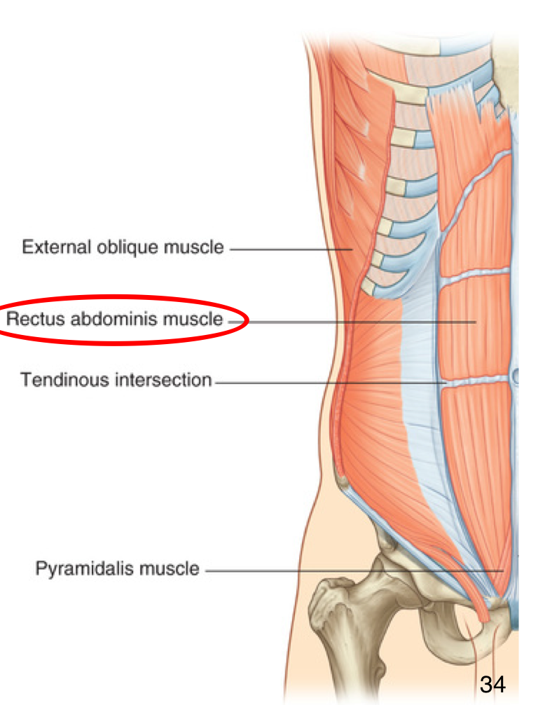
External oblique
Compress abdominal conents
Unilaterally: each muscle bends trunk ipsilaterally and turns abdomen contralaterally
Bilaterally: trunk flexion

Internal oblique
Compress abdominal contents
Unilaterally: each muscle bends trunk and turns abdoment ipsilaterally
Bilaterally: trunk flexion

Transversus Abdominis
Compress abdominal contents
draws in and increases intra-abdominal pressure (along with IO), supports abdominal contents (along with IO) and tenses thoracolumbar fascia

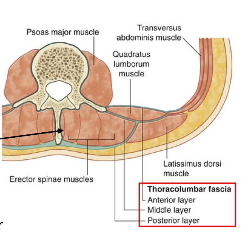
Thoracolumbar Fascia
connective tissue covering lumbar region
had anterior, middle, posterior layers
contains erector spinae, quadratus lumborum and multifidus
has attachments to latissimus dorsi, gluteus maximus, transversus abdominis, and internal oblique
stabilizes spine against anterior shear and felxion moments
helps with transmission of extension forces during lifting
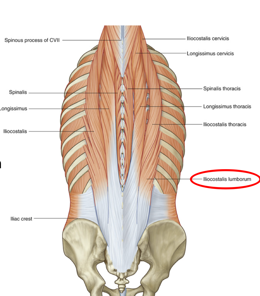
Iliocostalis Lumborum
Primary extensors of vertebral column
Unilaterally: slight side flexion and rotation of the vertebral column ipsilaterally
Bilaterally: extend the back
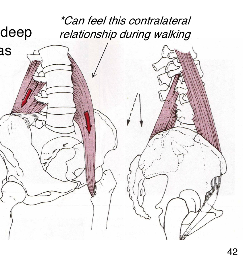
Relationship between Deep spinae and ilipsoas
work together contralaterally and ipsilaterally to stabilize the lumbar spine
anchor vertebrae to ilium and check against rotary forces
Lumbar Multifidus (A) (deep transversospinales muscles)
Unilaterally: contralateral rotation
Bilaterally: extend the vertebral column
thickended and prominent in lumbar region
attach to capsules of z-joints
Antiflexion: controls flexion during bending
Anti-shear: places a posterior and inferior directed force on lower lumbar levels to counter anterior shear
Increases spinal stability

Quadratus Lumborum
Depress and stabilize rib 12 and some lateral bending of the trunk

QL components
Medial portion:
iliotransverse part extends from iliac crest to lumbar TPs
costotransverse part extends from lumbar TPs to 12th rib
provides segemental stability bilaterally
Lateral portion:
iliocostal part extends from iliac crest to 12th rib (vertically)
works unilaterally to hike hip or to cause lumbar side flexion
