Medical Biology 2 L13
 Call Kai
 Learn
 Practice Test
 Spaced Repetition
 Match
 Flashcards
 Knowt Play
1/35

There's no tags or description
Looks like no tags are added yet.
36 Terms
ADHERENCE
The enhanced ability of a microbe to attach to a cell or surface
Examples
SLIME LAYERS
CAPSULE (also function to enable the bacterium to avoid phagocytosis)
FIMBRIAE (used for attachment)
SPECIAL PROTEINS
BIOFILMS
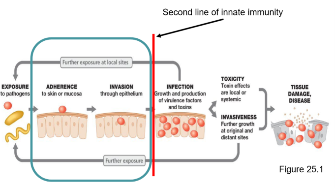
Invasion
THE ABILITY OF A PATHOGEN TO ENTER INTO A HOST CELL
The actual mechanism of entry is provided by the host
Pathogens will activate these mechanisms allowing them to enter the cells
They then produce proteins called INVASINS that rearrange the host cell structure to force the cell to uptake the pathogen

Invasion: portal of entry: MUCUS MEMBRANES
Inhalation, ingestion, urogenital tract
Invasion: portal of entry: SKIN
Attacks the skin layer, or burrows through the skin
Invasion: portal of entry: PARENTERAL
Enters through a wound in the skin or directly deposited under the skin
Second line of defense
used when the first line of defense is broken
Specialized cells can destroy a pathogen or its products
Previous exposure is not required
NO MEMORY and NO SPECIFICITY (the adaptive immunity has both memory and specificity)

Second line of defense: PHAGOCYTOSIS
Ingestion of microbes or particles by a cell
THE MICROBE IS ENGFULFED INSIDE A PHAGOCYTIC CELL AND KILLED
FAST RESPONSE
ALLOWS DEVELOPMENT OF ADAPTIVE IMMUNE RESPONSE
Second line of defense: PHAGOCYTOSIS Step 1: CHEMOTAXIS
Chemical attraction of phagocytes to microbes due to microbial products, components of white blood cells, damaged tissue cells, and the release of other chemicals
Second line of defense: PHAGOCYTOSIS Step 2: ADHERENCE
Attachment of the phagocyte plasma membrane to the surface of the microbes through the interaction of PAMPs (pathogen associated molecular patterns) on the pathogen with receptors on the phagocytes.
This step can be enhanced by OPSONIZATION
Second line of defense: PHAGOCYTOSIS Step 3: INGESTION
Pseudopods form around microbe and a PHAGOSOME is made (phagocytic vesicle)
Second line of defense: PHAGOCYTOSIS Step 4: DIGESTION
phagosome unites with a lysosome, forming a PHAGOLYSOSOME.
Microbe is digested by hydrolytic enzymes from lysosomes (10 - 30min)
Second line of defense: PHAGOCYTOSIS Step 5: FORMATION OF RESIDUAL BODIES
Phagolysosomes containing indigestible material is moved to the membrane and the contents are EXPELLED
Second line of defense: INFLAMMATION
Characterized by redness, pain, heat, and swelling, sometimes also with loss of function
FUNCTIONS TO CONTINE AND DESTROY INVASIVE AGENTS AND REPAIR DAMAGED TISSUES
Second line of defense: INFLAMMATION: VASODILATION AND INCREASED PERMEABILITY-
of blood vessels due to release of various chemicals. This permits defensive substances to leave the blood and enter injured areas
Second line of defense: INFLAMMATION: PHAGOCYTOSIS OF BACTERIA AND DEBRIS
Phagocytes appear and start to destroy microbes. (stimulated by chemokines)
When macrophages engulf large numbers of microbes and damaged tissue, they may also die leading to the formation of pus
Second line of defense: INFLAMMATION: TISSUE REPAIR
Occurs once all harmful substances have been removed or neutralized
regeneration time depends on the tissue itself
Second line of defense: ANTIMICROBIAL SUBSTANCES
The complement system
Interferon
The complement system
Consists of >30 serums
Acts in a cascade, one reaction triggers another, which triggers another
The purpose of a cascade is for amplification (1 protein activates 10 which activate another 10 etc)
The complement system: CLASSICAL PATHWAY
an antibody-antigen complex binds to a specific complement protein which eventually leads to the activation of C3 protein
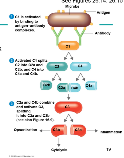
The complement system: ALTERNATIVE PATHWAY
An antigen binds to a specific complement protein which directly leads to the activation of C3 protein
Interferon
Compounds that interfere with virus multiplication

Interferon: GAMMA INTERFERON
Produced by lymphocytes and induce neutrophils and macrophages to kill bacteria
Interferon: ALPHA AND BETA INTERFERON
Produced by virus infected host cells
Diffuse to uninfected neighbouring cells
These cells are induced to produce antiviral proteins which disrupts viral multiplication
limit spread of infection
Invasions leads to infections
INFECTION happens when an organism gets through our natural barriers and starts to multiply
Occurs when a NON-NATIVE MICROBE IS ESTABLISHED AND GROWIN IN A HOST, disease may or may not result from infection
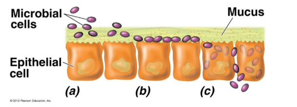
Infection: How a host can be harmed by microbes
COMPETITION FOR NUTRIENTS: pathogens can outcompete the host cell, which results in less growth in host cells
DIRECT DAMAGE:
multiplication: host cells are crowded out
production of metabolic by-products which can damage host cells (changes in pH) allowing other microbes through after penetration
PRODUCTION OF TOXINS
Substances produced by certain microbes and are poisonous
Some diseases are caused by toxins and not by the pathogen themselves
TOXICITY
Aspects of disease caused by toxins.
Eg
Tetanus caused by C. tetani. The organism DOES NOT LEAVE the wound site where it is growing, but releases the tetanus toxin which moves around the body.
INVASIVENESS
Ability to grow in host tissue and inhibit function.
Not necessarily dependent on a toxin molecule for disease
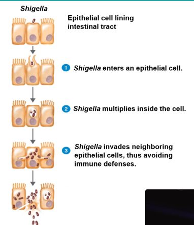
Toxicity and Invasiveness
Most pathogens use a combination of toxicity and invasiveness to cause disease
Enzymes that help with invasiveness
HYALURONIDASE
COLLAGENASE: Breaks down collagen
Toxins
molecules produced by pathogen that can harm cells/tissues or trigger damaging immune responses
The toxicity of an organism refers to its ability to cause disease by forming toxins
EXOTOXINS: released from cell
ENDOTOXINS: attached to the cell - LPS, LPS protein complexes
Enterotoxins: exotoxins that act in the small intestine - associated with food poisoning
Mechanisms of exotoxins
EXOTOXINS are produced in bacteria as part of their growth and then secreted out of the cell
Usually made of soluble proteins, and often are enzymes
only a SMALL AMOUNT is required to be considered toxic as enzymes can repeat toxic effects
More specific in their effects and targets
Can be produced by GRAM Positive and Negative

Mechanisms of endotoxins
Produced by GRAM Negative bacteria
Occurs when these cells lyse and the LPS PART of the outer membrane is released as an ENDOTOXIN
LARGER AMOUNTS are required to be toxic
Will vary in severity and in extreme cases cause death
Antibiotic treatment worsens the symptoms and then gets better. The antibiotics lyse the cells which increases the amount of toxins released
Endotoxins - Effects and examples
Released during bacterial MULTIPLICATION and CELL LYSIS
All endotoxins lead to similar unspecific symptoms such as chills, fever, weakness, aches, pains, shock
VIRULENCE FACTORS
Cellular components used to help establish and maintain disease
adhesion factors
redirecting nutrients for pathogen use (less for host cell)
enzymes that cause physical damage to cells or tissue
antiphagocytic factors
toxins
uses an assortment of virulence factors to enhance its pathogenicity
Symptoms vs Signs
SYMPTOMS: specific and subjective changes in the body (fatigue, weakness)
SIGNS: objective measurable changes in the body (temperature)
Stages of a disease: Specific sequence when host defenses are overcome
INCUBATION PERIOD: time between infection and appearance of disease signs and symptoms, can vary in length
ACUTE PERIOD: disease is at its height, with signs and symptoms
DECLINE PERIOD: disease signs and symptoms subside and host starts to feel better. Some can decline quickly some can slowly
CONVALESCENT PERIOD: patients regain strength and returns to normal