The Cell Cycle
 Call Kai
 Learn
 Practice Test
 Spaced Repetition
 Match
 Flashcards
 Knowt Play
1/31

Earn XP
Description and Tags
Gen Bio
32 Terms
Cell Division
The 1st important step of the cell cycle
3 main types - Binary fission, mitosis, meiosis
Functions of unicellular organism cell division
Reproduction (binary fission or mitosis)
Functions of multicellular organisms cell division
Reproduction (meiosis)
Growth & repair (mitosis)
Cells genetic information
Genome
Prokaryotes: One DNA molecule
Eukaryotes: Numerous DNA molecules packaged into chromosomes
Binary Fission
Chromosome replicates at the origion of replication
Two daughter chromosomes actively move apart
Plasma membrane pinches inward, dividing the cell into two
What do Eukaryotic chromosomes consist of
Chromatin
A complex of DNA and protein that condenses during cell division
Somatic Cells
Non reproductive body cells
Two sets of chromosomes
Gametes
Reproductive cells (eggs and sperm)
Have half as many chromosomes as somatic cells
Sister chromatids
Joined copies of the original chromosome, which seperate during cell division
Centromere
The narrow waist of the duplicated chromosome where the two chromatids are attach
Eukaryotic cell division process
Two sister chromatids seperate and move into two nuclei
Once seperate, the chromatids are called chromosomes
Eukaryotic cell division consists of
Mitosis, Meiosis, and Cytokensis
Mitosis
The division of the genetic material in the nucleus
Cytokinesis
The division of the cytoplasm. Usually, occurs simultaneously with telophase
Diploid (2n)
Normal number of chromosomes in somatic cell
Haploid (n)
Normal number of chromosomes in gamete
2 Major parts of the cell cycle
Interphase (cell growth and copying of chromosomes in preparation for cell division. G1, S, G2)
Mitotic phase (mitosis and cytokinesis)
Interphase
Makes up 90% of the cell cycle and has 3 phases
G1- Cell grows and makes proteins and organelles
S- DNA is duplicated
G2 - Cells prepare for division
Cytoplasmic contents double. Two centrosomes form

Stages of Mitosis
Prophase
Prometaphase
Metaphase
Anaphase
Telophase
Prophase
Chromosomes condense and the mitotic spindle begins forming in the cytoplasm. The microtubules rapidly grow out from the centrosomes.

Prometaphase
The nuclear envelope breaks apart, allowing spindle fibers to attach to the chromosomes. Microtubules extend from the centrosomes into the nuclear reigon
Some spindle microtubules attach to the kinetochores

Metaphase
The mitotic spindle is fully formed.
Chromosomes line up in the middle of the cell
Kinetochores of sister chromatids face opposite poles of the spindle.
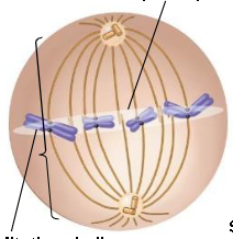
Anaphase
Sister chromatids separate and move to opposite sides
Daughter chromosomes are moved to opposite poles of the cell

Telophase
Two new nuclei envelopes form around the separated chromosomes
Chromatin uncoils
Mitotic spindle disappears

Mitotic Spindle
A system of fibers that separate the daughter chromosome
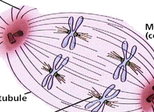
Microtubules
Tiny protein tubes that make up the spindle fibers

Centrosomes
Structures that order the microtubules during cell division

Spindle poles
Two opposite ends of the spindle
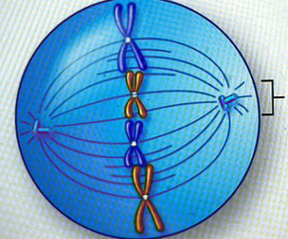
Kinetochore
A protein structure located at the centromere of a chromosome. It’s also where spindle microtubules attach
Animal cell division
Divide through cleavage furrow
Contracting ring of microfilaments are protein fibers that tighten around the middle of the cell, helping it pinch in two
Plant cell division
Build a cell plate in the middle
Use vesicles to build the plate because of their rigid cell wall