BIOL324 Exam 3
 Call Kai
 Learn
 Practice Test
 Spaced Repetition
 Match
 Flashcards
 Knowt Play
1/102

There's no tags or description
Looks like no tags are added yet.
103 Terms
What percentage of body weight is your skin
20%
5 functions of skin
1. Sensory- by housing receptors
2. Homeostasis- temperature regulation, pH, sweating, etc.
3. Protection- physical/chemical barrier, also houses phagocytes to rid foreign particles
4. Excretion- of sweat, electrolytes, waste, etc.
5. Lesser functions including calcium regulation (via vitamins D & B), and signaling molecules for attracting opposite gender
2 main layers of skin
- Epidermis: thin keratinized stratified squamous epithelium
- Dermis: thick conn tissue, also littered with smooth muscle, vessels, follicles, glands, etc
- Hypodermis is beneath dermis, although it isn't considered a main layer
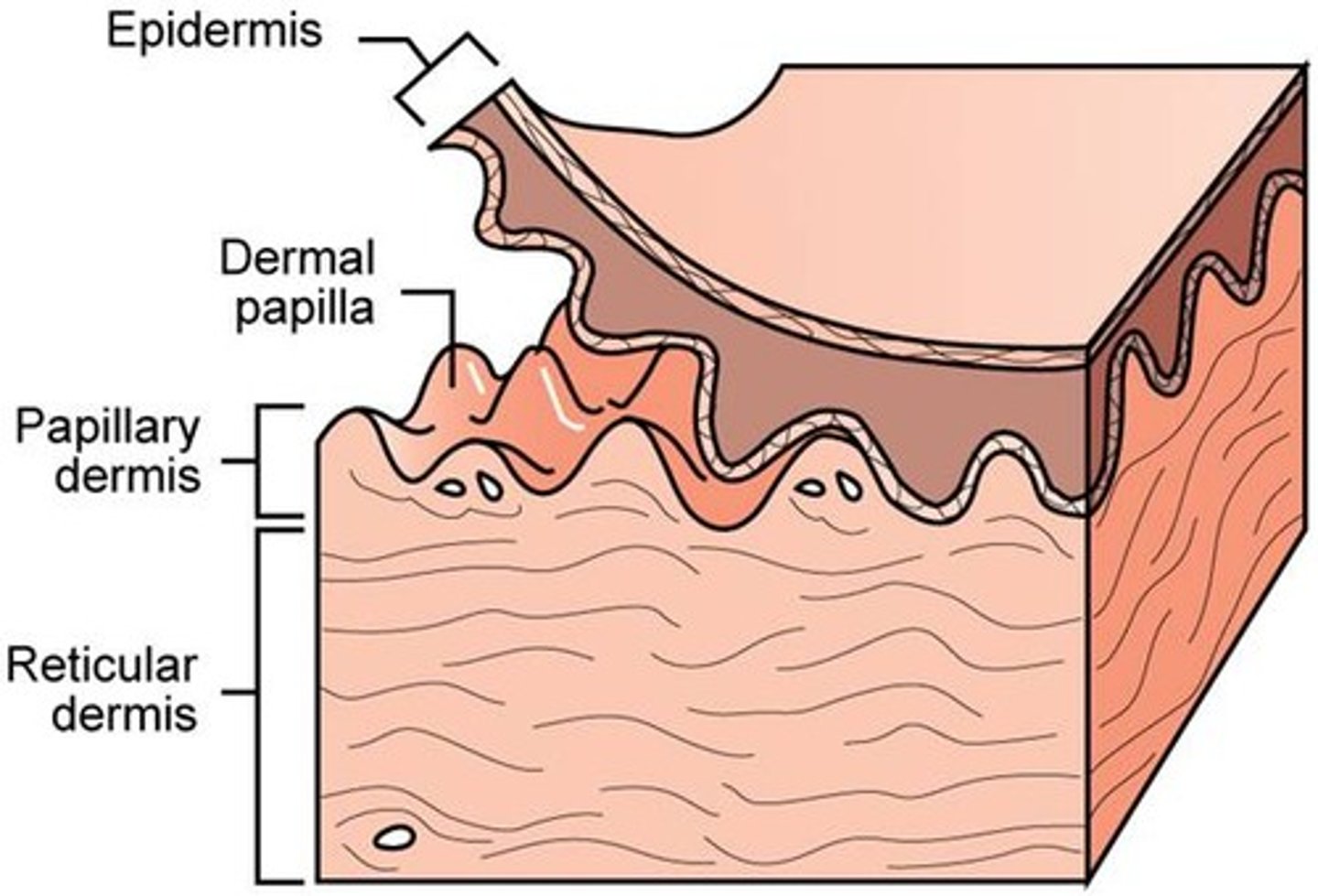
Name and describe the 2 layers of the dermis
1. Papillary layer- protrude into the epidermis, composed of loose conn tissue w/ elastic fibers
2. Reticular layer- under the dermal papillae, denser conn tissue with collagen and elastin higher in abundance
Difference in the weave of CT in the papillary/reticular layers
- Papillary: loose CT with randomly woven collagen and elastin- "feltlike"
- Reticular: denser CT runs in parallel lines called Langer's lines, made of collagen, elastic fibers, and reticular fibers
Hypodermis (subcutaneous) layer
- Layer of skin under the dermis, not considered a main layer of the skin
- Main function is insulation and energy storage for muscles via white fat
5 layers of epidermis
(Environment)
1. Stratum corneum
2. Stratum lucidum
3. Stratum granulosum
4. Stratum spinosum
5. Stratum basale
(Dermis)
Stratum basale (function, what type of cells?)
- Bottom-most layer of the epidermis
- Germ layer aka area of cell division (outward growth)
- One cell layer thick, those cells vary btwn simple columnar or cuboidal
- Where melanocytes are found
Stratum spinosum (how many layers? what is the cellular structure?)
- Layer of epidermis right above the stratum basale
- Stratified
- The cells are compartmentalized because they are inside "white halos" of extracellular space, makes it look like there is a rigid spine (spinosum) holding together it together
- Desmosomes connect the different cells together
Nodes of Bizzozero
- Groupings of desmosomes
- Known for being apparent in the stratum spinosum, connecting surrounding cells
Malpighian layer
An alternate name for both the stratum basale and the stratum spinosum together
Stratum granulosum (thickness, composition)
- Middle layer of the epidermis
- 2-4 cells thick of squamous epithelium (stratified squamous)
- Cells contain keratohyaline granules, as you move towards the surface, each cell layer of the stratum granulosum contains more granules
Stratum granulosum (function, reactants?)
- Cells in this layer are mature keratinocytes, and secrete a lot of keratin
- The two primary reactants to create keratin: keratohyalin + cytokeratin
Are the stratum granulosum, the stratum lucidum, and the stratum spinosum basophilic or acidophilic?
- Granulosum: basophilic
- Spinosum: acidophilic
- Lucidum: acidophilic
Stratum lucidum (appearance, cells, where in the body)
- Layer of the epidermis second from the top
- Cells are more flat than the granulosum (squamous) and appear transparent
- Cells in this layer are filled with eleidin
- Only found in thick skin, not thin skin
Thick vs thin skin
- Thick: palms of hands and feet, NO stratum lucidum
- Thin: everywhere else ex. arms, eyelids, etc; HAS a stratum lucidum
**Note: the skin on our back is the thickest on body, but is thin skin
**Note: Skin is the dermis + the epidermis. Thick and thin skin only refers to the epidermis
Stratum corneum (makeup)
- Layers of dead keratinocytes
- Varies in thickness
- Calluses can form which is a vast buildup of these dead cells
Cytokeratin => Tonofibrils
- Cytokeratin binds together to make up a tonofilament in the stratum basale
- These tonofilaments are bound together in the stratum spinosum by filaggrin to make tonofibrils
**Remember: tonofibrils/filaments help support desmosomes in the stratum spinosum
How big do kertohyaline granules become? Why? Membrane?
- Between 1 and 5 micrometers (um)
- Granules absorb tonofilaments (10 nm each) and tonofibrils that were made in the stratum basale/spinosum
- Not membrane-bound and don't leave the cells of the stratum ganulosum (non-secretory)
Desmosomes and Hemidesmosomes, what is the difference and function?
- Structures that counter the effects of friction on the skin
- Tonofilaments and tonofibrils reinforce these bindings
- Desmosomes bind cells of the epidermis tg, hemidesmosomes bind cells of the epidermis to cells of the dermis (through the basement mem)
What percentage of cells in the epidermis are keratinocytes
84%
Cytomorphosis
Cycle of a keratinocyte as it is differentiated in the stratum basale until it moves up and dies and falls off in the stratum corneum
Two major functions of the basal cells (stratum basale)
1. Differentiate (mitosis) into young keratinocytes that push up into the stratum spinosum
2. Their ribosomal activity produces cytokeratin proteins that form tonofilaments that are then later used in (hemi)desmosomes for support
What happens in the stratum granulosum and onward to produce keratin
- Tonofilaments/fibrils get absorbed into keratohyaline granules and they grow up to 5 micrometers in diameter
- Reacts with cytokeratin in the stratum granulosum to produce Eleidin (when the cells start producing eleidin, they are officially part of the stratum lucidum
- As the cells move up to the stratum corneum, this transforms into keratin
3 main components in the keratohyaline granules
1. Filaggrin
2. Phosphate-rich proteins
3. RNA
What indicates the switch of keratinocytes from the stratum lucidum to the stratum corneum
When the conversion of eleidin to keratin has been completed, the cells are officially part of the corneum
Role of loricrin and involucrin in the stratum granulosum
Insoluble proteins that are deposited throughout the keratinocyte's membrane
Act as a barrier between the "wet" internal environment and the "dry" external environment in the lucidum and corneum
Role of Serine proteases in the stratum granulosum/lucidum
- Inactive enzyme in the granulosum because of the neutral pH, exists between the cells
- In the stratum lucidum, the pH drops to ~4.5, the enzyme activates and targets desmosomal proteins
- The desmosomes break down and the flattened keratinocytes eventually separate from the corneum (sloughed off)
- The sloughing of these cells at the end of their life cycle maintains the uniform thickness of the epidermis as new cells differentiate in the basale
Lamellar bodies (where, membrane?, composition, size)
- Produced in the same stratum granulosum cells that form keratohyaline granules
- 0.1-0.5 micrometers
- Membrane-bound (secretory)
- Contain thin layers of lipids (lamellae) and serine proteases
- These cells eventually exocytose the lamellar body's contents into the extracellular space
Function of the contents inside a lamellar body
- Occurs in stratum granulosum
- Keratinocytes in granulosum release the contents of the lamellar body into the extracellular space
- Lipids inside the cell coat the membranes of other keratinocytes
- Serine proteases work on cleaning desmosomes to loosen up the keratinocytes so they can eventually flake off
What are the two mechanisms that keratinocytes use for water resistance in the stratum lucidum and the stratum corneum
- Lamellar bodies secrete lipids that line the outside of a keratinocyte's membranes
- Loricirin and Involucrin are insoluble proteins that line the inside of the keratinocyte's membrane
What percentage of epidermis cells are melanocytes
5% - this varies based on location though, the more exposed that part of the body is to the sun, the more melanocytes there are
- Also can vary for other reasons, male scrotums evolved to have more melanin (???)
Endoderm, ectoderm, mesoderm: which do keratinocytes, melanocytes, langerhans cells arise from
- Keratinocytes: ectoderm
- Melanocytes: ectoderm; neural crest
- Langerhans cells: mesoderm, blood cells in the bone marrow, then flow to the epidermis
Melanocytes (function, location, membrane?)
- Provide pigmentation (melanin) to keratinocytes to protect against UV radiation
- Found in stratum basale only
- Membrane-bound (secretory)
Melanocytes: desmosomes or hemidesmosomes?
Hemidesmosomes as anchors
Melanocyte vs melanosome
Melanocytes: cells producing melanin,
Mmelanosomes: organelles within melanocytes where melanin synthesis takes place
2 main components of melanosomes
1. Structural proteins
2. Enzymes
Melanosome stages I - IV
- S I: pre-melanosome, .25 micrometers in diameter
- S II: still a pre-melanosome, structural proteins secrete a grid-like framework which melanin will be deposited into
- S III: melanosome takes up AA tyrosine, subsequently converted to DOPA, and then melanin by the enzyme tyrosinase
- S IV: mature melanosome, melanin-filled melanosomes are transported to the cell processes where they are transported into adjacent keratinocytes via phagocytosis (process is called cytocrine secretion; takes place in the stratum spinosum, *think ab the processes extending off of the basal melanocytes into the layer above)
What percentage of epidermal cells are Langerhans Cells
~3%
Where are Langerhans cells located
In the stratum spinosum along epidermal ridges called rete ridges
Structure and alternate name for Langerhans cells
- Contain a cell body with extensions off, look much like a melanocyte
- Also called: Epidermal dendritic cells
Langerhans cells (function)
- Part of the mononuclear phagocytic system (MPS) ; essentially specialized macrophages
- Type of antigen-presenting cell (APC), they monitor the keratinocytes of the epidermis for antigens, when they find antigens, they will phagocytose it and display parts of it on the keratinocyte's surface
How are langerhans cells involved in immune response
- Cells will display part of antigens on keratinocyte surface as part of their duty as antigen presenting cells
- Immune cells will recognize this and next time it is detected it will illicit an immune response
- This delay in immune response is known as a delayed-type hypersensitivity reaction, like contact dermatitis
Birbeck granules
- Small "tennis-racket shaped" membranous granules found only in langerhans cells
- Function is unknown
What percent of epidermal cells are merkel cells (tactile cells)
~8%
Structure of Merkel cells (tactile cells)
- Modified basal cells (stratum basale)
- Attach to surrounding keratinocytes via desmosomes unlike other epidermal cells
- have cell processes similar to melanocytes and Langerhans cells
Relevance of dense-core neurosecretory granules
- Granules in merkel cells (tactile cells); less than 100 nm in diameter
- Contain catecholamines - function as neurotransmitters
Function of merkel cells (tactile cells)
- Specialized for sensing light touch
- Merkel cells connect to pseudo-unipolar afferent neurons to relay sensory information to CNS
- The catecholamines that the granules house act as the neurotransmitter to relay this information
- The neuron and the merkel cell together are called a merkel's corpuscle, a type of mechanoreceptor
Catecholamines
- Aka hormones like epinephrine and norepinephrine
- In the scope of this class, these hormones are secreted by dense-core neurosecretory granules in Merkel cells to relay light touch information to the CNS
Free nerve endings
- Non-capsulated endings of a nerve that penetrate up into the epidermis from below as far as the stratum granulosum
- Capable of relaying a broad range of feelings (ex. nociceptors for pain)
- Most numerous receptor in epidermis
Pacinian Corpuscles
- Encapsulated receptor found deep in dermis, almost at the hypodermis
- Very deep, therefore lots of pressure needed to stimulate them, therefore not as sensitive
Meisner's Corpuscles
- Encapsulated receptor located in the dermal papillae
- Closer to surface, therefore less pressure needed to stimulate, therefore more sensitive
2 major portions of respiratory system based on interaction with air
1. Conducting portion
2. Respiratory portion
Conducting portion of respiratory system
- From the nasal cavity to the terminal bronchioles
- Conditions incoming air before gas exchange by humidifying it and "cleansing" it
Respiratory portion of respiratory system
- Respiratory bronchioles and alveoli
- Function in gas exchange
Vestibule of the nasal cavity
- Just inside the nostrils, first point of entry
- Has vibrissae (hairs) that catch material coming in with the air, acting as a filter
What is the nasal fossa made up of?
- Point just after the vestibule (initial entrance)
Superior conchae
medial conchae
inferior conchae
Venous plexus (aka swell bodies) of the nasal cavity
- Plexus of veins under the epithelium in the nasal cavities
- One side of the plexus engorges (from back to front) every 20-30 mins, and then it switches to the other side
- Only 60%/40% shunting, not shutting off the whole half of the nose
- This helps air use all of the nasal cavity, and heats/humidifies air as well as it travels through the cavity (countercurrent heat exchange)
Epithelium of the respiratory system (starting outside the nose)
1. Outside the nose- keratinized stratified squamous epithelium (epidermis)
2. Vestibule- non-keratinized stratified squamous epithelium
3. Small amount of transitional epithelium
4. Nasal fossa- Respiratory epithelium (ciliated pseudostratified columnar epithelium
Function of cilia in respiratory epithelium
- Starts at nasal fossa (conchae) and extends down into bronchioles
- Moves mucous and trapped foreigners out of the airway
Immotile cilia syndrome (aka kartagener's), how does it relate to dyenin
- Congenital deficiency of motor proteins called dyenin in cilia, causing them to not work properly
- Mucous and anything trapped within are stagnant, acting as a growth medium for bacteria, causing frequent respiratory infections
Goblet cells (respiratory)
- Specialized columnar cells abundant in respiratory epithelium
- Produce boatloads of mucous
Brush cells (respiratory)
- Columnar cells with acting as sensory cells
- Have blunt microvilli on their apical surface, and synapse with afferent neurons (pseudo-unipolar) on their basal surface
- These neurons lead into the trigeminal nerve, mostly used for facial sensation (generates sneezes)
What do the basal cells of respiratory epithelium do?
- Act as stem cells regenerating lost tissue
- Work in the basement membrane
Small granule cells (respiratory) (define, two subpopulations)
- Cells are a part of a body-wide system of cells called Diffuse Neuro Endocrine System (DNES)
- Cells contain dark secretory granules ranging btwn 100-300nm in diameter
- These small granule cells are further separated into two different populations based on the composition of those granules
Small granule cells (respiratory): explain the two subpopulations based on granule composition
1. Catecholamines: secrete hormones like dopamine, epinephrine, etc. Produced/secreted by process called APUD
2. Other categories of chemicals: like serotonin and peptide hormones that are catecholamines
APUD
Amine precursor uptake and decarboxylation
- Catecholamines are synthesized and secreted
- Cells that secrete catecholamines are merkel cells (epidermis) and small granule cells (respiratory)
- Uptake of amino acids like Tryptophan, which are subsequently decarboxylated to synthesize hormones
What are two exceptions to the normal respiratory epithelium going down the respiratory tract
1. Olfactory epithelium in the superior turbinate of the nasal fossa
2. Non-keratinized stratified squamous epithelium at the larynx (aka epiglottis and vocal cords)
Olfactory epithelium
- Exception to the normal respiratory epithelium
- Exists in the nasal fossa, above the superior turbinate
- Still pseudostratified columnar, but is specialized to allow for smell
4 cells in Olfactory epithelium
1. Olfactory cells
2. Sustentacular cells
3. Basal cells
4. Brush cells
Olfactory cells
- One of the cells in olfactory epithelium
- Has cilia which act as odorants (detect smell)
- Cell body of the neuron is the cell in the pseudostrat epithelium, dendrite projects to apical surface connecting to the cilia, and it has a single axon that leaves through the basal surface to connect with the olfactory nerve (bulb)
- Neuron is bipolar
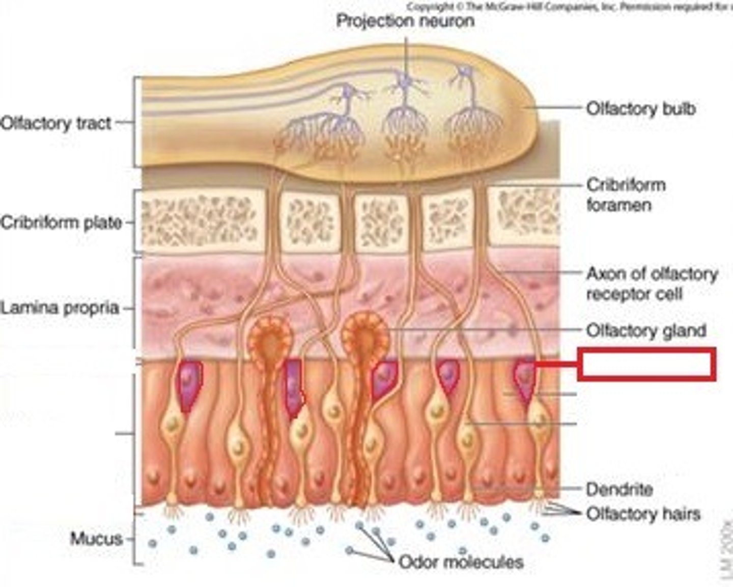
Sustentacular cell
- Supporting cell in olfactory epithelium (physical and metabolic support of the olfactory cells)
- Contain microvilli
- Produce a odorant binding protein- this carrier molecule facilitates the odorant binding to the non-motile cilia of an olfactory cell
3 types of neurons relevant to this class
1. Multipolar- 1 axon and 2+ dendrites (MOST abundant), are motor neurons
2. Bipolar- 1 axon and 1 dendrite (rare), found in the 5 senses (in the brain), sensory
3. Pseudo-unipolar- 1 axon with NO dendrites, only sensory neurons (afferent)
Bowman's Glands (aka olfactory glands) (location, classification, function)
- Found in respiratory epithelium near the olfactory cells (nasal fossa)
- The gland itself is under the epithelium in the lamina propria (CT) and the duct penetrates through to the surface
- Classified as simple serous acini
- Secrete serous solution (very watery) onto epithelial surface which keeps surface wet and acts as a solvent for odorants
- Constantly secretions wash away old odorants
Paracrine vs endocrine hormones
- Paracrine: localized secretion and effect
- Endocrine: Secreted somewhere, hormone used circulatory system to act on a part of the body farther away
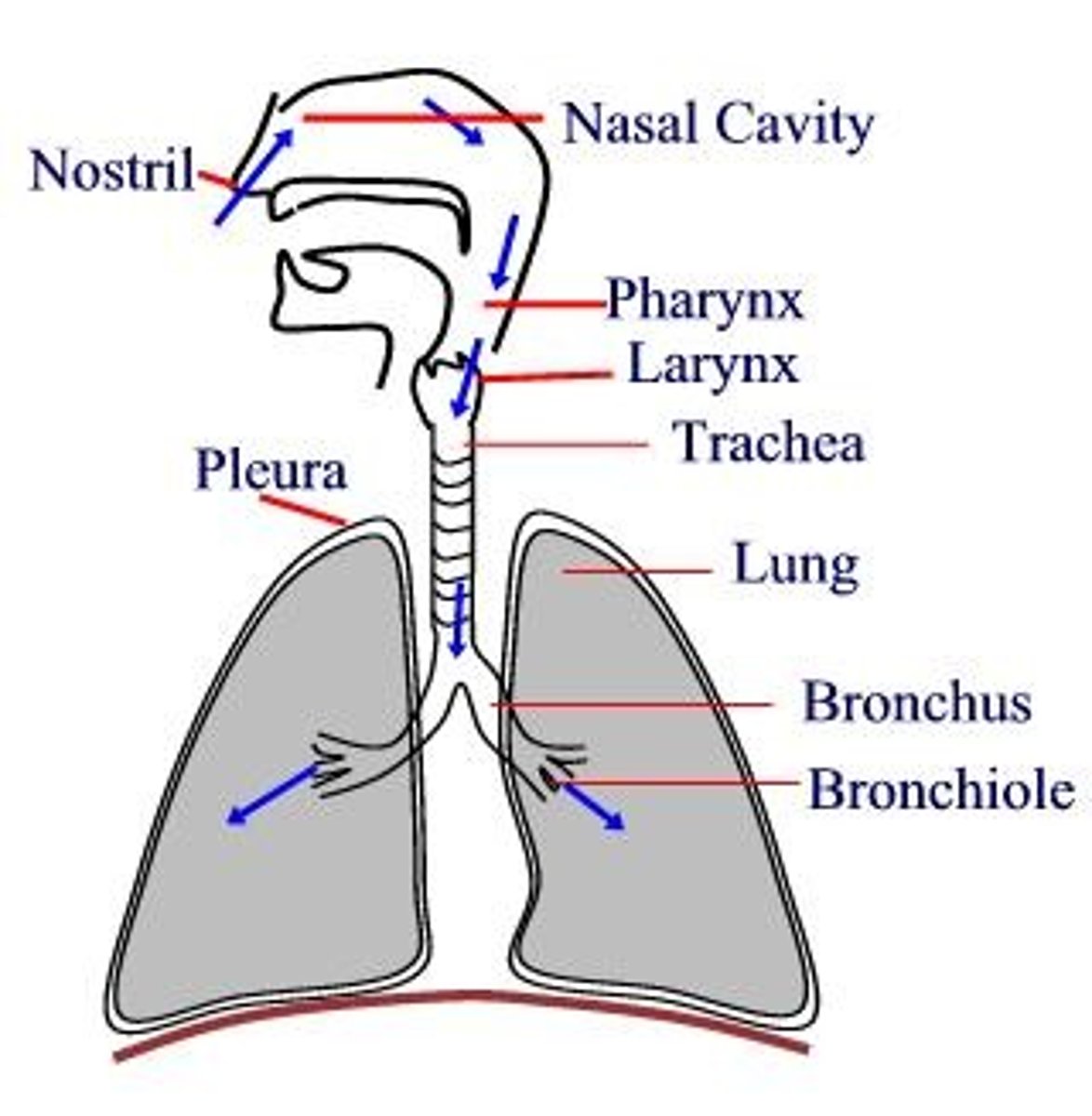
Path of respiratory system from nose to alveoli
1. Nasal vestibule
2. Nasal fossa/conchae
3. Pharynx
4. Larynx (voice box/epiglottis)
5. Trachea
6. 2 primary bronchus
7. Branch into bronchi (5 mm in diameter)
8. Terminal bronchioles
9. Respiratory bronchioles
10. Alveoli
Don't really needa memorize, more for context
Terminal bronchioles
- Last part of the conducting portion of the respiratory system
- Epithelium changes from ciliated pseudo. strat. columnar to ciliated simple columnar
- Lined with Club cells (aka bronchiolar exocrine cells)
Club cells (bronchiolar exocrine cells) (structure, location, secretions)
- Line terminal bronchioles
- Dome-shaped and non-ciliated
- 2 secretions: Surface-active agent (a slippery lipoprotein) and Club cell secretory protein (regulates inflammatory process of bronchioles)
What cells are typically found in bronchioles of smokers and not non-smokers
- Goblet cells
- Found in smoker's bronchioles
- Their epithelium reacts to the constant irritants, thinks it needs more lubricant
Metaplastic change
The transformation of one differentiated cell type to another differentiated cell type
- Ex. Goblet cells being in smoker's bronchioles, which used to be club cells. This can be a precursor to cancer
Function of smooth muscle in the bronchioles (what nerve does it run thru)
- Found in the lamina propria (CT) of terminal bronchioles
- Contraction: parasympathetic fibers
- Dilation: sympathetic fibers (means more oxygen entering)
- Fibers run through the vagus nerve
Respiratory bronchioles and alveoli (epithelium?)
- After terminal bronchiole comes the respiratory bronchiole lined with alveolar sacs/ducts
- Terminal and respiratory bronchioles : ciliated simple columnar epithelium
- Alveoli: simple squamous epithelium - easy gas exchange
3 cells in the alveolar epithelium
1. Type 1 pneumocyte- simple squamous cells of the epithelium; 95% of the layer
2. Type II pneumocyte (aka great alveolar cell)- 3% of the epithelial cells. Contain lamellar bodies with secretory granules which secrete surfactant, proteins, leukocytes, etc.
3. Brush cells - type of sensory neuron/cell with microvilli on apical surface
3 organs in the abdominaopelvic cavity that aren't part of digestive system
1. Spleen
2. Kidney (x2)
3. Adrenal glands (x2)
6 accessory digestive organs/structures
1. Tongue
2. Teeth
3. Salivary glands
4. Liver
5. Gall bladder
6. Pancreas
4 layers of alimentary canal
(most internal)
1. mucosa
2. submucosa
3. muscularis externa
4. serosa
(most external)
Mucosa layer of the alimentary canal
- Consists of a mucous layer covering the epithelium which covers the lamina propria (loose ct) which covers the muscularis mucosa (very thin dual muscle layer)
- Mucous is secreted by various glands
- Only places along canal not covered by mucous is the mouth opening and the anal opening
Considering the mucosa (alimentary canal), what are 3 things contained in the lamina propria?
1. Vessels and nerve fibers
2. Small glands
3. MALT
MALT vs GALT
GALT- Gut-associated lymphoid tissue - any lymphoid tissue of any layer of the alimentary canal
MALT- Mucosal-associated lymphoid tissue - a subsection of the GALT that only affects the mucosal layer of the alimentary canal
Muscularis mucosa and muscularis externa: what is the orientation of the two layers? What different plexuses innervate them?
Inner layer of SM wraps around the tube circularly; outer layer of SM runs longitudinally down the length of the tube
- Muscularis mucosa: innervated by miessner's plexus of the submucosa
- Muscularis externa: innervated by myenteric (aka Auerbach) plexus - from btwn the two layers of SM muscle, NOT the submucosa
Submucosa layer of the alimentary canal
- Dense connective tissue layer under the mucosal layer
- Acts as a pathway for nerves, vessels, and lymphatics
- "Holds" structures like glands (specifically brunner's glands in the duodenum) and GALT (specifically peyer's patches)
What plexus runs through the submucosa of the alimentary canal
- Miessner's Plexus
- Innervates the muscularis mucosa with parasympathetic fibers only
Peyer's patches
large collections of lymphoid tissue found in the submucosa of the small intestine (accumulation of lymphocytes)
Muscularis externa: what is its function? Plexus? (Para)Sympathetic?
- Function: controls peristalsis
- Innervated by myenteric plexus (aka auerbach plexus)
- Sympathetic and parasympathetic
Serosa of the alimentary canal: definition, what is it composed of, what does it arise from
- The outermost layer; simple squamous epithelium and thin layer of CT
- The epithelium is called a mesothelium because it arises from the mesoderm
- Encloses the whole system from the rest of the contents in the body cavity and has a fine layer of liquid on the outside
3 types of mucosa in the oral cavity (based on functionality)
1. Masticatory mucosa- exposed to the most chewing
2. Non-masticatory mucosa- little chewing
3. Specialized mucosa- specialized function, may be exposed to chewing
Masticatory mucosa (define, where)
One of the three types of mucosa in oral cavity
- Exposed to the most chewing
- Locations: gingiva and hard palate
- 3 different patterns of this mucosa appear
What are the 3 patterns of masticatory mucosa?
1: on the gums and raphe (line that runs down palates)
2: Fatty zone of hard palate (more towards front teeth)
3: Glandular zone of the hard palate (more towards back of mouth)
Non-masticatory mucosa (1. define 2. where 3. epithelium 4. composition)
One of the three types of mucosa in oral cavity
- Exposed to little chewing
- Found on cheeks, ventral surface of tongue, floor of mouth, and soft palate
- Epithelium for this mucosa: non-keratinized stratified squamous
- Composed of: Incomplete mucosa: epithelium, lamina propria, submucosa with salivary glands (no muscularis mucosa/externa or serosa)
what are the three salivary glands, what mucosa/layer do they run through?
1. parotid
2. submandibular
3. sublingual
Excrete saliva through glands running through non-masticatory mucosa layers