30 nonunion and bone defect
 Call Kai
 Learn
 Practice Test
 Spaced Repetition
 Match
 Flashcards
 Knowt Play
1/30

There's no tags or description
Looks like no tags are added yet.
31 Terms
جمعبندی مفهومی: در nonunion، ابتدا باید مشخص کنیم مشکل غالب، بیولوژی است یا مکانیک؛ سپس alignment، اندازه defect، وضعیت بافت نرم و احتمال عفونت را وارد تصمیمگیری کنیم. در nonunion عفونی، کنترل عفونت معمولاً مقدم است؛ در atrophic nonunion، بیولوژی و پایداری هر دو اهمیت دارند؛ و در hypertrophic nonunion، اصلاح پایداری و alignment محور اصلی درمان است.
در برخی تعاریف کلاسیک، Delayed union در حدود ۳ ماه و Nonunion در ۶ تا ۸ ماه مطرح میشود؛ اما این اعداد مطلق نیستند.
تفاوت سلولی
Nonunion: توقف فرایند ترمیم پیش از ایجاد اتصال استخوانی.
Delayed union: ممکن است پاسخ ترمیمی پریوست متوقف شود، درحالیکه پاسخ اندوستال هنوز بهطور کامل از بین نرفته باشد.
جمعبندی نهایی مخصوص شب امتحان
این ۱۰ نکته را حتماً به خاطر بسپار:
Nonunion یعنی شکست ترمیم و احتمال پایین جوشخوردن بدون مداخله جدید؛ زمان بهتنهایی کافی نیست.
Atrophic = مشکل بیولوژیک؛ Debridement و اغلب Bone graft.
Hypertrophic = مشکل مکانیکی؛ افزایش پایداری، معمولاً بدون Bone graft.
Oligotrophic = مشکل احتمالی ترکیبی؛ اصلاح تماس، پایداری و در صورت لزوم بیولوژی.
Pseudoarthrosis = حرکت مزمن و تشکیل شبهکپسول سینوویال؛ Debridement و بازکردن کانال مدولاری.
دیابت، سیگار، آسیب پرانرژی، آسیب بافت نرم و عفونت از عوامل مهم Nonunion هستند.
CT حساس است، اما اختصاصیت محدود آن میتواند به تشخیص اشتباه Nonunion منجر شود.
در Nonunion عفونی، کنترل عفونت و Debridement معمولاً مقدم بر ترمیم قطعی است.
در Malaligned nonunion، اصلاح بدشکلی باید بخشی از برنامه درمان باشد.
در انتخاب درمان، مشکل اصلی را پیدا کن: Biology، Mechanics، Infection، Alignment یا Bone defect.
جمعبندی بورد
Dynamization نسبتاً کمتهاجمی و کمهزینه است و گاهی اجازه Weight bearing فوری میدهد؛ اما درمان قطعی و قابلاعتماد همه Nonunionها نیست.
بخش سوم: ترکیب Plate و Nail برای Nonunion
در شکستگی حاد، ترکیب Nail و Plate بهصورت تاریخی با احتیاط استفاده میشد؛ زیرا Plate ممکن است سازه را بیشازحد سفت کند و Micromotion لازم برای Secondary bone healing را کاهش دهد.
موارد کاربرد
Distal femoral nonunion
Femoral shaft nonunion پس از IM nailing اولیه
Humeral nonunion پس از IM nailing
Augmentative plating

نکته مفهومی: در Stiff hypertrophic nonunion، مشکل عمدتاً مکانیکی است؛ اما در Mobile atrophic nonunion، مشک

Author’s preferred treatment for nonunions
رمز حفظی: «هایپرتروفیک = تثبیت؛ بدراستایی = اصلاح راستا»

الگوریتم


الگوریتم


.۳. خارج کردن یا حفظ ایمپلنت؟
این تصمیم به دو عامل اصلی بستگی دارد:
آیا ایمپلنت پایداری کافی ایجاد میکند؟
آیا احتمال کنترل عفونت با حفظ آن وجود دارد؟
.
مواردی که removal ترجیح دارد
ایمپلنت پایداری ناچیز یا ناکافی دارد.
کنترل عفونت دشوار است.
bone grafting موردنیاز است.
احتمال تشکیل biofilm و تداوم عفونت زیاد است
Taylor Spatial Frame (TSF)
یک circular external fixator ششمحوره است که امکان اصلاح همزمان deformity را فراهم میکند.
بخش دهم: Bone Graft در Nonunion
اصول کلی
Bone graft اتولوگ همچنان استاندارد مرجع برای بسیاری از Nonunionها، بهویژه Atrophic nonunion، محسوب میشود
Autogenous Bone Graft
اندیکاسیونهای رایج:
Atrophic nonunion
بعضی Oligotrophic nonunionها
بعضی Pseudoarthrosisها


Three humeral shaft fractures treated nonoperatively developed different types of nonunions: hypertrophic (A), oligotrophic (B), and atrophic (C).
Final radiographs after management of the nonunions with plate fixation alone for the hypertrophic (D) and oligotrophic (E) nonunions and plate fixation supplemented with bone graft for the atrophic nonunion (F).

Postoperative radiograph showing fibular fracture gap. B: At 3 months postoperatively, the gap persists. C:Nonunion repair with bone grafting yields union.

Hypertrophic nonunion resulting after IM nailing of a distal tibial shaft fracture (A, B) that was treated with plate and screw fixation (C, D) healed without adjuvant bone graft (E, F).

An oligotrophic femoral shaft nonunion after initial IM nailing (A) treated with exchange nailing (B) healed uneventfully (C), probably as the result of bone graft generated by reaming and improved stability

Pseudoarthrosis 2 years after fracture. B, C: Nonunion repair with ORIF and ICBG.
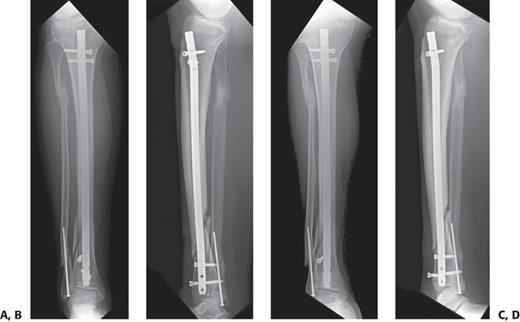
A, B: Radiographs showing a healing tibia fracture after IM nailing. C, D: One month later a fractured distal interlock confirms fracture nonunion.

: A comminuted clavicle shaft fracture treated with rigid fixation (B, C) yielded fracture healing without callus

Radiographs (A, B) and CT (C) showing atrophic nonunion 6 months after nonoperative management of a humeral shaft fracture.
After debridement of the nonunion, healthy long oblique surfaces without significant defect existed. D, E: Repair proceeded with ORIF without any bone graft, despite the atrophic nature of the nonunion. F, G: Six months after nonunion repair the fracture is united.

Plates can be used to treat nonunions at almost any part of any long bone.
A: A proximal femur nonunion was repaired with a proximal femoral locking plate with adjuvant ICBG and an intramedullary fibular strut. Nonunions of the midshaft (B) and distal (C) portions of the femur were repaired with distal femoral locking plates

A: An open distal femur fracture treated with debridement and lateral plating resulted in a large segmental defect. High varus stresses on the eccentrically placed plate (B) resulted in plate fracture prior to union despite bone grafting (C). D:Nonunion repair with revision plating and additional autologous bone graft led to fracture union.

Exchange nailing of a malaligned tibia nonunion.
A: An undersized nail was used to treat an open tibial shaft fracture leading to an atrophic nonunion in slight valgus alignment. B: Exchange nailing was performed without specific consideration of the malalignment resulting in an almost identical amount of valgus. C: A persistent nonunion, although now oligotrophic, with fractured interlocking screws has resulted.

An AP pelvis shows nonunion of an intertrochanteric femur fracture with substantial limb shortening. B: Nonunion that is confirmed with a CT. C: The nonunion and limb shortening are both managed with total hip arthroplasty.

comminuted pilon fracture (A) was initially treated with ORIF (B, C) and adjuvant ceramic bone void filler.
D, E: Nine months after ORIF, the patient was referred for management of nonunion associated with substantial ankle arthrosis, plate fracture, and varus malalignment. Staged management was initially with removal of hardware, debridement, and multiple cultures. F, G: After final negative cultures, reconstruction was with ankle fusion using ICBG harvested with acetabular reamers and expanded with allograft cancellous chips and a hindfoot fusion nail. H, I: Six months later, the fusion is nicely consolidated.

An infected nonunion of the distal femur (A) is treated with removal of failed implants, debridement, and implantation of an antibiotic-impregnated cement spacer according to the Masquelet technique (B).
C: Healing is accomplished after appropriate antibiotic therapy followed by nonunion repair with removal of the cement spacer, iliac crest bone grafting of the defect, and plate stabilization.

Schematic diagram of monofocal Ilizarov lengthening technique.
Increased length (L2–L1) is accomplished by increasing distance at one location (D2–D1
Schematic diagram of bifocal Ilizarov technique


An infected femoral nonunion is managed initially with removal of implants (A), debridement, and insertion of an antibiotic-impregnated IM “nail” (B).

درمان نان یونیون با وزن کذاری نسبی
A: AP radiograph showing a nonunion in a 27-year-old man 6 months after intramedullary nailing of a mid-diaphyseal femur fracture. At this point, the patient was fully weight bearing with minimal pain and an ultrasound external bone stimulator was applied 20 minutes daily. B: Six months later and without further surgical intervention, the nonunion is healed.