06sensorimotor system
 Call Kai
 Learn
 Practice Test
 Spaced Repetition
 Match
 Flashcards
 Knowt Play
1/31

There's no tags or description
Looks like no tags are added yet.
32 Terms
[A] WHat are the main functions of the sensorimotor system and what does it guide?
Main functions
movement
Posture, balance
Communication
Guide
internal representation of self and world
Detect changes in the environment (external&internal)
[A] What are the three fundamental principles of sensorimotor function?
Senso system = Hierarchical Organized: Association cortex operates at the highest level; muscles and receptors operate at the lowest level.
Motor output = guided by Sensory Input: Sensory feedback plays an essential role in controlling and refining movement.
Learning Alters Locus of Control: Experience and practice shift control from conscious, cortical levels to automatic, lower levels of the hierarchy.
[A] What are the four structural features of the general model of the sensorimotor system?
Hierarchical structure
Functional segregation
Parallel descending pathways
Feedback circuits
[A] What are the main tasks, inputs, and outputs of the Posterior Parietal Association Cortex (PPC)?
Main Tasks: Integrates spatial information for localizing body parts to be moved and external objects in space ("Where am I?" and "Where is the object I want to interact with?").
Main Inputs: Visual, auditory, and somatosensory systems.
Main Outputs: Secondary motor cortex, Dorsolateral prefrontal association cortex, and Frontal eye field (eye movement control).

[A] What does damage may lead to at the posterior parietal association cortex?
deficits in perception and Memory of spatial relationships
Inaccurate reaching and grasping
Problems with the control of eye movement
Attention deficits
[A] Compare Reflexes and Voluntary Actions across the 6 key dimensions from the lecture (Link to stimulus, Time delay, Synapses, Action, Response, Neuronal structures).
Link to stimulus: Reflex = Always | Voluntary Action = Sometimes
Time delay: Reflex = Short | Voluntary Action = Long
Synapses: Reflex = Few | Voluntary Action = Many
Action: Reflex = Simple | Voluntary Action = Complex
Response: Reflex = Stereotypical | Voluntary Action = Variable
Neuronal structures: Reflex = Spinal cord | Voluntary Action = Brain

[A] What two major clinical deficits result from damage to the Posterior Parietal Association Cortex?
Apraxia: A disorder of voluntary movement where patients have difficulty executing specific movements when requested, yet can perform them automatically in natural contexts e.g. wipe mouth (not due to paralysis (single motor deficit), aphasia (deficit in comprehension), or lack of motivation (e.g. fatigue, depression)).
Contralateral Neglect: A disturbance in the ability to respond to stimuli on the side of the body opposite (contralateral) to the brain lesion ("one side of the world does not exist"). Often accompanied by anosognosia (lack of awareness of the deficit)
[A] What is the main task, input, and output of the Dorsolateral Prefrontal Association Cortex (DLPFC)?
Main Task: Initiation of voluntary movements; mediates higher cognitive functions (working memory, decision rules, movement prediction).
Main Input: Posterior parietal association cortex.
Main Output: Secondary motor cortex, primary motor cortex, and frontal eye field.

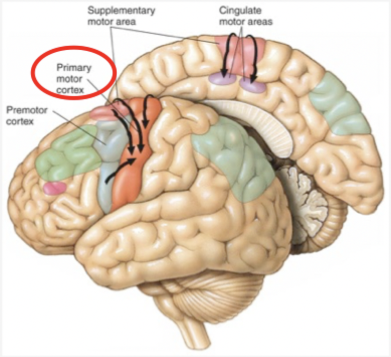
[A] What are the three sub-areas of the Secondary Motor Cortex, and what is its primary function?
3 Sub-Areas:
Supplementary Motor Area (SMA)
Premotor Cortex
Cingulate Motor Areas
Primary Function: Receives general instructions from association cortex and programs specific, complex patterns of movements.
Main Input: Association cortex
Main Output: Projects mainly to the Primary Motor Cortex

[A] What are Mirror Neurons, where were they discovered, and what is their proposed significance?
Definition: Specialized neurons that fire both when an individual performs a specific goal-directed movement OR when they observe another individual performing the same goal-directed movement.
Discovery: Discovered in the early 1990s by Giacomo Rizzolatti in the premotor cortex of macaque monkeys.
Proposed Function: Thought to underlie social cognition, action understanding, and empathy.

[A] Where is the Primary Motor Cortex located, and what are the effects of lesions?
Location: Precentral gyrus of the frontal lobe (major point of convergence for cortical sensorimotor signals).
Elicits movements
No direct connection to muscle but via spinal cord
Lesion Effects: Disrupts fine independent digit control(e.g. one finger), reduces movement speed, accuracy, and force of patient, and can cause astereognosis (inability to recognize objects by touch alone)
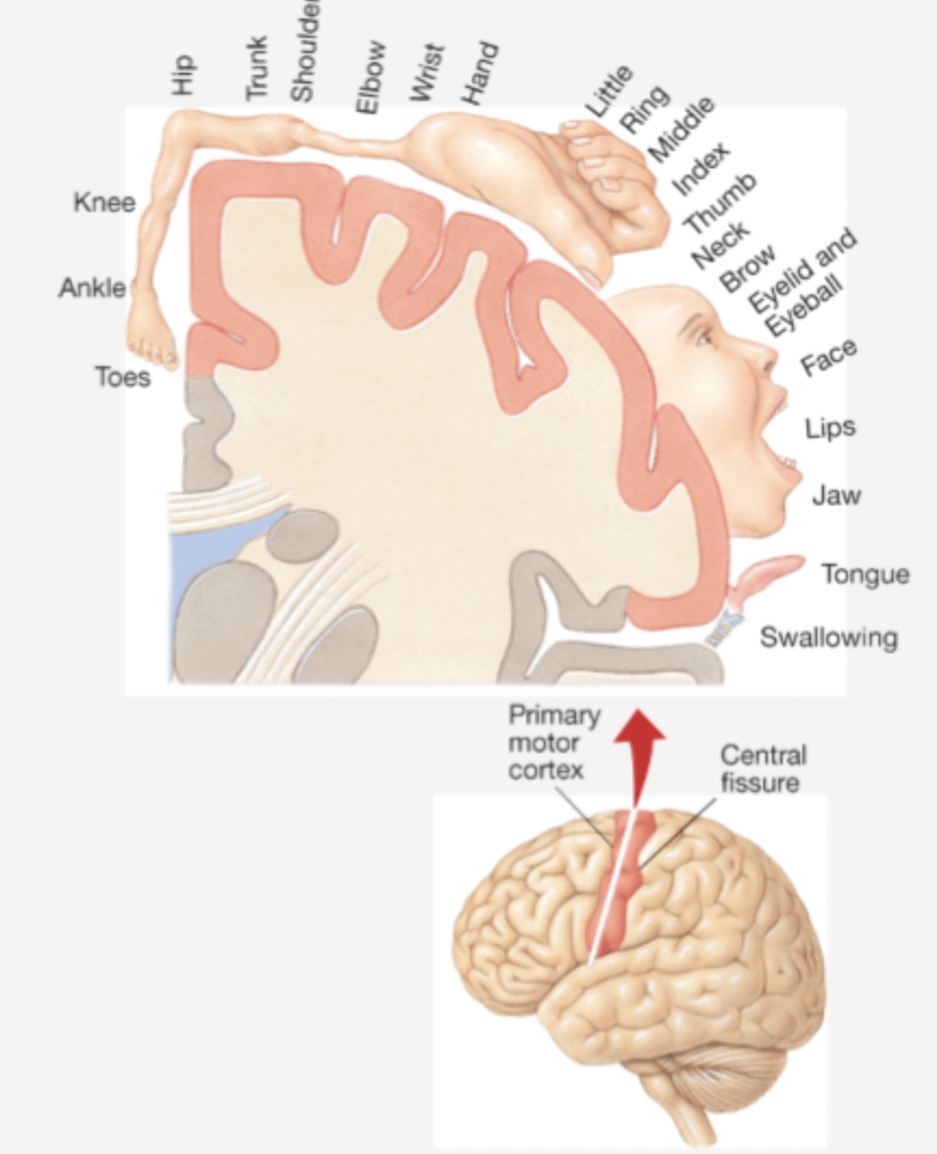
[A] How is the Primary Motor Cortex organized?
Motor Homunculus: Organized somatotopically; body parts capable of intricate, fine motor control (hands, face, mouth, tongue) are allocated disproportionately large cortical areas.
Electrical stimulation of various sites in the primary motor cortex elicits simple movements in the indicated parts of the body. (Penfield & Rasmussen)
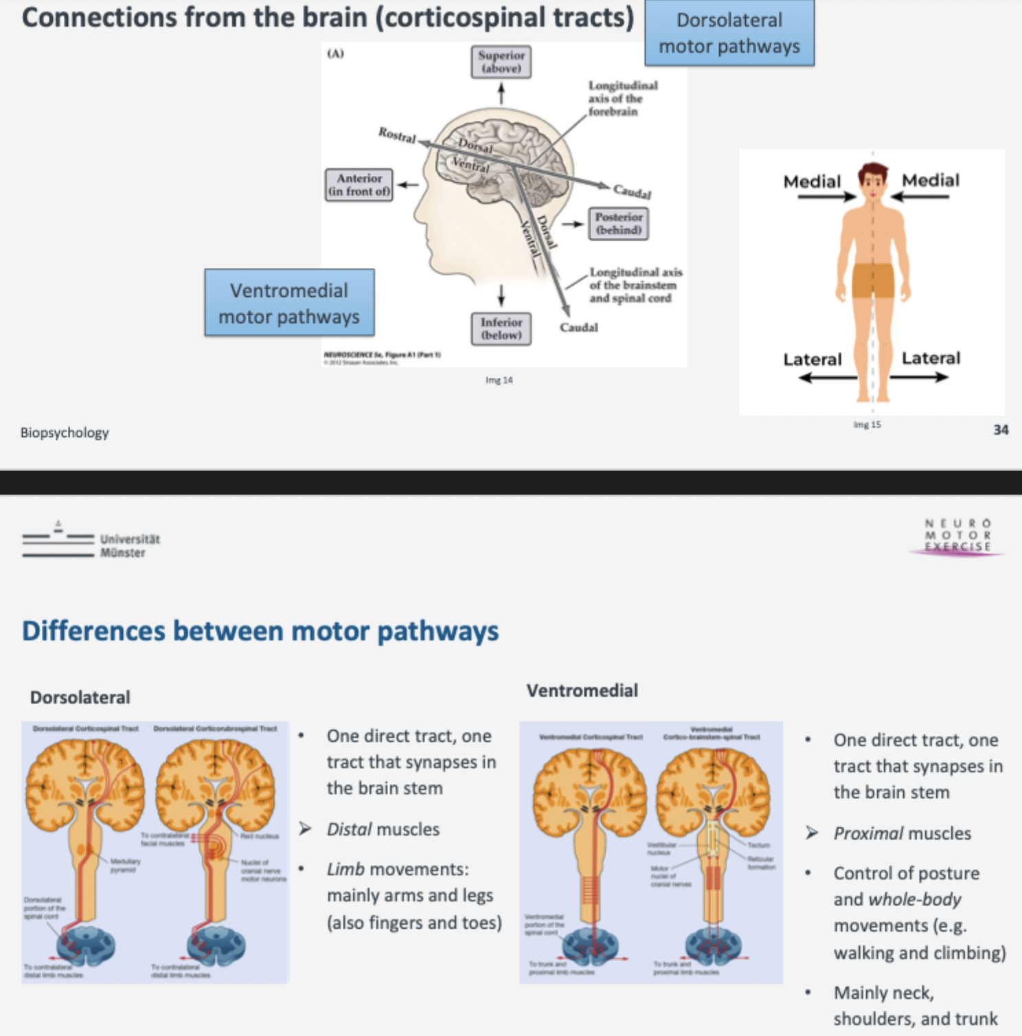
[A-ExQ]Compare the Dorsolateral and Ventromedial motor pathways regarding the body parts and movements they control.
Dorsolateral Pathways (Corticospinal & Corticorubrospinal):
Control distal muscles (limbs: arms, legs, fingers, toes).
Responsible for independent, fine limb movements. (Explicitly tested in sample exam questions!)
Ventromedial Pathways (Corticospinal & Cortico-Brainstem-Spinal):
Control proximal and trunk muscles (neck, shoulders, trunk).
Responsible for posture, balance, and whole-body movements (e.g., walking, climbing)
(Both: one direct tract, one tract that synapses in the brain stem)
[A] Compare Golgi Tendon Organs and Muscle Spindles in terms of location and what muscle property they monitor. (Receptor organs of tendons and muscles)
Golgi Tendon Organs: Embedded in muscle tendons; monitor muscle tension (provide CNS with that); act as a protective "brake" against excessively vigorous contractions to prevent muscle/tendon damage.
Muscle Spindles: Embedded within muscle tissue itself; monitor muscle length (stretch) (provide CNS with that); maintain muscle tone and convey information about body part positions

[A] Define Stretch Reflex, Withdrawal Reflex, and Reciprocal Innervation
Stretch Reflex (Knee-jerk / Patellar reflex): Monosynaptic reflex; tapping a tendon stretches muscle spindles → fires afferent sensory neurons → directly excites alpha motor neurons → muscle contracts.
Withdrawal Reflex: Polysynaptic reflex; painful cutaneous stimulus triggers rapid flexor muscle contraction to pull a limb away from harm.(e.g. stepping on glass)
Reciprocal Innervation: Neural wiring where excitation of an agonist muscle (e.g., flexor) automatically triggers inhibitory interneurons to relax the antagonist muscle (e.g., extensor)
[B] Innervation of muscle fibers (Motor Units/pool)
Motor units: a single motor neuron and all the individual skeletal muscle fibers it innervates
Motor pool: all alpha motor neurons that innervates a single muscle
Acetylcholine released at neuromuscular junction → activates motor end-plate → leads to contraction

[B] Contrast fast vs. Slow skeletal muscle fibers
Fast Muscle Fibers: Contract and relax quickly, generate great force, fatigue rapidly, poorly vascularized (few blood vessels).
Slow Muscle Fibers: Contract slowly, capable of sustained contraction for long periods, fatigue-resistant, highly vascularized
Note: Individual percentages of ft and st fibers vary between people depending on genetics and training
[B] What are two primary therapeutic approaches used to treat contralateral neglect caused by posterior parietal cortex damage?
Visual Scanning Therapy: Systematic visual search training (e.g., digital/tablet-based exercises) that forces the patient to actively direct their gaze and attention toward the neglected (contralateral) side of space.
Sensory Stimulation of the Affected Side: Providing targeted visual, auditory, or tactile input on the neglected side (e.g., speaking to the patient from their affected side or presenting objects/pictures there) to draw conscious awareness back to that half of space.
[A] What is the difference between the anatomical terms "contralateral" and "ipsilateral"?
Contralateral: Located on or affecting the opposite side of the body relative to a structure or brain lesion (e.g., a right hemisphere lesion causing neglect of stimuli on the left arm/leg).
Ipsilateral: Located on or affecting the same side of the body (e.g., right arm and right leg)
[C] Types of muscle
All animal movement depends on muscle contraction
3 types
cardiac
Skeletal
Smooth

[B] Why is the distinction between reflexive (involuntary) and voluntary action considered a continuum rather than a strict binary?
Differentiation between non-reflexive vs. Reflexive is sometimes blurry
Continuum Characteristics: Many movements contain both reflexive and voluntary elements.
Examples: Swallowing can be initiated voluntarily or triggered reflexively (and inhibited only within limits); walking is initiated voluntarily, but individual adjustments for bumps in the road or arm-swinging occur automatically
[A] Describe the direct and indirect pathways of the Basal Ganglia
Structures: Caudate + Putamen (= Striatum), Globus Pallidus, Substantia Nigra, Subthalamic Nucleus.
Connection
input from Cortex (frontal, prefrontal and parietal)
Output via motor thalaus to cortex (especially SMA)
Function
Direct Pathway: Disinhibits the motor thalamus → enhances / facilitates desired movement.
Indirect Pathway: Inhibits the motor thalamus → suppresses unwanted movement.

[A-ExQ] Basal Ganglia and their involvement in Parkinson's and Huntington's disease.
Disorders:
Hypokinetic (Parkinson's Disease): Loss of dopaminergic neurons in Substantia Nigra → increased inhibition of motor thalamus → rigidity, tremor, difficulty initiating movement. (Tested in sample exam question!)
Hyperkinetic (Huntington's Disease): Degeneration of striatal neurons → decreased basal ganglia output → overactivity of motor thalamus → involuntary, uncontrollable movements (chorea)
[A] Main tasks of the Basal ganglia
Selection and initiation of voluntary movements
Modulatie motor output → e.g. sequencing of movements
Involved in cognitive and emotional function
Reward based learning (skills and habits)
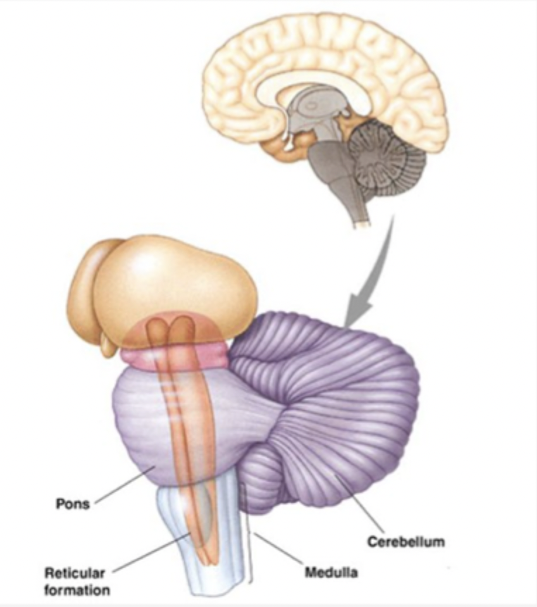
[A] What is the primary function of the Cerebellum?
Primary Function: Acts as a predictive model / comparator; compares intended motor commands (from motor cortex and brainstem) with actual sensory/vestibular feedback to correct ongoing movements and drive motor adaptation (specific form of motor learning)
Volume/Neurons: Contains ~10% of total brain volume but ~50% of all neurons.
[A] What symptoms result from cerebellar damage?
Damage may lead to: loss of precise direction/force/amplitude control, and loss of the ability to adapt to patterns of motor output to changing conditions
difficulites in standing, balance and gait disturbances, slurred speech, and inaccurate eye movements (tested clinically via finger-to-nose or tandem gait tests)
[A] What Symptoms of cerebellar damage resemble those of alcohol intoxication?
Clumsiness, slurred speech, inaccurate eye movements, difficulties which balance and coordination
in fact, alcohol interferes with regulat cerebellum activity
Chronic alcohol consumption can leave long term effects on the cerebellum
[B] Basal Ganglia and Cerebellum
Subcortical systems that interact with different levels of the sensorimotor hierarchy
[B] What are reflexes and what functions to they have?
reflexes are automatic, subconscious responses to changes within or outside the body
Functions
maintain homeostasis: breathing, blood pressure regulation, and heartbeat
Carry out automatic things we don’t need to think about to perform (swallowing, sneezing, coughing, vomiting)
Maintain our posture and trunken balance
! Some reflexes occur only in specific periods of development (newborn/infant reflexes)!
[A] What are the three sources of input to an alpha motor neuron in the spinal cord?
Sensory input from muscle spindles: Provides feedback regarding muscle length/stretch (essential for spinal stretch reflexes).
Input from upper motor neurons in the brain: Originates from motor cortex and brainstem to initiate and control voluntary movement.
Input from spinal interneurons: Mediates spinal reflex programs, reciprocal innervation, and motor pattern generation (can be excitatory or inhibitory)

[A] What are antagonistic muscles, why are they necessary, and how do flexor and extensor muscles differ?
Definition: Opposing sets of muscles that act in opposition to one another across a joint.
Why Needed: Moving a leg or an arm in more than one direction must be controlled by more than one muscle because muscles can only contract (pull), not push.
Flexor Muscle: Flexes or raises the limb (e.g., biceps flexing the arm).
Extensor Muscle: Extends or straightens the limb (e.g., triceps extending the arm)

[B] What are synergistic muscles, and what function do they serve in motor execution?
Definition: Any two muscles whose contraction produces the same movement, whether flexion or extension.
Function & Necessity: Synergist muscles stabilize muscle movements to keep them even, safe, and desired (acting as "helping hands", e.g., the brachioradialis and brachialis assisting the biceps brachii)